


Introduction
In his second major work, Salammbô, Flaubert (Reference Flaubert1862) portrayed the Carthaginians as a heartless people who sacrificed their children to gain favour with their gods Tanit and Ba'al Hammon. In this fictional account, a priest places these innocents—tied hand and foot, and cloaked to mask the horror ahead—first individually, and then en masse, in the hands of a huge brass statue of Ba'al, whose arms are then raised until the bodies fall into a pyre between its legs. Throughout, musicians play loudly to smother the wails of the victims. Flaubert's critics chastised him for embracing Graeco-Roman tales of rampant Carthaginian infant sacrifice too literally (Gras et al. Reference Gras, Rouillard and Teixidor1991).
Flaubert's scenario preceded, by 59 years, the first interpretation of urns bearing the burnt remains of humans and animals from a distinct cemetery (the ‘Tophet’) at the Carthaginian city of Motya (Motzia), Sicily, as evidence of sacrifice (Whitaker Reference Whitaker1921), which Poinssot (Poinssot & Lantier Reference Poinssot and Lantier1923) then applied to the Carthage Tophet.
Prominent French scholars rejected Whitaker's claim, which they saw as derivative of Flaubert and his reliance on what they considered to be inaccurate Graeco-Roman descriptions of Carthaginian infant sacrifice (Gras et al. Reference Gras, Rouillard and Teixidor1991). As Saumange wrote in 1922 (translated and quoted in Gras et al. Reference Gras, Rouillard and Teixidor1991: 151):
The imagination of the public, haunted by Flaubert's memory, has promptly dramatized the discovery: these children [. . .] are the victims of cruel holocausts which Carthage offered to Moloch. This is an imprudent and grave step to take lightly. Imprudent because it is important to know the excavation perfectly and in all details before advancing such a thing even hypothetically. Grave because one compromises the rehabilitation which the religious reputation of Carthage has benefited from among a good number of our best historians.
May we be permitted to ask ourselves whether the object of the wish was not simply to erect the image of the [. . .] god himself, and whether the presence of ashes of children could not have been intended to render the place of the betyl [sacred stone] forever untouchable, by burying bones.
In the end, however, Poinssot and Lantier (Reference Poinssot and Lantier1923) succeeded in suppressing interpretations of Tophets that argued against sacrifice as the sole cause for the presence of human remains in the Carthage Tophet and others.
Although an ‘all-sacrifice’ conception of Tophets was not universally accepted (Harden Reference Harden1927; Soliel et al. Reference Soliel, Muller and Richard1958; Richard Reference Richard1961), this interpretation has garnered a following that seeks support from several sources: Graeco-Roman tales by Diodorus and others; the reinterpretation of grave-marker inscriptions as parental vows to sacrifice their offspring to Ba'al and Tanit; the argument that animals were sacrificed and therefore humans were too; the interpretation of images on a particular stele as representing a priest carrying a sacrificial infant; and claims that only sacrifice warranted the use of scarce plant resources for pyres (see references in Smith et al. Reference Smith, Avishai, Greene and Stager2011, Reference Smith, Stager, Greene and Avishai2013; also Xella 2010; Quinn Reference Quinn and Gruen2011; Xella et al. Reference Xella, Quinn, Melchiorri and van Dommelen2013; Stager Reference Stager2014).
There is also significant support for a ‘not-all-sacrifice’ hypothesis, which does not rule out sacrifice, but takes into consideration other factors (see references in Schwartz et al. Reference Schwartz, Houghton, Macchiarelli and Bondioli2010, Reference Schwartz, Houghton, Bondioli and Macchiarelli2012): Tophets lie outside city limits and house the remains of prenatal, neonatal and young postnatal humans, which are absent from the main cemeteries in city centres; only Tophet burials are cremations; and the age range of humans buried at Tophets is consistent with natural causes of death. As for the ‘seminal’ stele (see Smith et al. Reference Smith, Avishai, Greene and Stager2011: fig. 1d), the fully outlined, erect adult figure (with ear, open eye, upright shoulders, upraised right arm with forward-facing hand, distinct manual digits and a bent right leg, well forward of the left leg) contrasts with the minimally outlined small individual that it holds (with down-turned, featureless head, torso, slumped shoulder and a curved line hinting at a limp, handless left arm). Observed without preconception, the adult holds an inanimate (deceased, perhaps cloaked) infant.
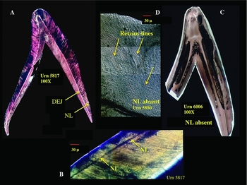
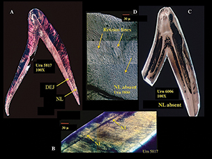
Figure 1. Presence vs absence of neonatal lines in deciduous incisors from the Carthage Tophet. A) At 100×, the dentino-enamel juncture is more visible than the neonatal line, which is clearly identifiable at 30μ (B). C & D) Neonatal line absent; D illustrates the uninterrupted field of Retzius lines (incremental growth lines in the enamel). Note the level of magnification required to visualise these structures.
Nevertheless, proponents of an ‘all-sacrifice’ interpretation reject any alternatives, reiterating that Graeco-Roman depictions of specific events actually reflect a widespread Carthaginian practice; that their interpretations of the stele are correct; and that the presence at other Tophets of the cremated remains of humans who ‘must’ have been sacrificed also means that the Carthage Tophet humans were sacrificed (Smith et al. Reference Smith, Avishai, Greene and Stager2011, Reference Smith, Stager, Greene and Avishai2013; also Xella 2010; Quinn Reference Quinn and Gruen2011; Xella et al. Reference Xella, Quinn, Melchiorri and van Dommelen2013; Stager Reference Stager2014). Furthermore, they denounce alternative theories as ‘revisionist’ (Lancel Reference Lancel1995; Quinn Reference Quinn and Gruen2011; Stager Reference Stager2014).
Prior osteological analyses of Carthage Tophet urn contents
In 1922 and then 1923, Graeco-Roman accounts of infant sacrifice at Carthage seemed validated when de Prorok and colleagues, and Poinssot and Lantier, respectively, discovered urns at the Carthage Tophet containing burnt bones, most of which were identified as belonging to human infants or children (Stager & Wolff Reference Stager and Wolff1984).
Although Kelsey (Reference Kelsey1926) assumed that most of the Carthage Tophet urns he excavated (more than 1000) contained human remains, neither he nor his successor Harden (Reference Harden1927) claimed that the Carthaginians engaged in rampant, sanctioned infant sacrifice. Between 1934 and 1936, Chabot and Lapeyre unearthed more than 1000 urns at the Tophet, most of which they assumed to contain the remains of either one or two human infants and/or children, or a young human and some animal remains (Lapeyre & Pellegrin Reference Lapeyre and Pellegrin1942).
After Charles-Picard and Cintas (Charles-Picard Reference Charles-Picard1945) excavated the Carthage Tophet, they gave forensic expert Richard (Reference Richard1961) the contents of 42 urns. Upon combining these remains with those from 138 urns from the Hadrumentum Tophet, Richard identified one or more humans in 88 urns, human and lamb in 59, and lamb in 29.
What the bones tell us
In 1976, L.E. Stager, director of the team excavating the Tophet, invited one of the authors here (Schwartz) to oversee both the on-site processing and preliminary assessment, and the subsequent detailed laboratory analysis, of the contents of 348 urns from the Carthage Tophet (dated to around the early eighth century to 146 BC) (Schwartz Reference Schwartz1993). Using the accepted MNI approach (minimum number of individuals as reflected in the number of the same tooth or skeletal element; Hesse & Wapnish Reference Hesse and Wapnish1985), Schwartz and Houghton documented evidence of 540 humans. Yet only urns with a MNI of one or two housed sufficient skeletal material to conclude that entire individuals were represented (Schwartz et al. Reference Schwartz, Houghton, Macchiarelli and Bondioli2010). When an urn's MNI indicated three or more individuals, there was insufficient skeletal material to argue for that number of entire individuals (e.g. there could be three incompletely represented individuals, or one or two skeletally well-represented individuals and only a few elements from other individuals).
As no single criterion can accurately assess age at death—nor in determining sex (Schwartz 2007)—Schwartz and Houghton used the standard multifactorial (multi-morphological/metric) approach (Fazekas & Kósa Reference Fazekas and Kósa1979; Lovejoy et al. Reference Lovejoy, Meindl, Mensforth and Barton1985; Schwartz 2007). Specifically, we (Schwartz et al. Reference Schwartz, Houghton, Macchiarelli and Bondioli2010) based estimates of age at death on: the state of crown and root formation (teeth being the most frequently preserved and analysable elements); age-related changes in basisphenoids and petrosals; and measurements of petrosals, lateral occipital parts, basiocciputs, pubes and ischia, which shrink minimally, if at all, when cremated (Krogman Reference Krogman and Levinson1949). Through numerous re-analyses, we refined the percentages of individuals in each age category.
From accepted age-estimation criteria using tooth and cranial-bone formation, we concluded that pre-/peri-/neonates together constituted ≥50% of the sample (Schwartz et al. Reference Schwartz, Houghton, Macchiarelli and Bondioli2010). Ages based on basicranial- and pelvic-bone measurements were determined by comparison with Fazekas and Kósa's (Reference Fazekas and Kósa1979) measurements of these bones in individuals of known age. We sought to avoid the criticism that cremated bones may have shrunk by incrementally increasing each linear measurement to allow for the possibility of 25% shrinkage, well beyond the shrinkage recorded for experimentally cremated human or animal remains (see references in Schwartz et al. Reference Schwartz, Houghton, Macchiarelli and Bondioli2010). Even at 25% shrinkage, each measurement classified some number of individuals as ‘prenatal’.
From these ageing criteria, we concluded that 24% were prenatal, 15% peri-/neonatal, and 17% less than 1 month of age. We tested these results via neonatal line (NL) analysis.
Identifiable in enamel only at high magnification (microns), a NL presents as a distinct, morphologically homogeneous line devoid of cross striations (Figure 1). It reflects a stress-induced disruption of enamel deposition coincident with the transition from in- to ex-utero, which may not correspond to full-term birth. NLs are always present in deciduous teeth (Antoine et al. Reference Antoine, Hillson and Dean2009), which begin developing by the twentieth intrauterine week (Antoine et al. Reference Antoine, Hillson and Dean2009; also Ten Cate Reference Ten Cate and Cate1989). Since first permanent molar crowns (lower M1 or upper M1) mineralise late in the third trimester, their NLs lie closer to the dentino-enamel juncture (DEJ) (Antoine et al. Reference Antoine, Hillson and Dean2009). Given the periodicity of enamel deposition and the lag between a stress and a response to it, an individual must survive around 7, or even 10–15, post-uterine days for a NL to form (Schwartz et al. Reference Schwartz, Houghton, Macchiarelli and Bondioli2010).
For NL analysis, Schwartz and Houghton sent co-authors Bondioli and Macchiarelli 50 deciduous tooth crowns from the ‘perinatal’ category (Schwartz et al. Reference Schwartz, Houghton, Macchiarelli and Bondioli2010). Twenty-six crowns lacked a NL. In the 24 crowns with NLs, enamel thickness indicated survival of at least two weeks post-NL-formation. Upon comparing NL with our morphological/metrical age estimates (M) of the same individuals, M>NL in only 10% of the sample. As M typically equalled or over-estimated NL-derived ages, we concluded that M-based age estimates were reliable (Schwartz et al. Reference Schwartz, Houghton, Macchiarelli and Bondioli2010). Taking everything into account, we concluded that: “the Carthaginian Tophet, and by extension other Tophets, were cemeteries for the remains of human prenates and infants who died from a variety of causes” (Schwartz et al. Reference Schwartz, Houghton, Macchiarelli and Bondioli2010: 10).
When our analyses consistently identified pre- and neonates, Stager demanded return of the sample, which he sent to P. Smith (Stager Reference Stager2014). Subsequently, Smith, Stager and others have since dismissed our work through misrepresentation of it, and have also rejected our results and defended the ‘all-sacrifice’ theory through erroneous assumption and incorrect criteria. Given that these allegations and misinformation appeared in this journal, we present the reader with the relevant background, arguments and correct analytical information.
A refutation?
Smith et al. (Reference Smith, Avishai, Greene and Stager2011: 860–61) not only mischaracterised us (Schwartz et al. Reference Schwartz, Houghton, Macchiarelli and Bondioli2010) as asserting that the Carthage Tophet was solely “a cemetery for the burial of aborted or stillborn infants”, but also criticised us for not accounting for tooth-crown and bone shrinkage, for estimating age primarily on the basis of an inappropriate combination of petrosal length and width measurements, and for using questionable long-bone measurements and NL analysis to estimate age. They also dismissed our analysis because the percentages of identified pre-/perinates were not the same in all cranial and pelvic-bone metric analyses. As most bioarchaeologists know, however, multiple criteria will not yield exactly the same estimate, but, together, they provide a more realistic approximation of age than using one criterion. Smith et al. (Reference Smith, Stager, Greene and Avishai2013) subsequently reiterated these objections and defended their age estimates based on tooth-crown height corrected for purported shrinkage, which identified most individuals as one to two postnatal months of age. From this, they asserted that the Carthage humans were alive and available for sacrifice. We address these and other ‘bones of contention’ individually.
Only sacrifice can account for individuals in the Carthage Tophet
Regardless of whether some Carthage Tophet humans were sacrificed, it does not preclude death by natural causes (Schwartz et al. Reference Schwartz, Houghton, Bondioli and Macchiarelli2012): for example, spontaneous abortion, which is common enough today for perinatologists to refer to it as ‘reproductive wastage’ (Durfee Reference Durfee and Wharton1987). Furthermore, if we accept that sanitary conditions at Carthage were as poor as at contemporaneous Pompeii, Ostia and Rome (Scobie Reference Scobie1986), all Carthaginians (pregnant women included) were susceptible to cholera, dysentery, gastroenteritis, infectious hepatitis, leptospirosis, typhoid and parasitic intestinal infestations, most of which result in severe dehydration, which remains a primary cause of peri- and postnatal death (Behrman & Shiono Reference Behrman, Shiono, Fanaroff and Martin1997). Also relevant to considering infant mortality are: low birth weight (Behrman & Shiono Reference Behrman, Shiono, Fanaroff and Martin1997); severe viral infections and malaria leading to premature birth and perinatal mortality; infectious diseases (smallpox, vaccinia, listeriosis) resulting in stillbirth; and non-infectious diseases (cholestasis, hypertension, toxemia, renal disease) causing stillbirth, abortion and preterm delivery (Taylor & Pernoll Reference Taylor, Pernoll, Pernoll and Benson1987). As these are today major causes of peri- and postnatal death, they must have constituted natural—and probably more prevalent—elements of Carthaginian life. As Becker (Reference Becker, Lally and Moore2011: 24) commented in light of the high incidence of perinatal mortality in non-industrial societies and its unexpected increase in modern industrialised societies: “The last months of a pregnancy, the process of parturition, and various stresses during the months after birth provide a frightening trio of challenges that lead to [. . .] ‘infant mortality’”.
Bone shrinkage
Our age estimates (pace Smith et al. Reference Smith, Stager, Greene and Avishai2013) did not derive primarily from measurement of the petrosal bone and an inappropriate combination of its length and width. We did measure the petrosal, but we also measured other cranial and several pelvic bones, and compared each measurement individually to those in Fazekas and Kósa's (Reference Fazekas and Kósa1979) ageing tables. Moreover, even though these bones shrank little, if at all, when we incrementally increased our measurements to compensate for an unrealistic shrinkage of 25%, prenates were still identified. We did not measure long bones.
Tooth-crown shrinkage
Even if cremated teeth shrink, their morphology and relative states of development do not change—a fact that others have used when comparing Carthage Tophet crowns with unburned crowns (e.g. Smith et al. Reference Smith, Avishai, Greene and Stager2011, Reference Smith, Stager, Greene and Avishai2013; Figure 2B). Thus, the estimates we achieved using relative states of crown formation are viable (Schwartz et al. Reference Schwartz, Houghton, Macchiarelli and Bondioli2010).

Figure 2. Shrinkage and neonatal lines. A) Developing teeth from the Carthage Tophet illustrating unaltered crown shape via continuity between mature (smooth) and maturing (uneven, due to loss of carbonate and water, not shrinkage) enamel. B) A Carthage Tophet molar (left) and an uncremated molar (right), which Smith et al. (Reference Smith, Stager, Greene and Avishai2013) correctly identified as being at the same developmental age, thereby demonstrating that heat does not affect tooth morphology or relative states of development; note continuity of mature and maturing enamel on both specimens. C) Dentino-enamel junctures of Carthage Tophet and uncremated molars; Smith et al. misinterpreted the difference in dentino-enamel juncture clarity as indicating heat-induced elimination of the neonatal line. D) The dentino-enamel junctures of Carthage Tophet molars and uncremated molars; the dark band delineates the dentino-enamel juncture, which Smith et al. (Reference Smith, Avishai, Greene and Stager2011) misidentified as a neonatal line. The 2mm scale Smith et al. used is insufficient to demonstrate neonatal line presence/absence. A, C & D are from Smith et al. (Reference Smith, Avishai, Greene and Stager2011); B is from Smith et al. (Reference Smith, Stager, Greene and Avishai2013). Black and white (B) and blue and white labelling (C, D) added by present authors.
Although Smith et al. (Reference Smith, Stager, Greene and Avishai2013) cited Krogman (Reference Krogman and Levinson1949) as demonstrating marked heat-induced shrinkage, he actually questioned whether this occurred, as unerupted, jaw-embedded developing teeth (as most of the Carthage Tophet teeth were) are less susceptible to the effects of heat than erupted teeth, which crack and/or split (see also Krogman 1962). They also claimed (Smith et al. Reference Smith, Avishai, Greene and Stager2011, Reference Smith, Stager, Greene and Avishai2013) that both Shipman et al. (Reference Shipman, Forster and Schoeninger1984) and Buikstra and Swegle (Reference Buikstra, Swegle, Bonnischen and Sorg1989) demonstrated heat-induced crown shrinkage. Only Shipman and colleagues cremated teeth experimentally, however, and these were erupted sheep molars, which differ from enamel-capped human teeth in having vertical plates of enamel separated by occlusally exposed fields of soft dentine. Not unexpectedly, it was not the crystalline enamel but the hydrated dentine that shrank.
Smith et al. (Reference Smith, Avishai, Greene and Stager2011) compared crown heights of specimens from the Carthage Tophet with similarly formed, unburned teeth from an unidentified sample. As the Carthage crowns were 0.6mm shorter, they increased their age estimates by around four weeks. Human populations differ markedly in their tooth dimensions (Coughlin Reference Coughlin1967; Yuen et al. Reference Yuen, So and Tang1997; Hanihara & Ishida Reference Hanihara and Ishida2005; Anfe et al. Reference Anfe, Arakaki, Nakamura and Viera2012), however, so any effect that heat might have on tooth size can be determined only by measuring the same teeth, pre- and post-cremation, as Deutsch and colleagues (Deutsch & Shapira Reference Deutsch and Shapira1987; Mayer et al. Reference Mayer, Schneider, Sydney-Zax and Deutsch1990; Sydney-Zax et al. Reference Sydney-Zax, Mayer and Deutsch1991) and Soliel et al. (Reference Soliel, Muller and Richard1958) did. These studies revealed that shrinkage is at best negligible—a fact that Soliel et al. used to identify pre- and perinates in their Carthage Tophet sample. As for Deutsch et al.’s studies, crown weight decreased as a result of loss of water and carbonates (see Figure 2A & B). (See also Smith et al. Reference Smith, Stager, Greene and Avishai2013 for incorrectly comparing measurements of cremated teeth from some, with uncremated teeth from other, studies as demonstration of shrinkage.)
Correcting for their inflation of ages, it is striking that Smith and colleagues’ mortality distribution is virtually identical to ours when ‘fetal’ and ‘perinatal’ categories are combined (Figure S1 in online supplementary material (OSM)). This is consistent with present-day fetal/infant mortality profiles (Chalmers & Macfarlane Reference Chalmers, Macfarlane and Wharton1980; Taylor & Pernoll Reference Taylor, Pernoll, Pernoll and Benson1987; Saunders & Barrans Reference Saunders, Barrans, Hoppa and Fitzgerald1999): in other words, a large proportion of spontaneously aborted or stillborn fetuses (Durfee Reference Durfee and Wharton1987), and of peri- and neonates, who are at serious risk of death during the first two postnatal weeks (Chalmers & Macfarlane Reference Chalmers, Macfarlane and Wharton1980) (also see our NL estimates above).
Neonatal lines
Smith et al. (Reference Smith, Avishai, Greene and Stager2011, Reference Smith, Stager, Greene and Avishai2013) claimed that heat can alter the internal structure of teeth and eliminate evidence of NLs. They also claimed that NLs may be present in permanent, but not deciduous, teeth. From these assertions, they concluded that age estimates based on NL presence/absence are unreliable.
The magnifications used by Smith et al. are insufficient to identify NLs: i.e. millimetres rather than microns (1μ = 0.001mm). Consider their case for asserting that a NL can be present in uncremated, and absent in cremated, teeth. In their fig. 3d and 3e, they present a sectioned, uncremated M1 that purports to show a NL and a Carthage Tophet M1 that supposedly lacks this feature (see Figure 2C & D here). In all cases, however, Smith et al. illustrate the always-present DEJ, which can be identified at low magnification. In Figure 2C, the DEJ is better defined in the uncremated M1 than in the Carthage Tophet M1. In Figure 2D, the DEJ of the Carthage Tophet M1 appears as expected (as it also does in the specimens in Figure 2C), while in the uncremated tooth, separation of dentine and enamel created a dark band that Smith et al. incorrectly identified as a NL. To reiterate: NLs develop not between enamel and dentine, but in enamel, and cannot be visualised in magnifications as low as millimetres.
In support of their contention that deciduous teeth may lack NLs, Smith and colleagues (Reference Smith, Stager, Greene and Avishai2013: 1195) cite Antoine et al. (Reference Antoine, Hillson and Dean2009) as being able to “locate this line in only one of five teeth they examined”. Antoine et al. (Reference Antoine, Hillson and Dean2009: 49) actually wrote that NLs can always be identified in deciduous crowns, which begin to form months before birth, but may be difficult to locate in the less fully developed M1 crowns of pre-, peri- and neonates because they lie close to the DEJ. This accounts for their being able to identify a NL in only one of the five permanent molars that they analysed (a M1 vs 3 M1s and 1 M1). As we only scrutinised deciduous teeth for NLs, we stand by our results—26 of 50 crowns lacked a NL—which confirms the presence of some number of prenates.
Do other studies support the ‘all-sacrifice’ theory?
Richard (Reference Richard1961) analysed the contents of 42 urns from the Carthage Tophet together with remains from 138 urns from the Hadrumentum Tophet. From long-bone measurements representing 41 individuals, he identified 5 (12.2%) prenates (1 of 7.5 fetal months, and 4 of 8–9 fetal months), 20 (48.8%) perinates, and 16 (39%) postnates (9 of ≤1 month, 3 of 1–3 months, and 4 of 2–3 years). Conservatively, he suggested 1 prenate, 33 perinates and 7 postnates. Using developing teeth representing 147 humans, Richard identified 16 prenates (10.9%), 118 perinates (80.3%) and 13 postnates (8.8%; 6 of a few months of age, 7 of 3–6 years). His conservative estimate was 13 postnates, from 2–16 prenates, and the rest as perinates. Recognising that his results were consistent with expected mortality, Richard tentatively suggested that 5% were prenatal, 75% perinatal and 20% postnatal (pace Smith et al. Reference Smith, Avishai, Greene and Stager2011, Reference Smith, Stager, Greene and Avishai2013). As 26 teeth that we classified as ‘perinatal’ lacked a NL, Richard's perinatal category probably included prenates.
Based on a small number of bones from 16 Carthage Tophet urns, Gejvall (Reference Gejvall1949) suggested that 1 human was neonatal to 3 months of age, 7 were greater than 3 months, and 3 were 3–4 postnatal months. Given Gejvall's small sample, his conclusions are not inconsistent with ours (pace Smith et al. Reference Smith, Avishai, Greene and Stager2011, Reference Smith, Stager, Greene and Avishai2013).
Additionally, Muller et al. (Reference Muller, Depreux, Muller and Fontaine1952) analysed the contents of 44 Carthage Tophet urns and those of 31 urns from the Sousse Tophet. They identified humans in 32 urns, animals in 2 urns and commingled human/animal remains in 38 urns. They determined age using sphenoid development/coalescence, long-bone length, Haversian canal configuration and semicircular canal orientation: 65 individuals (around 87% of the sample) were near birth, 1 of 21 days, 2 of 30 postnatal days, and the rest of 2–3 postnatal months. Some of the 65 individuals were probably prenatal.
Recently, Xella (Reference Xella2009) defended the ‘all-sacrifice’ theory via his ‘new’ approach, which relied solely on classical Greek and Roman writings. Subsequently, Xella and colleagues (Xella 2010; Quinn Reference Quinn and Gruen2011; Xella et al. Reference Xella, Quinn, Melchiorri and van Dommelen2013) included the interpretations of archaeologists, historians and epigraphers, which, they concluded, tell the same story: Tophets were cemeteries for sacrificed children. Smith et al.’s claims fit their interpretation. They (Xella et al. Reference Xella, Quinn, Melchiorri and van Dommelen2013) rejected our results on the grounds that analyses by Docter et al. (Reference Docter, Smits, Hakbijl, Stuijts and van der Plicht2003), Melchiorri (Reference Melchiorri2010) and Ciasca et al. (Reference Ciasca, Di Salvo, Castellino and Di Patti1996) contradicted ours.
Docter et al. studied 6 Carthage Tophet urns. Using tooth and skeletal development, they identified 3 newborns in 1 urn, 2 newborns in each of 2 urns, 1 newborn in each of 2 urns, and one 6–9-year-old child in 1 urn. Given our results, it is probable that 3 complete individuals were not represented in that first urn, and that ‘newborn’ includes prenates.
Melchiorri analysed 72 Sulci Tophet (Sardinia) urns, and identified 52 humans, ranging in age from prenatal to 4–5 years. Of the 30 classified as neonatal, some were probably prenatal.
From the Motya Tophet, Ciasca et al. (Reference Ciasca, Di Salvo, Castellino and Di Patti1996) identified human remains in 132 urns, and comingled human and animal remains in 303 urns. They reported that the humans ranged in age from birth (the majority) to 6 months. For 112 of these individuals, they determined that 95 (84.8%) were neonates, 8 were 1 month of age, 4 were 2 months, 1 was 2–3 months, 3 were 4 months, and 1 was 6 months of age. ‘Neontatal’ undoubtedly included prenates.
Animal remains
Another defence of the ‘all-sacrifice’ theory is that as all Carthage Tophet animals must have been sacrificed, all humans must also have been sacrificed (see references in Smith et al. Reference Smith, Avishai, Greene and Stager2011, Reference Smith, Stager, Greene and Avishai2013; see also Xella 2010; Xella et al. Reference Xella, Quinn, Melchiorri and van Dommelen2013).
We identified lamb or kid alone in some urns, and lamb or kid, and infrequently bird and fish, commingled with human bones in others (Schwartz et al. Reference Schwartz, Houghton, Macchiarelli and Bondioli2010). Never, however, was there enough bone to suggest the interment of an entire animal (see Table S1 in OSM). This pattern is consistent with then contemporaneous practice: although an animal may have been killed sacrificially, only part of it (that was less desirable for eating) was burned as an offering to the gods; the rest was consumed (Detienne Reference Detienne, Detienne and Vernant1989). Given the skewed representation of animal skeletal elements, it appears that the circumstances leading to their presence in the Carthage Tophet differed from those involving humans.
Advocates of the ‘all-sacrifice’ theory also specify when this event occurred. Gejvall (Reference Gejvall1949) and Docter et al. (Reference Docter, Smits, Hakbijl, Stuijts and van der Plicht2003) thought that all lamb/kid remains from the Carthage Tophet represented spring-born neonates, and Stager (Reference Stager2014) extrapolated from this, and from the rare occurrence of commingled bird and human remains, to the notion of a regular springtime ritual. Our data, however, demonstrate that the low incidence of lamb/kid/bird remains, whether alone, or commingled with human bones (Table S1), cannot support the claim: if all humans were sacrificed, it was always in the spring. More broadly, given the number of potential natural causes of death (see above), it is unlikely that Carthaginians, whether or not interred in the Tophet, died only at certain times of the year.
Yet another justification of the ‘all-sacrifice’ theory?
As deforestation began with the founding of Carthage (van Zeist et al. Reference van Zeist, Bottema and van der Veen2001), it has been argued that nothing less profound than sacrificial cremation would warrant the use of dwindling wood resources otherwise needed for building ships and habitation (Smith et al. Reference Smith, Avishai, Greene and Stager2011, Reference Smith, Stager, Greene and Avishai2013). In reality, Carthage Tophet pyres typically comprised thin branches (Schwartz Reference Schwartz1993; Docter et al. Reference Docter, Smits, Hakbijl, Stuijts and van der Plicht2003), mostly from cultivated small trees (e.g. Prunus) and bushes (e.g. Ligustrum) (Docter et al. Reference Docter, Smits, Hakbijl, Stuijts and van der Plicht2003). Furthermore, charcoal of the largest plant present (Quercus) suggests that only scraps—probably left over from large-scale construction—were used (Docter et al. Reference Docter, Smits, Hakbijl, Stuijts and van der Plicht2003).
Conclusion
In summary, Tophets housed the remains of primarily pre-/perinates, newborns and children ≤5 years of age, and lay outside the city, while main cemeteries lay within city limits and typically contained humans ≥5 years. Only Tophet individuals were cremated and interred in urns. The disparity between urns in the presence of entire individuals, as well as often marked differences between urn contents in degree of incineration (including skeletal elements still in articulation), probably reflects differences at different times and on different occasions in acts of cremation and efforts to recover remains (Schwartz et al. Reference Schwartz, Houghton, Macchiarelli and Bondioli2010). Furthermore, cloth adhering to the internal side of a perinate's barely burned ilium suggests that, after cooling, bones were collected and either wrapped or placed in sacks before being placed in an urn (Schwartz Reference Schwartz1993; Figure S2 in OSM).
That Carthaginians maintained two different cemeteries is compatible with the Punic-derived, Roman-Carthaginian practice of not considering offspring as ‘persons’ until they had survived a certain number of years (Norman Reference Norman2002; Stuckey Reference Stuckey2009). Furthermore, as Becker (Reference Becker, Lally and Moore2011) reviews in detail from study of Cazzanello and other south Etrurian sites (e.g. Tarquinia), the burial of prenates, perinates and even children up to five years of age in cemeteries apart from the main cemetery was apparently commonplace in Etruscan culture.
From this perspective, rather than conceiving of the Carthage Tophet as a sanctuary solely for the sacrificed, it is not unreasonable to perceive it as a cemetery for humans (with or without attendant animal sacrifices) who, having died prior to formal acceptance into society, were returned to the gods through the smoke of cremation (Stuckey Reference Stuckey2009). Moreover, given that the Carthage Tophet and Cazzanello's children's cemetery are similar in both housing perinates and children, but only Carthage Tophet individuals were cremated, the difference between these two cemeteries is reasonably attributed to cultural differences in burial, not sacrificial, practice (cf. Becker Reference Becker, Lally and Moore2011).
While Xella (Reference Xella2009, 2010; Xella et al. Reference Xella, Quinn, Melchiorri and van Dommelen2013) and others (e.g. Quinn (Reference Quinn and Gruen2011) defend the ‘all-sacrifice’ theory by giving priority to non-osteological sources—“archaeology, historical and especially epigraphic evidence” (Xella et al. Reference Xella, Quinn, Melchiorri and van Dommelen2013: 1206)—none can falsify osteological evidence: the teeth and bones of a prenate are the teeth and bones of a prenate. As Becker (Reference Becker, Lally and Moore2011: 31) cautioned in considering infant vs main cemeteries in Rome and Etruria: “Direct evidence suggest[s . . .] that historians and others might wish to consider how the biological evidence relates to their interpretations of the written records”.
Although questions remain regarding the reality and extent of Carthaginian infant sacrifice, as well as the identity of those who buried their offspring in Tophets (probably not the poor who, unable to afford formal burial, would seek other avenues for disposing of the dead; cf. Becker Reference Becker, Lally and Moore2011), it seems prudent to think beyond the bizarre and inhumane, and to consider all potential aspects of daily life, in which the unspectacular and mundane are also important: Tophets were cemeteries for the very young, regardless of how they died.
Acknowledgements
Thanks to Bruno Maresca and Amélie Vialet for help in translation, and to Marshall Becker and Thomas Plummer for supporting our endeavour.
Supplementary material
To view supplementary material for this article, please visit https://doi.org/10.15184/aqy.2016.270


