Hostname: page-component-848d4c4894-x5gtn Total loading time: 0 Render date: 2024-06-02T01:16:16.220Z Has data issue: false hasContentIssue false

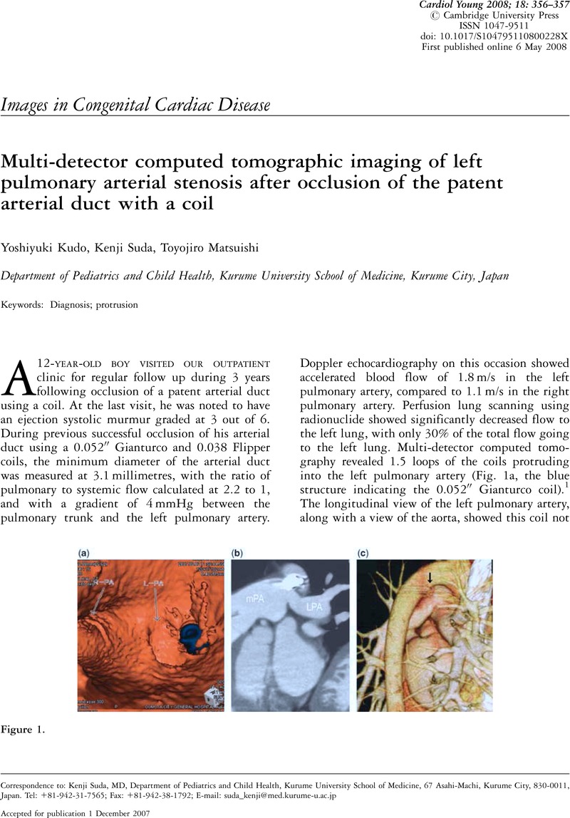
Multi-detector computed tomographic imaging of left pulmonary arterial stenosis after occlusion of the patent arterial duct with a coil

Published online by Cambridge University Press:  01 June 2008

Yoshiyuki Kudo
Affiliation:
Department of Pediatrics and Child Health, Kurume University School of Medicine, Kurume City, Japan
Kenji Suda*
Affiliation:
Department of Pediatrics and Child Health, Kurume University School of Medicine, Kurume City, Japan
Toyojiro Matsuishi
Affiliation:
Department of Pediatrics and Child Health, Kurume University School of Medicine, Kurume City, Japan
*
Correspondence to: Kenji Suda, MD, Department of Pediatrics and Child Health, Kurume University School of Medicine, 67 Asahi-Machi, Kurume City, 830-0011, Japan. Tel: +81-942-31-7565; Fax: +81-942-38-1792; E-mail: suda_kenji@med.kurume-u.ac.jp

Abstract

Image of the first page of this content. For PDF version, please use the ‘Save PDF’ preceeding this image.'
Type
Images in Congenital Cardiac Disease
Copyright
Copyright © Cambridge University Press 2008

Access options

Get access to the full version of this content by using one of the access options below. (Log in options will check for institutional or personal access. Content may require purchase if you do not have access.)

References

1.Hayabuchi, Y, Mori, K, Kagami, S. Virtual endoscopy using multidetector-row CT for coil occlusion of patent ductus arteriosus. Cathet Cardiovasc Interv 2007; 70: 434439.CrossRefGoogle ScholarPubMed