


Introduction
Calodium hepaticum Moravec 1982 (Trichinellida: Capillaridae) (syn. Capillaria hepatica) is a zoonotic nematode with a worldwide distribution (Schmidt, Reference Schmidt2001; Fuehrer, Reference Fuehrer2014a,Reference Fuehrerb). Although the host range of C. hepaticum includes a wide spectrum of mammals (including humans) (Fuehrer et al., Reference Fuehrer, Igel and Auer2011; Fuehrer, Reference Fuehrer2014a,Reference Fuehrerb), this parasite is predominantly associated with the families Muridae (Murinae, Deomyinae) and Cricetidae (Arvicolinae, Neotominae, Cricetinae, Sigmodontinae, Gerbilinae and Cricetomyinae) (Schmidt, Reference Schmidt2001; Fuehrer, Reference Fuehrer2014a). Of these rodents, the Norway rat Rattus norvegicus, Berkenhout, 1769, seems to be the most important host species worldwide, with high prevalence reported in several continents (Duque et al., Reference Duque, Aranzazu, Agudelo-Florez, London, Quiroz and Rodas2012; Moreira et al., Reference Moreira, Giese, Silva, Melo, Furtado, Maldonado and Santos2013; Fuehrer, Reference Fuehrer2014a), followed by the black rat Rattus rattus, Linnaeus, 1758 (Almeida-Silva et al., Reference Almeida-Silva, Fava, Potenza, Reis and Campos2011; Moreira et al., Reference Moreira, Giese, Silva, Melo, Furtado, Maldonado and Santos2013), and the house mouse Mus musculus, Linnaeus, 1758 (Gotardo et al., Reference Gotardo, Andrade and Andrade2000; Fuehrer, Reference Fuehrer2014a).
In the New World, the predominant Muroidea rodents are Sigmodontinae (Cricetidae), comprising approximately 84 genera and nearly 380 species (Patton et al., Reference Patton, Pardiñas and D'Elía2015). Sigmodontine rodents are predominantly distributed in South America with about 85 endemic genera (D'Elía & Pardiñas, Reference D'Elía, Pardiñas, Patton, Pardiñas and D'Elía2015). At least 108 species of sigmodontine rodents have been recorded in Argentina, including representatives of nine tribes and some incertae sedis taxa (Patton et al., Reference Patton, Pardiñas and D'Elía2015; Teta et al., Reference Teta, Abba, Cassini, Flores, Galliari, Lucero and Ramírez2018; Formoso & Teta, Reference Formoso and Teta2019).
The potential role that Sigmodontinae play as hosts of C. hepaticum has been little investigated, but a recent report showed that they might be important for parasite dynamics, at least in South America (Fantozzi et al., Reference Fantozzi, Robles, Beldomenico and Monje2018). Several Sigmodontinae species from Argentina were found to be infected by C. hepaticum, with high prevalence in Akodon azarae Fischer, 1829 (Fantozzi et al., Reference Fantozzi, Robles, Beldomenico and Monje2018).
Adults of C. hepaticum are found in the liver. Hepatic infections occur following ingestion of embryonated eggs by a mammal. After hatching, the larvae penetrate through the intestinal wall, migrate into the hepatic portal system, and develop as adults in the hepatic parenchyma (Spratt & Singleton, Reference Spratt, Singleton, Samuel, Pybus and Kocan2001), where gravid females lay immature eggs which are then exposed to the next host after the death and decomposition of the infected host or when it becomes prey (Spratt & Singleton, Reference Spratt, Singleton, Samuel, Pybus and Kocan2001; Camargo et al., Reference Camargo, Camargo, de Souza Vera, Barreto, Tourinho and de Souza2010). Moreover, it has been suggested that cannibalism may be important in the transmission among rats (Rothenburger et al., Reference Rothenburger, Himsworth, Chang, LeJeune and Leighton2014).
Several examination techniques are available to detect the presence of C. hepaticum eggs. The most widely used approach for the C. hepaticum infection has been based on morphology, morphometry and histopathology (Ferreira & Andrade, Reference Ferreira and Andrade1993; Andrade & Andrade, Reference Andrade and Andrade2004; Almeida et al., Reference Almeida, Caldas, Corrêa, Rodrigues-Silva, Siqueira and Machado-Silva2012; Duque et al., Reference Duque, Aranzazu, Agudelo-Florez, London, Quiroz and Rodas2012; Fantozzi et al., Reference Fantozzi, Sanchez, Ciorciari, Peña, Canal and Beldomenico2019). In turn, a novel procedure for taxonomic species identification through morphological and morphometric characteristics of parasite eggs in capillariids has recently been proposed (Borba et al., Reference Borba, Martin, Machado-Silva, Xavier, de Mello and Iñiguez2021b).
The aetiological diagnosis of helminth infections is usually based on the finding of characteristic eggs, larvae, or proglottids in biological samples. Most of the helminths living in the intestinal tract or in associated organs produce eggs. Each helminth species produces eggs that display intraspecific variation in size/shape, but which are mostly highly uniform in their morphological phenotype, and size and shape are therefore used as criteria for egg identification of biological samples, that is, the shape and size of helminth eggs are characteristic features of most helminth species, being commonly used as diagnostic criteria. Furthermore, eggshells are usually smooth and may vary considerably in thickness, depending on the helminth species (Ash & Orihel, Reference Ash and Orihel2010). Thus, various shell structure particularities, such as the mucoid polar opercula present in Trichuris, Calodium or Capillaria, are also important in identification. Concretely, eggs of Trichuris species are associated with prominent mucoid polar opercula and Capillaria and Calodium species eggs are associated with inconspicuous mucoid polar opercula. The subjectivity of the terms such as prominent or inconspicuous highlights the need to use markers that allow this characteristic to be quantified.
Adults of C. hepaticum are found in the liver and are identified through ultrasound, biopsy or necropsy. Only in spurious cases, have the eggs been observed in faeces (coproanalysis). In those cases, eggs are identified by means of morphology and morphometry. Classical morphometric techniques are, unfortunately, not able to characterize the features of the egg mucoid polar opercula, let alone assert host species influence on the parasite egg. For this reason, new tools should be applied to advance in diagnostics. Furthermore, morphological analyses are necessary to complement molecular techniques (Valero et al., Reference Valero, Darce, Panova and Mas-Coma2001, Reference Valero, Perez-Crespo, Periago, Khoubbane and Mas-Coma2009, Reference Valero, Perez-Crespo, Khoubbane, Artigas, Panova, Ortiz, Maco, Espinoza and Mas-Coma2012; García-Sánchez et al., Reference García-Sánchez, Rivero, Callejón, Zurita, Reguera-Gomez, Valero and Cutillas2019, Reference García-Sánchez, Reguera-Gomez, Valero and Cutillas2020; Reguera-Gomez et al., Reference Reguera-Gomez, Valero and Oliver-Chiva2021).
Regarding the morphometric characterization of Trichinellida representatives, geometric morphometrics has proven to be a useful tool to differentiate male adults (García-Sánchez et al., Reference García-Sánchez, Rivero, Callejón, Zurita, Reguera-Gomez, Valero and Cutillas2019) and eggs (García-Sánchez et al., Reference García-Sánchez, Reguera-Gomez, Valero and Cutillas2020) of Trichuris spp. populations infecting non-human primates and Sus scrofa domestica that can complement molecular data. Nevertheless, hitherto, no studies of this kind have been carried out in representatives of other genera such as Calodium. Furthermore, these new morphometric concepts provide adequate tools to characterize the morphological phenotype of Calodium eggs to assess the influence of different factors, such as the host species.
The objectives of this work are: (i) to propose a new analytical methodology; and (ii) to characterize morphologically eggs of C. hepaticum from natural infection of three species of sigmodontine rodents from Argentina (A. azarae Fischer, 1829, Calomys callidus Thomas, 1916 and Oligoryzomys flavescens Waterhouse, 1837). To this end, C. hepaticum eggs from adult parasites that had previously been genetically characterized were used.
Material and methods
Study sites
The study area is located in central Argentina, in the Espinal Ecoregion, characterized by a warm and humid climate (Matteucci, Reference Matteucci, Morello, Matteucci, Rodriguez and Silva2018). There are four well marked seasons (delimited by solstices and equinoxes), with abundant rains in autumn and summer (annual precipitation of 900 to 1200 mm) and with an average annual temperature of 18°C (Matteucci, Reference Matteucci, Morello, Matteucci, Rodriguez and Silva2018).
Sigmodontine rodents were captured in riparian environments. The localities were Esperanza (ES), Santa Fe Province (31° 24′ 20″ S–60° 58′ 16″ W) and La Picada (LP), Entre Ríos Province (31° 44′ 06″ S–60° 21′ 53″ W), about 80 km one from the other (fig. 1). Both sites are dominated by grasslands and situated in close proximity to important water courses that are tributaries of the Paraná River basin. ES is located on the El Salado River, and LP is located on the Las Conchas stream.

Fig. 1. Localities studied in the El Espinal ecoregion, Argentina: (A) Esperanza in Santa Fe Province; and (B) La Picada in Entre Ríos Province.
Sample collection
Rodents were captured between June 2014 and September 2016, in three-night trapping sessions carried out every five weeks. Eight trapping sites were selected (four fixed grids in ES and four in LP) consisting of 0.5 ha where 50 Sherman-type live-traps were placed and baited with pelletized commercial dog food. Plots were located in places with natural grassland. Traps were checked every morning and trapped rodents were transported to a field laboratory, anaesthetized by inhalation of isoflurane and sacrificed by cervical dislocation. The following data were taken and recorded from each individual: weight (g); total length (head + body + tail (cm)); length hind foot (mm); ear length (mm); and sex. Body condition was evaluated by estimating through palpation the degree of fat cover over the vertebral column and the pelvic bones, giving a score between 2 and 10, following Burthe et al. (Reference Burthe, Telfer, Lambin, Bennett, Carslake, Smith and Begon2006). Rodents were identified at species level by assessing their cranial morphology (Massoia & Fornes, Reference Massoia and Fornes1965; Massoia, Reference Massoia1973; Goncálvez & De Oliveira, Reference Goncálvez and De Oliveira2004; Udrizar Sauthier et al., Reference Udrizar Sauthier, Formoso, Andrade, Podestá and Teta2020), according to Patton et al. (Reference Patton, Pardiñas and D'Elía2015) for rodent taxonomy.
Four hundred and nine individual Sigmodontinae livers were weighed and examined, immediately after the hosts had been euthanized, under a magnifying lens to find evidence of nematodes. A total of 64 (15.6%) livers were found to be infected, recovering in all cases female nematodes. Eggs of C. hepaticum were obtained from liver disintegration.
This study has its limitations. Previous studies of Fasciola hepatica egg size showed characteristic morphometric traits to be related to their definitive host species (Valero et al., Reference Valero, Panova, Comes, Fons and Mas-Coma2002, Reference Valero, Perez-Crespo, Periago, Khoubbane and Mas-Coma2009). Nevertheless, this phenomenon has not been previously described in the species of the genus Calodium. Calodium hepaticum eggs used in this work were recovered from wild Sigmontinidae rodents from Argentina and not from experimental host samples. The prevalence of infected rodents in the geographical area of study has been previously cited by the authors, detecting C. hepaticum infection in 64 of 409 rodents (15.6%) (Fantozzi et al., Reference Fantozzi, Robles, Beldomenico and Monje2018). The role of the main reservoir or secondary reservoir of a helminth species is marked by the prevalence of infection. Consequently, the low prevalence present in C. callidus and O. flavescens makes it difficult to plan a study design with an adequate number of samples.
Molecular analysis of C. hepaticum adults from liver samples
Genomic DNA from individual worms was extracted using the AccuPrep® Genomic DNA Extraction Kit (Bioneer) according to the manufacturer's protocol. Genomic DNA concentration and purity were assessed using the SPECTROstar Nano and the MARS Data Analysis Software (BMGLabtech, Germany). The 18S rRNA partial gene was amplified by polymerase chain reaction (PCR), using primers 18S 965 and 18S 1573R (Powers et al., Reference Powers, Neher, Mullin, Esquivel, Giblin-Davis, Kanzaki, Stock, Mora and Uribe-Lorio2009). The PCR was performed in an Ivema T-18 thermocycler (Ivema Desarrollos, Argentina) using 5 μl of 5× Phire® reaction buffer, 200 μM dNTP, 0.4 pM of each primer, 100 ng of genomic DNA, and 0.5 μl of Phire® Hot Start II DNA polymerase (Finnzymes, Finland) and completed to 25 μl with molecular grade water. PCR conditions were as follows: 1 cycle at 98°C for 3 min, followed by 35 cycles at 98°C for 10 s, 57°C for 20 s, 72°C for 35 s and 1 final cycle at 72°C for 5 min (Fantozzi et al., Reference Fantozzi, Robles, Beldomenico and Monje2018). A negative control was included in each amplification run. Homologies were performed using the Basic Local Alignment Search Tooln program from the United States National Center for Biotechnology Information website (http://www.ncbi.nlm.nih.gov/BLAST).
Phenotypic analyses: proposal for a new analytical methodology
Measurement techniques
A total of 314 eggs of C. hepaticum were analysed (218 eggs from 11 individuals of A. azarae, 57 eggs from three individuals of C. callidus and 39 eggs from two individuals of O. flavescens). Initially, each egg was isolated with a micropipette, thoroughly washed with distilled water and immediately placed between microscope slides and coverslips with a water-based mounting medium to improve their image and preservation.
With the image analysis software, lineal biometric characters, areas and ratios of the eggs were obtained. Only well-preserved eggs were used for measurements which were made with a microscope (Nikon Eclipse E 800) at 100× magnification and using image analysis software (ImagePro Plus, version 5.1 for Windows, Media cybernetics, Silver Spring, USA) for images captured by a digital camera (Nikon Coolpix 5400). Representative images of the photographed eggs are shown in fig. 2.

Fig. 2. Eggs of Calodium hepaticum in different sigmodontine hosts: (A) Akodon azarae; (B) Calomys callidus; (C) Oligoryzomys flavescens (optical microscopy, original magnification = ×400). Scale bars: 20 μm.
Nineteen measurements were used. To identify the ones that give some valuable information about the egg morphology and were, therefore, suitable for principal component analysis (PCA), an iterative process of measurement combinations was performed.
Measurements were specifically assayed in this study due to the lack of standardized parameters. These measurements consider the characteristic shape of these eggs, with their two mucoid polar opercula of different size. In order to properly describe each of the two independently, the one of the larger size is denominated A, while the smaller one is denominated B.
Apical width mucoid polar opercula (L1 in A, L5 in B). Width measured from the top of the mucoid polar opercula.
Medium width mucoid polar opercula (L2 in A, L6 in B). Width measured at the middle of the mucoid polar opercula.
Basal width mucoid polar opercula (L3 in A, L7 in B). Measurements made inside the eggshell. Egg basal width mucoid polar opercula.
Length mucoid polar opercula (L4 in A, L8 in B). Length measured between the top and the end of the mucoid polar opercula.
Wall thickness at its midpoint (L9). Wall thickness width measured at the central axis of the egg.
Egg length excluding mucoid polar opercula length (L10). Measurements made inside the eggshell. Egg length without considering the mucoid polar opercula length.
In addition, other measurements classically considered in other helminth eggs were also applied:
Egg area (EA). Area included in the figure defining the egg outline.
Egg perimeter (EP). Length of the egg outline.
Egg roundness (ER). Defined as (perimeter2/4π × area). It is a measurement of how circular an object is (the expected perimeter of a circular object divided by the actual perimeter). A circular object will have a roundness of 1.0, while more irregular objects will have larger values (Valero et al., Reference Valero, Perez-Crespo, Periago, Khoubbane and Mas-Coma2009, Reference Valero, Bargues, Calderón, Artigas and Mas-Coma2018).
Egg length (EL). Feret diameter along major axis of egg.
Egg width (EW = L11). Feret diameter along minor axis of egg.
Size ratio (SR). Size length over size width (EL/EW).
Other measurements obtained from each egg were available in the options of the image analysis software:
Diameter (maximum). The maximum segment that passes through the centre and joins two opposite points of a circle.
Diameter (minimum). The minimum segment that passes through the centre and joins two opposite points of a circle.
Diameter (mean). The mean between minimum and maximum diameter.
Numerical methods
Biometrical data were examined using multivariate analysis. Current statistical techniques in morphometrics make it possible to test the null hypothesis of conspecific populations being simply the allometric extension of each other, provided that a common allometric trend is identifiable (Rohlf & Marcus, Reference Rohlf and Marcus1993). Morphological differences were quantified by geometric morphometrics, which includes validated methods for analysing variation in organismal shape (Klingenberg, Reference Klingenberg, Marcus, Corti, Loy, Naylor and Slice1996). These procedures focus on the retention of geometric information throughout a study and provide efficient, statistically powerful analyses that can readily relate abstract, multivariate results to the physical structure of the original specimens (Slice, Reference Slice2007).
The PCA is an estimator of certain parametric structure characteristics of the population. It may also be used as a dimension-reduction technique that identifies orthogonal linear combinations of the original variables that most efficiently reproduce sample variability. The latter use is particularly important in morphometric research as the number of shape variables to be analysed can be very large and often exceeds reasonable sample sizes (Slice, Reference Slice2007). For the analysis, we built a two-dimensional morphospace plot based on the first two principal components.
A way of correcting the problem of the existence of variation in size due to ontogeny or other causes would be to remove statistically the effect size present within the sample of each group and then apply canonical discriminant analysis to the samples already corrected for within-group differences in size. Thus, size-free canonical discriminant analysis was used on the covariance matrix of measurements transformed into logarithms to assess morphometric variation among samples. This technique consists of removing the effect of within-group ontogenetic variation, regressing each character separately on the within group first principal component (a multivariate size estimator) (Periago et al., Reference Periago, Valero, El Sayed, Ashrafi, El Wakeel, Mohamed, Desquesnes, Curtale and Mas-Coma2008; Valero et al., Reference Valero, Bargues, Calderón, Artigas and Mas-Coma2018).
The resulting ‘allometry-free’ (effect of size on morphological variation), or size-free, variables were submitted to a canonical variate analysis (CVA) and Mahalanobis distances were derived (Mahalanobis, Reference Mahalanobis1936). The Mahalanobis distance is a statistical technique that can be used to measure how distant a point is from the centre of a multivariate normal distribution, that is, it measures the separation of two groups. Mahalanobis distances were used to quantify shape divergence between groups. In the present analysis, the degree of shape divergence between C. hepaticum eggs populations was assessed through pairwise Mahalanobis distances (Ashrafi et al., Reference Ashrafi, Valero, Peixoto, Artigas, Panova and Mas-Coma2015; García-Sánchez et al., Reference García-Sánchez, Reguera-Gomez, Valero and Cutillas2020).
The PCA, CVA and Mahalanobis distances were performed using both the Collecting Landmarks for Identification and Characterization package and the available online XYOM software (https://xyom.io/) and tested by non-parametric permutation tests with 1000 iterations each. Values were considered statistically significant when P < 0.05 (Dujardin & Dujardin, Reference Dujardin and Dujardin2019; García-Sánchez et al., Reference García-Sánchez, Rivero, Callejón, Zurita, Reguera-Gomez, Valero and Cutillas2019; Reguera-Gomez et al., Reference Reguera-Gomez, Valero and Oliver-Chiva2021). Nevertheless, to avoid matrix singularities, some measurements were discarded because of their partial overlap with other measurements.
The following seven non-redundant measurements (i.e. one is not included in another) were considered in the egg analysis: L2 medium width mucoid polar opercula width A; L4 length mucoid polar opercula A; L6 medium width mucoid polar opercula B; L8 length mucoid polar opercula B; L9 wall thickness at its midpoint and L10 egg length excluding mucoid polar opercula length; and L11 egg width (see fig. 3).

Fig. 3. Standardized measurements for the morphometric phenotyping of Calodium hepaticum eggs in sigmodontine species. The mucoid polar opercula larger in size is denominated A, while the smaller one is denominated B. L1 apical width mucoid polar opercula A; L2 medium width mucoid polar opercula A; L3 basal width mucoid polar opercula A; L4 length mucoid polar opercula A; L5 apical width mucoid polar opercula B; L6 medium width mucoid polar opercula B; L7 basal width mucoid polar opercula B; L8 length mucoid polar opercula B; L9 wall thickness at its midpoint; L10 egg length excluding mucoid polar opercula length; and L11 egg width.
Statistical analyses
Statistical analyses were conducted using the R version 4.0.0 software (The R Project for Statistical Computing; http://www.rproject.org). Bivariant correlations (Spearman's rank correlation) were calculated for body host variables (body weight, liver weight, rodent length and rodent body condition) and egg size (principal component 1 (PC1)). Also, generalized linear mixed models (GLMM) were calculated. All models included host identification as a random intercept to account for the lack of independence of observations. PC1 was used as response variable and host species (= host sample size effect), and body condition and liver weight were used as independent variables. Season and year of capture were also included in the models to control for potential confounding. The ‘glmer’ function of the lme4 Package was used. The relevance of the terms was evaluated with the second order Akaike information criterion (AICc) to account for small sample sizes (Arnold, Reference Arnold2010; Burnham et al., Reference Burnham, Anderson and Huyvaert2011). When the inclusion of each term did not reduce AICc values in two or more units (ΔAICc < 2), it was dropped from the model. Results were considered statistically significant when P < 0.05.
To evaluate the possible influence of geographical location on the morphological phenotype of parasite eggs, a non-parametric test (Spearman correlation test) between the Mahalanobis distances and geographical altitudinal/latitudinal and geographical distances (km) was applied.
Results
Molecular criteria for species identification
The resulting sequences (excluding primers) were 100% identical (600/600) to the corresponding 18S rRNA sequence of C. hepaticum obtained from R. norvegicus (KT875351) and from A. azarae and C. callidus (MG686613) in Argentina (Fantozzi et al., Reference Fantozzi, Robles, Beldomenico and Monje2018). Complete identity was also observed with 18S rRNA sequences of C. hepaticum in the Old World from Arvicola terrestris (JX456635) (Guardone et al., Reference Guardone, Deplazes, Macchioni, Mag and Mathis2013), R. norvegicus (KT875351) and A. amphibious (JX456635-JX456636) in Switzerland (Bolukbas et al., Reference Bolukbas, Demirtas, Gurler, Inal, Acici and Umur2020).
Morphological characterization of C. hepaticum eggs from natural infection of three sigmodontine rodent species by means of the new analytical proposal
The overall size of C. hepaticum eggs obtained from geometric morphometric analysis samples is shown in table 1.
Table 1. Comparative morphometric data (extreme values, mean and standard deviation) of Calodium hepaticum eggs from three Sigmodontinae rodent species (Akodon azarae, Calomys callidus and Oligorizomys flavescens).

n = number of eggs.
The comparison of measurements of C. hepaticum eggs coming from three Sigmodontinae rodent species by GLMM analysis is shown in table 2, presenting significant differences between eggs from C. callidus and A. azarae.
Table 2. Significant differences in measurements between Calodium hepaticum eggs coming from three Sigmodontinae rodent species (Akodon azarae, Calomys callidus and Oligorizomys flavescens) by generalized linear mixed models analysis (P < 0.05).
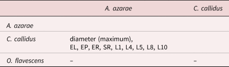
All models included host identification as a random intercept to account for the lack of independence of observations.
Also, the study of egg phenotypical characteristics was carried out by PCA, where PC1 and principal component 2 together, contributed 76% of the total variation. The resulting factor maps (fig. 4) clearly illustrate global size differences in the parasite eggs from the three Sigmodontinae species analysed and illustrate three areas corresponding to: (a) subgroup covering A. azarae and O. flavescens eggs; (b) subgroup covering A. azarae, C. callidus and O. flavescens eggs; and (c) subgroup covering A. azarae and C. callidus eggs (fig. 4). Interestingly, the results show that C. hepaticum eggs in A. azarae and O. flavescens share larger size values and that smaller specimens are present mainly in A. azarae and C. callidus.

Fig. 4. Factor map corresponding to individual principal component analysis attributed to each egg of Calodium hepaticum derived from different sigmodontine host species: Akodon azarae; Calomys callidus; and Oligoryzomys flavescens. Each sample is projected onto the first (PC1, 60%) and second (PC2, 16%) principal components. The horizontal axis is PC1, the vertical axis is PC2. Together, they contributed 76% to the total variation. Each group is represented by its perimeter. Circle, square and triangle represent the centroid in each group.
The degree of similarity between egg populations was assessed through pairwise Mahalanobis distances. These distances were calculated comparing eggs from three host species with each other. Malahanobis distances are displayed in table 3. When comparing C. hepaticum eggs from A. azarae with respect to eggs from C. callidus, and eggs from A. azarae with respect to eggs from O. flavescens; the detected distances were smaller than in the comparison of C. hepaticum eggs from C. callidus with respect to eggs from O. flavescens. Summing up, the largest distances were detected between parasite eggs from C. callidus and O. flavescens.
Table 3. Mahalanobis distances between the three Calodium hepaticum egg groups according to the three host species: Akodon azarae; Calomys callidus; and Oligorizomys flavescens.

* statistically different over 1000 permutations with Bonferroni correction (P < 0.05).
Host body variables and the intraspecific variability of parasite eggs
Significant correlations were obtained between each egg parasite PC1 (n = 314) and rodent corporal characteristics: weight (rho: 0.322, P < 0.001); liver weight (rho: 0.214, P < 0.001); rodent length (rho: 0.352, P < 0.001); and rodent body condition (rho: 0.310, P < 0.001). Summing up, positive correlations between egg parasite PC1 and host variables were detected.
The final GLMM showed C. callidus associated with PC1. Smaller C. hepaticum eggs are present mainly in C. callidus (table 4).
Table 4. Generalized linear mixed models showing the association between principal component 1 (PC1) and host variables.

The significant coefficients are shown in boldface type (P < 0.05). *Akaike information criterion (AICc) value increments if the variable is dropped.
Finally, there was no consistent correlation between the size-free pattern of variation and altitudinal/latitudinal and geographical distances (km). No significant values between Mahalanobis distances and geographical distances were detected (data not shown).
Discussion
According to previous C. hepaticum egg measurements described from R. rattus, length averaged 60 μm (range 40 μm to 75 μm) and 37 μm (range 27–41 μm) in width (Choe et al., Reference Choe, Lee, Seo, Chai, Lee, Eom and Chi1993; Fugassa et al., Reference Fugassa, Taglioretti, Gonçalves, Araújo, Sardella and Denegri2008; Carvalho-Costa et al., Reference Carvalho-Costa, Silva and de Souza2009; Resendes et al., Reference Resendes, Amaral, Rodrigues and Almeria2009; Buńkowska-Gawlik et al., Reference Buńkowska-Gawlik, Perec-Matysiak, Burzyńska and Hildebrand2017). The average length of the C. hepaticum eggs analysed herein (66.99 μm, range: 52.81–76.34 μm, in A. azarae; 60.36 μm, range 51.64–71.26 μm, in C. callidus, 65.76 μm, range 58.12–72.26 μm, in O. flavescens) (table 1) falls within this range although our minimum record is slightly smaller in C. callidus. The maximum width (44.82 μm) detected in the C. hepaticum eggs from C. callidus analysed in the present study is also remarkably larger than previously reported values (range 27–41 μm) (Choe et al., Reference Choe, Lee, Seo, Chai, Lee, Eom and Chi1993; Fugassa et al., Reference Fugassa, Taglioretti, Gonçalves, Araújo, Sardella and Denegri2008; Carvalho-Costa et al., Reference Carvalho-Costa, Silva and de Souza2009; Resendes et al., Reference Resendes, Amaral, Rodrigues and Almeria2009; Gonçalves et al., Reference Gonçalves, Ascaso, Santos, Serra, Juliao and Orlandi2012; Buńkowska-Gawlik et al., Reference Buńkowska-Gawlik, Perec-Matysiak, Burzyńska and Hildebrand2017). Summing up, previous descriptions showed a wide range in the measurements included, indicating the necessity of studies analysing the influence of the different factors involved such as geographical origin or definitive host influence.
Morphological characterization of C. hepaticum eggs from natural infection of three species of sigmodontine rodents applying the new analytical proposal
Our study provides the first geometric analysis of the egg morphometry of C. hepaticum from sigmodontine rodents from Argentina, PCA proving to be an efficient method to analyse C. hepaticum eggs. The measurements chosen to be included in the PCA have been proposed for the first time. Through the centroids obtained in PCA, parasite eggs from C. callidus, were differentiated from those from A. azarae and O. flavescens. Calodium hepaticum eggs from C. callidus appeared separated from A. azarae and O. flavescens. On the other hand, the factor map shows an overlapping area between the parasite eggs originating from the three rodent species. The subgroups observed in fig. 4 are likely to reflect ecological or geographical influences.
In previous studies, the influence of the geographical location or host species on the size of helminth adults and eggs has been reported, for example, in species of Fasciola (Valero et al., Reference Valero, Darce, Panova and Mas-Coma2001, Reference Valero, Panova, Comes, Fons and Mas-Coma2002, Reference Valero, Perez-Crespo, Periago, Khoubbane and Mas-Coma2009, Reference Valero, Perez-Crespo, Khoubbane, Artigas, Panova, Ortiz, Maco, Espinoza and Mas-Coma2012, Reference Valero, Bargues, Calderón, Artigas and Mas-Coma2018; Ashrafi et al., Reference Ashrafi, Valero, Peixoto, Artigas, Panova and Mas-Coma2015) or Trichuris (Garcia-Sánchez et al., Reference García-Sánchez, Rivero, Callejón, Zurita, Reguera-Gomez, Valero and Cutillas2019, Reference García-Sánchez, Reguera-Gomez, Valero and Cutillas2020). Nevertheless, these phenomena have so far not been described in the genus Calodium.
With respect to representatives of Nematoda eggs, previous analyses using geometric morphometry focused only on Trichuris species. García-Sánchez et al. (Reference García-Sánchez, Reguera-Gomez, Valero and Cutillas2020) used a total of eight lineal egg measurements (maximum width of polar opercula, minimum width of polar opercula, base width of polar opercula, length of polar opercula measured from exterior midpoint to the narrow midpoint, total length of polar opercula measured from the exterior midpoint to the base midpoint, wall thickness at its midpoint, wall thickness in contact with polar opercula and interior length of the egg) in the characterization of the size of Trichuris eggs. The study of interspecific variability enables the analysis of the morphological patterns in different Trichuris sp. eggs associated with different host species [(macaque (Macaca sylvanus), eastern black-and-white colobus (Colobus guereza kikuyuensis), grivets (Chlorocebus aethiops) and Brazza's monkey (Cercopithecus neglectus)]. In contrast to that study, herein seven measurements are used: L2 medium width mucoid polar opercula A; L4 length mucoid polar opercula A; L6 medium width mucoid polar opercula B; L8 length mucoid polar opercula B; L9 wall thickness at its midpoint; L10 egg length excluding mucoid polar opercula length; and L11 egg width. The different measurements applied, compared with the proposal by García-Sánchez et al. (Reference García-Sánchez, Reguera-Gomez, Valero and Cutillas2020), are based on the fact that herein the two mucoid polar opercula are characterized independent from each other. It is noteworthy that the asymmetry detected in Calodium eggs in polar opercula is more pronounced than that detected in polar opercula of Trichuris eggs.
The most frequently morphological parameters reported by other researchers are length and width (among others: Carvalho-Costa et al., Reference Carvalho-Costa, Silva and de Souza2009; Almeida et al., Reference Almeida, Caldas, Corrêa, Rodrigues-Silva, Siqueira and Machado-Silva2012; Macchioni et al., Reference Macchioni, Chelucci, Guardone, Mignone, Prati and Magi2013; Klisiowicz et al., Reference Klisiowicz, Reifur, Shimada, Haidamak, Cognialli and Ferreira2014; Sinniah et al., Reference Sinniah, Narasiman, Habib and Gaik Bei2014; Buńkowska-Gawlik et al., Reference Buńkowska-Gawlik, Perec-Matysiak, Burzyńska and Hildebrand2017). Several reports on Capillaridae eggs detected distinct morphotypes, for example, in Trichuris sp. (Petrželková et al., Reference Petrželková, Hasegawa, Appleton, Huffman, Archer, Moscovice, Mapua, Singh and Kaur2010; Klaus et al., Reference Klaus, Zimmermann, Röper, Radespiel, Nathan, Goossens and Strube2017; Garcia-Sánchez et al., Reference García-Sánchez, Reguera-Gomez, Valero and Cutillas2020; Kamani et al., Reference Kamani, Massetti, Olubade, Balami, Samdi, Traub, Colella and González-Miguel2021), or species of the genera Aonchotheca, Baruscapillaria, Capillaria, Calodium, Echinocoleus, Eucoleus, Pearsonema and Tridentocapillaria (Borba, Reference Borba2019; Borba et al., Reference Borba, Enoki, Lopes-Torres, Machado-Silva and Iñiguez2021a). Interestingly, C. hepaticum eggs from Meriones persicus and R. rattus are described as having a ‘very peculiar morphology’, that is, a punctuated and a radial eggshell ornamentation, with a thick eggshell (5.54 μm) (Borba et al., Reference Borba, Martin, Machado-Silva, Xavier, de Mello and Iñiguez2021b). Furthermore, Borba et al. (Reference Borba, Martin, Machado-Silva, Xavier, de Mello and Iñiguez2021b) recently reported a new methodology for Capillariid species identification based on artificial intelligence technology. In this case, despite having taken the measurements of the two mucoid polar opercula, the discriminant analysis between representatives of Capillariidae was carried out using only the length and width of the eggs as measurements (Borba et al., Reference Borba, Martin, Machado-Silva, Xavier, de Mello and Iñiguez2021b).
Host body variables and intraspecific variability of parasite eggs
The capacity of organisms to present different phenotypes under varying environmental conditions (phenotypical plasticity) has been studied since the Neo-Darwinian synthesis (Pigliucci, Reference Pigliucci2005). However, few studies have quantitatively assessed the influence that the host body characteristics exercises on egg morphology of helminth parasites. In some helminth species, previous studies show that the host animal body can strongly influence the phenotype of the egg stage. As pointed out by Poulin (Reference Poulin1997), the host mass correlates with the space available for parasites in various organs, which may put physical constraints upon helminth body size. Thus, in the case of F. hepatica, Valero et al. (Reference Valero, Panova, Comes, Fons and Mas-Coma2002) showed that the small host body mass of rats and mice was associated with diminished F. hepatica egg size.
The present study focuses on the intraspecific variability of C. hepaticum eggs associated with the host body characteristics. The PC1 of the egg samples can be interpreted as a measure of overall egg size, and positive correlations were detected between eggs PC1 and host rodent corporal characteristics (weight, liver weight, rodent length and rodent body condition). Furthermore, GLMM corroborate the association between egg PC1 and host variables. Herein, the phenotypical plasticity of C. hepaticum eggs related to host body characteristics is shown. Future research with an adequate sample size is needed to define this parasite–host relationship.
Fantozzi et al. (Reference Fantozzi, Robles, Beldomenico and Monje2018) documented the occurrence of C. hepaticum in two rodent assemblages from central Argentina, basing the diagnosis on morphological characteristics, histopathological examination and 18S ribosomal RNA sequences, showing a prevalence of 41.2% in A. azarae, 13.8% in C. callidus and 7.1% in O. flavescens. The high prevalence observed in A. azarae, associated with the wide size range of the parasite eggs evidenced by PCA, suggests A. azarae to be the Sigmodontinae host species that plays the most important role as reservoir host for C. hepaticum in the New World.
The present study proposes a new standardized approach able to characterize egg morphological features of C. hepaticum eggs. As a matter of fact, without having previously demonstrated this possibility, the results obtained in the present work are expected to lay the groundwork for descriptive or comparative morphometric studies. In the latter case, this tool allows the evaluation of the possible influence of the different factors on the morphological phenotype of parasite eggs such as the host species influence, geographical location or spurious infections in humans, among others.
Acknowledgements
The authors thank L. Antoniazzi (LeCen-ICIVET), A. Berduc (PEREB-La Picada), students of Capybara Group UNL, M.L. Reynoso, C.A. Fernandez, A. Fasano, I. Monsalvo, P. Hernán Capovilla, N. Mordini (UNL), T. Ricardo (FHUC), F. Peña (LeCen-ICIVET) and C. Palavecino (LeCen- ICIVET) for their collaboration during the fieldwork. The authors also thank P. Teta and U. Pardiñas for the identification of the rodents, C. Fantozzi for the graphic design of C. hepaticum eggs and P.F. Cuervo for the map of the localities studied.
Author contributions
MCF: methodology, investigation, formal analysis and writing – original draft; MRG: methodology and investigation; PMB: methodology, investigation, writing – original draft and funding acquisition; SMC: writing – review and editing, conceptualization and funding acquisition; MDB: conceptualization, formal analysis, writing – review and editing and funding acquisition; and MAV: conceptualization, formal analysis, writing – review and editing and funding acquisition.
All authors critically reviewed the manuscript and gave final approval for the version to be published.
Financial support
MCF received a fellowship of the Consejo Nacional de Investigaciones Científicas y Técnicas (CONICET, Argentina) and is currently the recipient of a fellowship of the Programa para la promoción de la investigación científica, el desarrollo tecnológico y la innovación en la Comunitat Valenciana, Generalitat Valenciana (CIAPOS/2021/166). This study was supported by grants from CONICET and Agencia Nacional de Promoción Científica y Tecnológica (ANPCyT), project number: PICT 2012-1552 (PMB-presenting support for this work). Valencia centre collaboration funded by PROMETEO Program, Programa de Ayudas para Grupos de Investigación de Excelencia, Generalitat Valenciana, Grants/Award Numbers: 2016/099, 2021/004; Red de Investigación de Centros de Enfermedades Tropicales, Grant/Award Number: RD16/0027/0023; ISCIII-Subdirección General de Redes y Centros de Investigación Cooperativa RETICS, Ministry of Science and Innovation; CIBER de Enfermedades Infecciosas, Grant/Award Number: CB21/13/00056; ISCIII, Ministry of Science and Innovation.
Conflicts of interest
None.
Ethical standards
Sample collection was carried out during fieldwork under official permits granted by the Dirección General de Recursos Naturales, Gobierno de Entre Ríos (autorización n° 002/14 and 001/16, Expediente 1.657.179) and Ministerio de Aguas, Servicios Públicos y Medio Ambiente, Gobierno de Santa Fe (autorización n° 193, Expediente 02101-0014145-3). This study was carried out in accordance with Protocol n°135/12 approved by the bioethics committee of Facultad de Ciencias Veterinarias, Universidad Nacional del Litoral. Specimens collected in live traps were humanely sacrificed (euthanized by cervical dislocation while being deeply anaesthetized with isoflurane), following the recommendations of the Ethics Committee for Research on Laboratory Animals, Farm and Obtained from Nature of the National Council of Scientific and Technical Research (CONICET), and subsequently by the National Agency for the Promotion of Science and Technology of Argentina (ANPCCyT). No endangered species were involved in this study.


 Open access