


Crossref Citations
This article has been cited by the following publications. This list is generated based on data provided by Crossref.
Guerrieri, Ania Naila
Montesi, Monica
Sprio, Simone
Laranga, Roberta
Mercatali, Laura
Tampieri, Anna
Donati, Davide Maria
and
Lucarelli, Enrico
2020.
Innovative Options for Bone Metastasis Treatment: An Extensive Analysis on Biomaterials-Based Strategies for Orthopedic Surgeons.
Frontiers in Bioengineering and Biotechnology,
Vol. 8,
Issue. ,
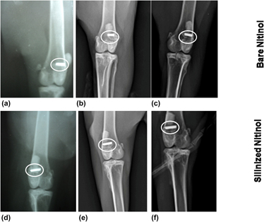
Sinha, Sarmita
Priyadarshani, Jyotsana
Bavya Devi, Karuppasamy
Kishore, Anyam Vijay
Das, Piyali
Chanda, Abhijit
Das, Soumen
Roy, Mangal
and
Nandi, Samit Kumar
2020.
In vivo performance analysis of silanized and coated nitinol wires in biological environment.
Journal of Materials Research,
Vol. 35,
Issue. 10,
p.
1262.
Ji, Hua
Wang, Yongqi
Li, Zhiyong
Huang, Zhaoxia
and
Chai, Mingxia
2021.
Electrolytic Polishing Test and Surface Properties of Nitinol Tube.
International Journal of Electrochemical Science,
Vol. 16,
Issue. 3,
p.
210364.
He, B.
Huang, X.
Zhao, C.
Zhao, G.
and
Hong, Q.
2021.
Successful removal of a trapped epidural catheter facilitated by using a nickel/titanium alloy (nitinol) suture as a guidewire.
Anaesthesia Reports,
Vol. 9,
Issue. 2,
Shanmuganantha, Lohashenpahan
Baharudin, Azmi
Sulong, Abu Bakar
Shamsudin, Roslinda
and
Ng, Min Hwei
2021.
Prospect of Metal Ceramic (Titanium-Wollastonite) Composite as Permanent Bone Implants: A Narrative Review.
Materials,
Vol. 14,
Issue. 2,
p.
277.
Sivakumar, Ponnurengam Malliappan
Yetisgin, Abuzer Alp
Sahin, Sevilay Burcu
Demir, Ebru
and
Cetinel, Sibel
2022.
Bone tissue engineering: Anionic polysaccharides as promising scaffolds.
Carbohydrate Polymers,
Vol. 283,
Issue. ,
p.
119142.