


No CrossRef data available.
Article contents
Morpho-functional Analysis of the Head Glands in Three Auchenorrhyncha Species and Their Possible Biological Significance
Part of:
Micrographia Collection
Published online by Cambridge University Press: 12 September 2022
Abstract
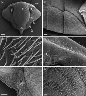
The Cicadomorpha Philaenus spumarius, Neophilaenus campestris, and Cicadella viridis are known transmitters of the bacterium Xylella fastidiosa. Here, we studied the ultrastructural organization of their cephalic glands. Our investigations with scanning, transmission, focused ion beam-scanning electron microscopes and light microscope revealed for the first time in Auchenorrhyncha the presence of two types of cephalic glands. Both belonged to the Class III epidermal glands, according to the Noirot and Quennedey classification. Type A glands were the most common, being mainly located around antennae, lorum, and gena. Moreover, these glands were observed also on the abdomen and thorax, always in association with sensilla trichoidea. The second type of glands (type B) were located exclusively at the apical part of the postclypeus in P. spumarius and N. campestris. The ultrastructural organization was similar in both types, being composed of a secretory cell and a conducting canal. Differences were observed in the width of the cuticular opening, being smaller in the type II glands. In addition, we have recorded the presence of a maxillary sensory pit in all species and described sensilla trichoidea ultrastructural organization. Finally, we discussed the ultrastructural organization of the glands and their potential biological role.
- Type
- Micrographia
- Information
- Copyright
- Copyright © The Author(s), 2022. Published by Cambridge University Press on behalf of the Microscopy Society of America
References
Ahmad, A, Kundu, A & Dey, D (2020). Wax glands ultrastructure and chemical composition of wax of giant mealybug Drosicha stebbingii (Green) (Hemiptera: Monophlebidae). J Asia Pac Entomol 23, 546–553.CrossRefGoogle Scholar
Ammar, E, Alessandro, RT & Hall, DG (2013). Ultrastructural and chemical studies on waxy secretions and wax-producing structures on the integument of the woolly oak aphid Stegophylla brevirostris Quednau (Hemiptera: Aphididae). J Microsc Ultrastruct 1, 43–50.CrossRefGoogle Scholar
Ammar, E, Hentz, M, Hall, DG & Shatters, RG (2015). Ultrastructure of wax-producing structures on the integument of the Melaleuca psyllid Boreioglycaspis melaleucae (Hemiptera: Psyllidae), with honeydew excretion behavior in males and females. PLoS One 10, e0121354.CrossRefGoogle Scholar
Barbier, R, Ferran, A, Le Lannic, J & Allo, M-R (1992). Morphology and ultrastructure of integumentary glands of Semiadalia undecimnotata Schn. (Coleoptera: Coccinellidae). Int J Insect Morphol Embryol 21, 223–234.CrossRefGoogle Scholar
Bartlet, E, Isidoro, N & Williams, IH (1994). Antennal glands in Psylliodes chrysocephala, and their possible role in reproductive behaviour. Physiol Entomol 19, 241–250.CrossRefGoogle Scholar
Betz, O (2010). Adhesive exocrine glands in insects: Morphology, ultrastructure, and adhesive secretion. In Biological Adhesive Systems, Byern, J & Grunwald, I (Eds.), pp. 111–152. Vienna: Springer.CrossRefGoogle Scholar
Billen, J (2009). Occurrence and structural organization of the exocrine glands in the legs of ants. Arthropod Struct Dev 38, 2–15.CrossRefGoogle ScholarPubMed
Billen, J & Ito, F (2018). Novel thoracic glands in the ant Myopias hollandi. Arthropod Struct Dev 47, 229–237.CrossRefGoogle ScholarPubMed
Bin, F, Wäckers, F, Romani, R & Isidoro, N (1999). Tyloids in Pimpla turionellae (L.) are release structures of male antennal glands involved in courtship behaviour (Hymenoptera: Ichneumonidae). Int J Insect Morphol Embryol 28, 61–68.CrossRefGoogle Scholar
Boullis, A & Verheggen, FJ (2016). Chemical ecology of aphids (Hemiptera: Aphididae). In Biology and Ecology of Aphids, Vilcinskas, A (Ed.), pp. 171–198. Florida: CRC Press Boca Ranton.Google Scholar
Chen, J & Qiao, G (2012). Wax gland plates in Hormaphidinae (Hemiptera: Aphididae): Morphological diversity and evolution. Entomol News 122, 27–45.CrossRefGoogle Scholar
Cornara, D, Morente, M, Markheiser, A, Bodino, N, Tsai, C-W, Fereres, A, Redak, RA, Perring, TM & Lopes, JRS (2019). An overview on the worldwide vectors of Xylella fastidiosa. Entomol Gen 39, 157–181.CrossRefGoogle Scholar
Cwikla, PS & Freytag, PH (1983). External morphology of Xestocephalus subtessellatus (Homoptera: Cicadellidae: Xestocephalinae). Ann Entomol Soc Am 76, 641–651.CrossRefGoogle Scholar
De Santis, F, Conti, E, Romani, R, Salerno, G, Parillo, F & Bin, F (2008). Colleterial glands of Sesamia nonagrioides as a source of the host-recognition kairomone for the egg parasitoid Telenomus busseolae. Physiol Entomol 33, 7–16.CrossRefGoogle Scholar
Di Giulio, A & Muzzi, M (2018). Two novel approaches to study arthropod anatomy by using dualbeam FIB/SEM. Micron 106, 21–26.CrossRefGoogle ScholarPubMed
Di Giulio, A, Rossi Stacconi, MV & Romani, R (2009). Fine structure of the antennal glands of the ant nest beetle Paussus favieri. Arthropod Struct Dev 38, 293–302.CrossRefGoogle ScholarPubMed
Evans, JW (1973). The maxillary plate of Hornoptera-Auchenorrhyncha. J Entomol Ser A Gen Entomol 48, 43–47.CrossRefGoogle Scholar
Evans, JW (1975). The external features of the heads of leafhoppers (Homoptera, Cicadelloidea). Rec Aust Mus 29, 407–439.CrossRefGoogle Scholar
Foldi, I & Pearce, MJ (1985). Fine structure of wax glands, wax morhoplogy and function in the female scale insect, Pulvinaria regalis Canard. (Hemiptera: Coccidae). Int J Insect Morphol Embryol 14, 259–271.CrossRefGoogle Scholar
Giglio, A, Ferrero, EA & Brandmayr, TZ (2005). Ultrastructural identification of the antennal gland complement in Siagona europaea Dejean 1826, a myrmecophagous carabid beetle. Acta Zool 86, 195–203.CrossRefGoogle Scholar
Hölldobler, B & Wilson, EO (2009). The Superorganism: The Beauty, Elegance, and Strangeness of Insect Societies. New York: WW Norton & Company.Google Scholar
Janse, JD & Obradovic, A (2010). Xylella fastidiosa: Its biology, diagnosis, control and risks. J Plant Pathol 92, 35–48.Google Scholar
Krall, BS, Bartelt, RJ, Lewis, CJ & Whitman, DW (1999). Chemical defense in the stink bug Cosmopepla bimaculata. J Chem Ecol 25, 2477–2494.CrossRefGoogle Scholar
Laumann, RA, Kavčič, A, Moraes, MCB, Borges, M & Čokl, A (2013). Reproductive behaviour and vibratory communication of the neotropical predatory stink bug Podisus nigrispinus. Physiol Entomol 38, 71–80.CrossRefGoogle Scholar
Liang, A (2005). A new structure on the subantennal process of Borysthenes species (Hemiptera: Fulgoromorpha: Cixiidae: Borystheninae). Proc Biol Soc Wash 118, 809–814.CrossRefGoogle Scholar
Liang, AP (2020). A new structure on the frons of male adults of the Asian rice spittlebug Callitettix versicolor (Hemiptera: Auchenorrhyncha: Cercopidae). Zootaxa 4801, 591–599.CrossRefGoogle Scholar
Lockwood, JA & Story, RN (1987). Defensive secretion of the southern green stink bug (Hemiptera: Pentatomidae) as an alarm pheromone. Ann Entomol Soc Am 80, 686–691.CrossRefGoogle Scholar
Lucchi, A & Mazzoni, E (2004). Wax production in adults of planthoppers (Homoptera: Fulgoroidea) with particular reference to Metcalfa pruinosa (Flatidae). Ann Entomol Soc Am 97, 1294–1298.CrossRefGoogle Scholar
Noirot, C & Quennedey, A (1974). Fine structure of insect epidermal glands. Annu Rev Entomol 19, 61–80.CrossRefGoogle Scholar
Noirot, C & Quennedey, A (1991). Glands, gland cells, glandular units: Some comments on terminology and classification. In Annales de la Société Entomologique de France, Berti, MN & Pluot-Sigwalt, MD (Eds.), vol. 27. pp. 123–128. Paris: Société entomologique de France.Google Scholar
Quennedey, A (1998). Insect epidermal gland cells: Ultrastructure and morphogenesis. Insecta 11, 177–207.Google Scholar
Rakitov, RA (2002). What are brochosomes for? An enigma of leafhoppers (Hemiptera, Cicadellidae). Denisia 176, 411–432.Google Scholar
Rakitov, RA (2009). Brochosomal coatings of the integument of leafhoppers (Hemiptera, Cicadellidae). In Functional Surfaces in Biology, Gorb, S (Ed.), pp. 113–137. Dordrecht: Springer.CrossRefGoogle Scholar
Rakitov, R & Gorb, SN (2013). Brochosomes protect leafhoppers (Insecta, Hemiptera, Cicadellidae) from sticky exudates. J R Soc Interface 10, 2–6.CrossRefGoogle ScholarPubMed
Riolo, P, Isidoro, N, Ruschioni, S, Minuz, RL, Bin, F & Romani, R (2016). Anatomy of the antennal dorsal organ in female of Neodryinus typhlocybae (Hymenoptera: Dryinidae): A peculiar sensory structure possibly involved in perception of host vibration. J Morphol 277, 128–137.CrossRefGoogle ScholarPubMed
Riolo, P, Ruschioni, S, Minuz, RL, Romani, R & Isidoro, N (2014). Female sex pheromone gland of the boxwood leafminer, Monarthropalpus buxi (Diptera: Cecidomyiidae): Morphological and behavioural evidence. Eur J Entomol 111, 75–81.CrossRefGoogle Scholar
Roell, T, Bianchi, FM, Kochenborger Leite, AP & Campos, AL (2020). External morphology of the abdominal glands in Asopinae (Hemiptera: Heteroptera: Pentatomidae). Arthropod Struct Dev 57, 100946.CrossRefGoogle Scholar
Romani, R, Isidoro, N, Riolo, P, Bin, F, Fortunato, A, Turillazzi, S & Beani, L (2005). A new role for antennation in paper wasps (Hymenoptera, Vespidae): Antennal courtship and sex dimorphic glands in antennomeres. Insectes Soc 52, 96–102.CrossRefGoogle Scholar
Sevarika, M, Rondoni, G, Conti, E & Romani, R (2021 a). Antennal sensory organs and glands of the harlequin ladybird, Harmonia axyridis. Entomol Exp Appl 169, 111–124.CrossRefGoogle Scholar
Sevarika, M, Rondoni, G, Ganassi, S, Pistillo, OM, Germinara, GS, De Cristofaro, A, Romani, R & Conti, E (2022). Behavioural and electrophysiological responses of Philaenus spumarius to odours from conspecifics. Sci Rep 12, 1–9.CrossRefGoogle ScholarPubMed
Sevarika, M, Rossi Stacconi, MV & Romani, R (2021 b). Fine morphology of antennal and ovipositor sensory structures of the gall chestnut wasp, Dryocosmus kuriphilus. Insects 12, 231.CrossRefGoogle ScholarPubMed
Stroiński, A, Gnezdilov, VM & Bourgoin, T (2011). Sub-brachypterous Ricaniidae (Hemiptera: Fulgoromorpha) of Madagascar with morphological notes for these taxa. Zootaxa 70, 1–70.CrossRefGoogle Scholar
Tabata, J & Ichiki, RT (2016). Sex pheromone of the cotton mealybug, Phenacoccus solenopsis, with an unusual cyclobutane structure. J Chem Ecol 42, 1193–1200.CrossRefGoogle ScholarPubMed
Tavella, L & Arzone, A (1993). Comparative morphology of mouth parts of Zyginidia pullula, Empoasca vitis, and Graphocephala fennahi (Homoptera, Auchenorrhyncha). Boll Zool 60, 33–39.CrossRefGoogle Scholar
Wang, R-R, Wan, X-Y, Liang, A-P & Bourgoin, T (2013). A SEM study of antennal and maxillary sensilla in Zema gressitti Fennah (Hemiptera: Fulgoromorpha: Tropiduchidae). Micron 44, 261–267.CrossRefGoogle Scholar
Wang R-R, Wan, X-Y & Liang, A-P (2014). Fine structure of sensory apparatus on the head of Cixiopsis punctatus. Journal of Insect Science 14, 99.Google Scholar
Zhang, L, Wang, S, Billen, J & Wei, C (2021). Morphology and ultrastructure of the epithelial femoral gland in cicadas (Hemiptera: Cicadidae). Arthropod Struct Dev 64, 101086.CrossRefGoogle Scholar