


Crossref Citations
This article has been cited by the following publications. This list is generated based on data provided by Crossref.
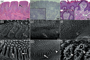
Melo, Lucas Inácio dos Santos
Matias de Oliveira, Radan Elvis
Freitas Caetano de Sousa, Ana Caroline
de Oliveira, Rysónely Maclay
Lima, Mariana Almeida
Fragoso, Ana Bernadete Lima
Silva, Flávio José de Lima
Attademo, Fernanda Loffler Niemeyer
Luna, Fábia de Oliveira
Pereira, Alexsandra Fernandes
and
de Oliveira, Moacir Franco
2024.
Antillean manatee (Trichechus manatus manatus Linnaeus, 1758) Tongue Morphology and Adaptive Herbivorous Implications.
Microscopy and Microanalysis,
Vol. 30,
Issue. 1,
p.
160.