


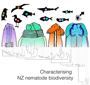
Introduction
Nematodes are extraordinarily ubiquitous, abundant and diverse. There are more than 40 000 species currently known, thus they represent one of the most speciose metazoan groups on earth (Zhang, Reference Zhang2013). The rate of descriptions is slowly increasing with about 400 new species descriptions per year (Hodda, Reference Hodda2022a). However, estimates suggest there are somewhere between 500 000 and 5 000 000 nematode species in total, which is 8–100 times more than that of the current known total (May, Reference May1988; Hugot et al., Reference Hugot, Baujard and Morand2001; Hodda, Reference Hodda2022b). These estimates and the variability between them make it clear that although considerable effort has been directed towards understanding nematode biodiversity within natural systems, much is left to be discovered. Addressing this deficiency is increasingly important now more than ever, as biodiversity loss is considered an urgent conservation priority across the globe (Butchart et al., Reference Butchart, Walpole, Collen, van Strien, Scharlemann, Almond, Baillie, Bomhard, Brown, Bruno, Carpenter, Carr, Chanson, Chenery, Csirke, Davidson, Dentener, Foster, Galli, Galloway, Genovesi, Gregory, Hockings, Kapos, Lamarque, Leverington, Loh, McGeoch, McRae, Minasyan, Hernández Morcillo, Oldfield, Pauly, Quader, Revenga, Sauer, Skolnik, Spear, Stanwell-Smith, Stuart, Symes, Tierney, Tyrell, Vié and Watson2010).
Anderson (Reference Anderson2000) estimated that approximately 33% of nematodes currently known are parasitic in vertebrate definitive hosts. Aside from this large contribution to nematode species richness, parasitic nematodes are well recognized for their zoonotic, economic and ecological importance within natural systems, especially in the marine environment. Various marine nematodes are zoonotic; humans who consume undercooked infected flesh can develop mild-to-severe allergic reactions, or acquire accidental infections, either of which can lead to death in severe cases (Audicana et al., Reference Audicana, Ansotegui, Fernández de Corres and Kennedy2002; Mattiucci et al., Reference Mattiucci, Fazii, De Rosa, Paoletti, Megna, Gielmo, De Angelis, Costa, Meucci, Calvaruso, Sorrentini, Palma, Bruschi and Nascetti2013). In their non-human hosts, nematodes can reduce the fecundity, growth and overall health of their host, also sometimes leading to death (e.g. Wiese et al., Reference Wiese, Davidson and Nettles1977; Vanstreels et al., Reference Vanstreels, Yabsley, Swanepoel, Stevens, Carpenter-Kling, Ryan and Pistorius2018). At a larger scale, such pathological infections may potentially cause secondary extinctions, especially if the host taxa are threatened. Although nematodes are not typically cited as the primary cause of mortality events, they can be associated as a contributing factor (e.g. Abollo et al., Reference Abollo, Gestal and Pascual2001a). For fish hosts that are commercially important, nematode infections can make fish undesirable for human consumption, reducing the marketability of fillets, and consequently posing a potential economic risk for some fisheries (Abollo et al., Reference Abollo, Gestal and Pascual2001b; EFSA, 2010). Contrary to these negative impacts, parasitic nematodes also play major roles within food webs and perform ecological functions within healthy natural systems (Singleton and McCallum, Reference Singleton and McCallum1990; Timi and Poulin, Reference Timi and Poulin2020). They can be used as biological tags for biomonitoring of host species (e.g. Melendy et al., Reference Melendy, McClelland and Hurlbut2005; Poulin and Kamiya, Reference Poulin and Kamiya2015), as indicators of ecosystem health or environmental change (Zarlenga et al., Reference Zarlenga, Hoberg, Rosenthal, Mattuicci and Nascetti2014) and, due to their high diversity, can be used as models for resolving evolutionary hypotheses regarding species radiation events (e.g. Mattiucci and Nascetti, Reference Mattiucci and Nascetti2008). Clearly, a wealth of knowledge concerning the structure and functioning of natural systems can be gained from investigating parasitic nematodes within marine ecosystems. Despite this however, they, along with other parasite taxa are often underrepresented or ignored completely in studies of biodiversity and ecology (Lafferty et al., Reference Lafferty, Allesina, Arim, Briggs, De Leo, Dobson, Dunne, Johnson, Juris, Marcogliese, Martinez, Memmott, Marquet, McLaughlin, Mordecai, Pascual, Poulin and Thieltges2008).
There are a few reasons why parasitic nematodes are underrepresented in studies of biodiversity. First, nematodes are usually small, making them seemingly insignificant, and live within their host, thereby going undetected. Second, nematodes are often more difficult to identify using traditional morphological features than other metazoan groups, as many exist as cryptic species complexes which lack distinguishable morphological characters between life stages, and between closely related species (Nadler and Pérez-Ponce de León, Reference Nadler and Pérez-Ponce de León2011). This makes it difficult to identify nematodes to low taxonomic levels for non-experts and sometimes even for experts. Lastly, the taxonomy of phylum Nematoda continues to change and evolve at varying taxonomic levels with the addition of new data and approaches (Hodda, Reference Hodda2021). Currently, inventories of nematode taxa that comprise ‘difficult to delineate taxa’ are incomplete (Mattiucci and Nascetti, Reference Mattiucci and Nascetti2008). However, molecular discrimination methods are increasingly incorporated in studies that aim to identify nematodes at a genotype, population and species level, especially for those groups that comprise cryptic species; these have led to significant advances in our understanding of nematode evolution and ecology (Shamsi et al., Reference Shamsi, Norman, Gasser and Beveridge2009b; Nadler and Pérez-Ponce de León, Reference Nadler and Pérez-Ponce de León2011; Mattiucci et al., Reference Mattiucci, Cipriani, Webb, Paoletti, Marcer, Bellisario, Gibson and Nascetti2014; Li et al., Reference Li, Lü, Nadler, Gibson, Zhang, Chen, Zhao and Guo2018).
In New Zealand, fewer than 10% of animals expected to host parasitic helminths have any records of infection (Bennett et al., Reference Bennett, Presswell and Poulin2022a). Furthermore, Bennett et al. (Reference Bennett, Presswell and Poulin2022a) reported that, of the nematodes recorded infecting New Zealand's marine animals, more than half require further study to confirm their species identity. Limited genetic data are available for most nematode–host associations that are known, hindering our ability to corroborate identifications from existing checklists, which include data from fish species (Hine et al., Reference Hine, Jones and Diggles2000), seabird species (McKenna, Reference McKenna2010, Reference McKenna2018) and marine mammals (Lehnert et al., Reference Lehnert, Poulin and Presswell2019). However, some studies focusing on delineation of nematode groups have included a few New Zealand animals previously (e.g. Anisakis as per Mattiucci et al., Reference Mattiucci, Nascetti, Cianchi, Paggi, Arduino, Margolis, Brattey, Webb, D'Amelio, Orcheta and Bullini1997, Reference Mattiucci, Cipriani, Levsen, Paoletti and Nascetti2018). The uncertainty of current data for New Zealand nematodes reflects a lack of taxonomic effort, with the sources of current records biased towards purposes such as fisheries management (Bennett et al., Reference Bennett, Presswell and Poulin2022a). Going forward, obtaining genetic data for New Zealand's marine parasitic nematodes from a wide range of host taxa should provide insights into (1) what nematode taxa are present in New Zealand, including unique genotypes, species complexes or species, (2) which free-living animals host nematodes, (3) identification of unknown potentially disease-causing nematodes and (4) the diversity of zoonotic species, as it is not yet understood which genotypes or species are infective to humans (Hochberg and Hamer, Reference Hochberg and Hamer2010). Genetic data would also provide a starting point for future comparison for monitoring changes in diversity or distribution of parasites (Mattiucci and Nascetti, Reference Mattiucci and Nascetti2008; Faltýnková et al., Reference Faltýnková, Georgieva, Kostadinova, Bray, Kimpel, Kuhn and Mehlor2017).
With this in mind, the aim of this study is to characterize genetically the biodiversity of parasitic nematodes that infect a range of New Zealand's marine animals. The data presented here result from an extensive parasite biodiversity survey carried out between 2019 and 2021. We took a broad, collaborative and opportunistic surveying approach by utilizing deceased marine animals from around New Zealand from a range of locations and sources. We primarily used molecular data and phylogenetic analysis to characterize the biodiversity of the main nematode clades infecting some of New Zealand's marine animals. It is not our intention to update the current evolutionary hypotheses or taxonomies regarding the nematode groups presented, but rather to use phylogenies to visually illustrate the diversity present in New Zealand waters. We update several unresolved nematode records and provide numerous new geographic and host records. Our findings also serve as a baseline for future studies aiming to understand changes in species composition or distribution of parasites in New Zealand waters.
Materials and methods
Host and parasite collection
Between June 2019 and August 2021, a total of 611 individuals of 94 animal species were dissected with the aim of molecularly characterizing the parasitic helminth biodiversity within some of New Zealand's marine animals. Helminths other than nematodes found during this exploration will be reported elsewhere. Marine animals examined for nematodes were obtained from a range of sources and locations around New Zealand. All were provided deceased as by-catch or as a by-product of other research, except for a few inter- or sub-tidal fish species that were collected using hand nets and euthanized under a University of Otago animal use protocol (permit AUP-19-190). All animals were obtained between June 2019 and August 2021; Supplementary material 1 details each host species investigated, with locality data where known. Note that in some cases location data were not provided when information pertained to confidential fishery by-catch pelagic seabirds; such records are reported as caught within New Zealand's exclusive economic zone (EEZ). The host taxa investigated include mostly vertebrates, including 39 seabirds, 40 teleost fish, 10 chondrichthyan and 1 marine mammal species. We also include data from 4 cephalopod species as they are known intermediate/paratenic hosts for some marine nematodes in New Zealand (Smith et al., Reference Smith, Roberts and Hurst1981). Other invertebrate species were also dissected during this survey, only 1 of which hosted parasitic nematodes; those results are presented in a checklist of parasites infecting New Zealand's marine invertebrates (Bennett et al., Reference Bennett, Poulin and Presswell2022b).
Marine animals were defrosted if frozen or dissected fresh. Organs within hosts examined for nematodes differed depending on host taxa. For teleost fish and chondrichthyan hosts, muscle tissue, gastrointestinal tract and internal organs were removed and dissected. For seabirds, gastrointestinal tracts, and in some cases lungs, were removed and dissected. For marine mammals, gastrointestinal tracts or fecal samples were investigated. Lastly, for cephalopods, internal cavities were dissected and investigated.
Molecular data
Representatives of each nematode species, when numbers and condition allowed, were chosen for DNA sequencing. Genomic DNA was extracted using the DNeasy® Blood and Tissue kit (Qiagen, Hilden, Germany) according to the manufacturer's protocol. First, a partial sequence of the small subunit rDNA 18S gene was amplified, primarily to identify nematodes to the lowest taxonomic level possible. Next, cytochrome c oxidase subunit 1 (cox1) gene was targeted for representatives from the genus Anisakis, internal transcribed spacers 1 (ITS1) and 2 (ITS2) genes were targeted for representatives of the genera Contracaecum and Hysterothylacium, and the large subunit rDNA 28S gene for species belonging to Acuariidae, to allow comparison between closely related species (choice of markers was based on previous studies: Cross et al., Reference Cross, Collins, Campbell, Watts, Chubb, Cunningham, Hatfield and MacKenzie2007; Shamsi et al., Reference Shamsi, Norman, Gasser and Beveridge2009b; Costa et al., Reference Costa, Graci, Cammilleri, Buscemi, Collura, Vella and Ferrantelli2018; Mutafchiev et al., Reference Mutafchiev, Gorgiev and Mariaux2020, respectively). Polymerase chain reaction (PCR) protocols for the 18S gene included primers Nem18SF and Nem18SR (Wood et al., Reference Wood, Wilmshurst, Rawlence, Bonner, Worthy, Kinsella and Cooper2013) and conditions consisting of 94°C for 5 min, 35 cycles of 94°C for 30 s, 54°C for 30 s and 72°C for 1 min, and 72°C for 10 min. PCR protocols for cox1 included a primer mix of LCO1490 and HCO (Folmer et al., Reference Folmer, Black, Hoeh, Lutz and Vrijenhoek1994) and conditions followed that of Prosser et al. (Reference Prosser, Velarde-Aguilar, León-Règagnon and Hebert2013). PCR protocols for ITS1 and ITS2 included primers SS1 N13r and SS2 NC2 (Shamsi and Suthar, Reference Shamsi and Suthar2016), respectively, under conditions of 94°C for 5 min, 30 cycles of 94°C for 30 s, 55°C for 30 s and 72°C for 30 s, and 72°C for 5 min. PCR protocols for 28S included primers T16 and T30 (Harper and Saunders, Reference Harper and Saunders2001) under conditions of 94°C for 5 min, 30 cycles of 94°C for 30 s, 45°C for 30 s and 72°C for 2 min, and 72°C for 7 min. PCR products were cleaned using EXOSAP™ Express PCR Product Cleanup Reagent (USB Corporation, Cleveland, OH, USA) following the manufacturer's instructions. Sanger sequencing by capillary electrophoresis was performed by the Genetic Analysis Service, Department of Anatomy, University of Otago (Dunedin, New Zealand), Macrogen Incorporated (Seoul, Republic of Korea) or by Massey Genome Services, School of Fundamental Science, Massey University (Palmerston North, New Zealand).
Sequences were imported into Geneious Prime® v1.2, trimmed using the trim function with default parameters and manually edited for incorrect and ambiguous bases. An alignment was created for each of the main nematode groups recovered, together with sequences of close relatives downloaded from GenBank following BLASTn searches. The alignments were as follows: Spirurina (18S), Acuariidae (18S), Hysterothylacium (ITS1 and ITS2), Anisakis (cox1), Contracaecum (ITS1 and ITS2) (Chromadorea: Rhabditida) and Capillariidae (18S) (Enoplea: Trichinellida). For the higher level Spirurina 18S alignment, NCBI searches were performed and a few representatives per suborder were selected manually. Although partial 28S was obtained for some acuariids, no phylogeny is presented; however, newly produced 28S acuariid sequences are listed in Supplementary material 2. One of each unique genotype produced here was selected for comparison in alignments, although all newly generated sequences are submitted to GenBank for future comparison (see Supplementary material 2). When required, the program Gblocks v0.91.1 (Castresana, Reference Castresana2000; Talavera and Castresana, Reference Talavera and Castresana J2007; Lemoine et al., Reference Lemoine, Correia, Lefort, Doppelt-Azeroual, Mareuil, Cohen-Boulakia and Gascuel2019) was used to refine nuclear gene alignments, removing poorly aligned regions. In total, 6 alignments and corresponding phylogenetic inferences were performed. Ingroups, outgroups and GenBank accession numbers for downloaded sequences are provided in each figure or figure captions. The program jModelTest v2.1.6 (Guindon and Gascuel, Reference Guindon and Gascuel2003; Darriba et al., Reference Darriba, Taboada, Doallo and Posada2012) was used to estimate the model of evolution for each alignment, restricted to 3 substitution models for compatible use with MrBayes. Models selected were as follows: GTR + I + G for Spirurina 18S alignment, HYK + I + G for Acuariidae 18S alignment, HYK + I + G for Hysterothylacium ITS1 and ITS2 concatenated alignment, GTR + I + G for Anisakis cox1 alignment, GTR + G for Contracaecum ITS1 and ITS2 concatenated alignment and HYK + I + G for Capillariidae 18S alignment. Bayesian inference was conducted for each alignment in MrBayes version 3.2.7a (Huelsenbeck and Ronquist, Reference Huelsenbeck and Ronquist2001) using the online interface: Cyberinfrastructure for Phylogenetic Research (CIPRES) Science Gateway (Miller et al., Reference Miller, Pfeiffer and Schwartz2010). Analyses performed had random starting trees for 2 runs (each with 1 cold and 3 heated chains), employing a Markov Chain Monte Carlo approach for sampling the joint posterior probability distribution across 10 000 000 generations at a heating chain temperature of 0.02°C. The first 25% of samples were discarded as burnin.
After each analysis, mixing and convergence estimates were evaluated through CIPRES output files and Tracer v1.6.0 (Rambaut et al., Reference Rambaut, Drummond, Xie, Baele and Suchard2018) to ensure appropriateness of each estimated phylogeny. Resulting trees were summarized in a 50% majority-rule consensus tree with clade credibility support values (Bayesian posterior probability, BPP) and branch length information. Trees were visualized in FigTree v1.4.4 (http://tree.bio.ed.ac.uk/software/figtree/) and edited in Inkscape v1.1 (https://inkscape.org). BPP higher than 0.8 was considered moderately supported, and greater than 0.95 was considered strong support for nodal positions. Uncorrected pairwise genetic distances were also estimated in MEGA v10 (Stecher et al., Reference Stecher, Tamura and Kumar2020).
Morphological data
Morphological data were gathered from representative nematode specimens to allow identification to the lowest taxonomic level possible. Nematodes were cleared with lactophenol as temporary mounts for light microscopy. Morphological identification was facilitated with the use of ImageJ software (Wayne Rasband, NIH, USA) from photographs obtained on an Olympus BX51 compound microscope mounted with DP25 camera attachment. Parasites were identified to the lowest taxon possible using the morphological keys of Anderson et al. (Reference Anderson, Chabaud and Willmott2009) and Gibbons (Reference Gibbons2010) and original species descriptions.
Results
In total, we found that 66% of host species investigated (62 out of 94) were infected with parasitic nematodes, and identified 23 species within 7 families (Acuariidae, Desmidocercidae, Hedruridae, Ascarididae, Anisakidae, Cucullanidae and Capillariidae) from 2 suborders (21 species of 7 families belonging to suborder Spirurina and 2 species of 1 family belonging to suborder Trichinellina) within phylum Nematoda. Nematodes were recovered from a range of infection sites, from lungs, gastrointestinal tracts, muscle tissues and other internal organs, including within 70% (28/40) of teleost fish species, 77% (30/39) of seabird species, 40% (4/10) of chondrichthyan species, 50% (2/4) cephalopod species and within the 1 marine mammal species investigated. The number of nematode species per infected host species ranged from 1 to 5 and on average each infected species hosted 1.5 nematodes. Of the fish host species investigated, mullet and sprat sp. 1 were infected with the highest number of nematode species, 4 and 5 species, respectively. The nematodes infecting seabirds ranged from 1 to 4 species per infected host (on average, 1.9 species per host), and red-billed gulls, spotted shags and white-chinned petrels all hosted 4 species each. All chondrichthyans, the leopard seal and all infected cephalopods hosted 1 nematode species each. Table 1 lists each of the nematode–host associations recovered in this study, including life stage data, host scientific names and which associations constitute new host records. Records identified using morphological data only are also included in this table. Below we present estimated phylogenies for each main clade of nematodes infecting New Zealand's marine animals, including suborder Spirurina (Fig. 1), family Acuariidae (Fig. 2), genera Hysterothylacium (Fig. 3), Anisakis (Fig. 4) and Contracaecum (Fig. 5) and family Capillariidae (Fig. 6) to illustrate the genetic diversity uncovered.

Fig. 1. Bayesian phylogenetic inference of nematodes from suborder Spirurina inferred from partial 18S sequence data. Black silhouettes represent definitive hosts. Infraorders or lower taxonomic levels of interest are depicted by shaded boxes to the right of the phylogeny. Coloured sequences represent species recovered in this study from New Zealand's marine animals. BPP denoted by black and black-outlined white squares. Scale represents substitution per base. Outgroups include representatives of suborder Tylenchina, family Aphelenchoididae (MH844706, JQ957895 and JQ975889).

Fig. 2. Bayesian phylogenetic inference of nematodes from family Acuariidae inferred from partial 18S sequence data. Black silhouettes represent definitive hosts and grey silhouettes represent intermediate hosts. BPP denoted by black and black-outlined white squares. Scale represents substitution per base. Each colour represents a species recovered in this survey from New Zealand's marine animals. Subfamilies are reported in brackets. Outgroups include representatives of family Desmidocercidae for 18S data (MW481211 and MW718120).

Fig. 3. Bayesian phylogenetic inference of nematodes of the genus Hysterothylacium (family Raphidascarididae) inferred from ITS1 and ITS2 data. Black silhouettes represent definitive hosts and grey silhouettes represent intermediate hosts. Coloured sequences represent species recovered in this study from New Zealand's marine animals. BPP denoted by black and black-outlined white squares. Scale represents substitution per base. Outgroups include representatives of Contracaecum (AJ250415-6 and MW481320).

Fig. 4. Bayesian phylogenetic inference of nematodes of the genus Anisakis (family Anisakidae) inferred from cox1 data. Black silhouettes with ‘A’ next to them represent accidental hosts and grey silhouettes represent intermediate hosts. BPP denoted by black and black-outlined white squares. Shaded areas represent species clades; coloured sequences represent those recovered in this survey from New Zealand's marine animals. Scale represents substitution per base. Outgroup includes a representative of Contracaecum (MW133972).

Fig. 5. Bayesian phylogenetic inference of nematodes of the genus Contracaecum (family Anisakidae) inferred from ITS1 and ITS2 data. Black silhouettes represent definitive hosts and grey silhouettes represent intermediate hosts. BPP denoted by black and black-outlined white squares. Each colour represents a unique genotype recovered in this survey from New Zealand's marine animals. Scale represents substitution per base. Outgroups include representatives of genus Hysterothylacium (MW370746 and MF680035).

Fig. 6. Bayesian phylogenetic inference of nematodes from family Capillariidae inferred from 18S data. Black silhouettes represent definitive hosts. Each colour represents a species recovered in this survey from New Zealand's marine animals. BPP denoted by black and black-outlined white squares. Scale represents substitution per base. Outgroups include representatives of Trichuridae, Trichinellidae and Habronematidae (AY851261, AY851265, AY702701 and EU004816).
Table 1. List of parasitic nematode species recovered from New Zealand's marine animals in this study including data regarding new geographic records, life stage (A = adult, L = larval), host and if the host–nematode association is new for New Zealand's exclusive economic zone (NZ EEZ)

Geographic records entered as ‘Unknown’ are those where we cannot be certain that our specimens belong to the same taxon as a similar one mentioned in the literature, or those that require further study to confirm their identity.
Suborder Spirurina
Our estimated 18S phylogeny of suborder Spirurina included 17 unique newly generated sequences, 3 outgroups and 71 downloaded sequences representing 6 infraorders within Spirurina, with some clades collapsed for clarity (Fig. 1). The collapsed clades comprise 16 Acuariid (5 newly generated and 11 downloaded: accessions presented in Fig. 2), 4 Hysterothylacium (2 newly generated and 2 downloaded: MF072709 and MF072705), 3 Anisakis (1 newly generated and 2 downloaded: MT246663 and MF072697) and 3 Contracaecum (1 newly generated and 2 downloaded: MT233442 and AY702702) sequences. The Oxyuridomorpha, Gnathostomatomorpha and Rhigonematomorpha were found to be monophyletic, and the positions of Oxyuridomorpha and Gnathostomatomorpha are strongly supported in the estimated phylogeny, congruent with what has previously been found for these groups (e.g. Laetsch et al., Reference Laetsch, Heitlinger, Taraschewski, Nadler and Blaxter2012) (Fig. 1).
Infraorder Spiruromorpha is displayed as paraphyletic in this reconstruction, comprising 3 clades within Spirurina, 2 of which form polytomies with members of Spirurina incertae sedis. Within the first Spiruromorpha clade, we recovered Desmidocercidae and Acuariidae representatives (Acuariidae displayed in Fig. 2). Within Desmidocercidae, the Diomedenema dinarctos 18S sequences produced here are identical to the one produced in the original species description by Presswell and Bennett (Reference Presswell and Bennett2021a) from Fiordland crested penguins (GenBank MW718120). Here, we provide additional gene sequences for the species, including cox1 and ITS1 (Supplementary material 2). Our Desmidocercella australis sequence also matched one produced by Presswell and Bennett (Reference Presswell and Bennett2021b) from spotted shags (GenBank MW481211).
Within family Hedruridae (infraorder Spirurina), Hedruris spinigera was found infecting sprat and mullet, and the sequences produced matched 100% to that of H. spinigera infecting amphipods in New Zealand from Luque et al. (Reference Luque, Vieira, Herrmann, King, Poulin and Lagrue2010).
Infraorder Ascaridomorpha occupies a polyphyletic position in our estimated phylogeny forming 2 separate clades, only 1 of which (clade including superfamily Ascaridoidea) showed high nodal support for its positioning as a monophyletic group within Spirurina (Fig. 2). Within superfamily Ascaridoidea, we recovered species belonging to genus Hysterothylacium (family Raphidascarididae), depicted in Fig. 3. We also recovered species from family Anisakidae, genus Neoterranova infecting elasmobranchs and genus Porrocaecum infecting seabirds (Fig. 1). Species recovered that belong to genera Anisakis and Contracaecum are detailed below and depicted in Figs 4 and 5, respectively.
The second unresolved Ascaridomorpha clade includes representatives of families Cosmocercidae (superfamily Cosmocercoidea), Quimperidae and Cucullanidae (superfamily Seuratoidea), of which we recovered 2 cucullanid species infecting fish definitive hosts (Fig. 1). Intraspecific uncorrected pairwise genetic divergence between our Dichelyne pleuronectidis sequence and that of the same species isolated from ridged-eye flounder caught in the South China Sea (GenBank KJ855210; Li et al., Reference Li, Xu and Zhang2014) is 2.47%. The genetic divergence threshold for delineation of species within Dichelyne is estimated at 2.04–3.39% meaning it is not possible to confirm whether or not these 2 sequences are indeed the same species (Li et al., Reference Li, Xu and Zhang2014).
Family Acuariidae
Acuariidae was the most diverse nematode family recovered from New Zealand's marine animals; all acuariids infect seabirds as definitive hosts, and all were found infecting at least 2 seabird species each. Overall, we recovered 6 species of acuariids from seabirds, all of which are depicted in Fig. 2, except for Viktorocara torea for which DNA amplification was not successful.
Our resulting 18S acuariid phylogeny includes 7 newly generated sequences and 13 sequences from the literature (including 2 outgroup sequences). It reflects the monophyletic nature of Acuariidae with high nodal support (as depicted in previous studies, e.g. Černotíková et al., Reference Černotíková, Horák and Moravec2011; Mutafchiev et al., Reference Mutafchiev, Gorgiev and Mariaux2020) (Fig. 2). Our 18S tree provides high support for the positioning of Pectinospirura argentata as a sister taxon to most other acuariid representatives, as well as the positioning of Seuratia shipleyi as a distinct sister taxon to most other acuariids (Fig. 2). Only 1 species recovered (S. shipleyi) appeared to exhibit intraspecific genetic variation at both 18S (genetic divergence 0.1%) and 28S (genetic divergence 0.8%; see Supplementary material 2 for GenBank accession numbers).
Genus Hysterothylacium
Hysterothylacium spp. were found infecting teleost fish as intermediate hosts, and teleosts and chondrichthyans as definitive hosts. We recovered 2 species, Hysterothylacium aduncum and Hysterothylacium deardorffoverstreetorum, of which H. deardorffoverstreetorum was only recovered from 1 definitive (giant stargazer) and 1 intermediate host (triplefin), whereas H. aduncum was found in 19 teleost and elasmobranch host species. Our ITS1 and ITS2 phylogeny exhibited high nodal support and resolution for each branch split (Fig. 3). Our sequences of H. aduncum were all identical showing no intraspecific variation in ITS1 and ITS2. Hysterothylacium aduncum sequences produced here exhibited 2.1% genetic divergence in ITS 1 and ITS2 from a specimen of H. aduncum from a whitespotted conger eel off the coast of China (GenBank MF539777). Although only 2 sequences were produced for H. deardorffoverstreetorum (1 from each host), they also showed no intraspecific genetic variation at ITS1 and ITS2. This species exhibited 1.15% genetic divergence from the closest representative on GenBank; H. deardorffoverstreetorum sequenced from a flounder off the coast of Brazil (GenBank MF539777; Knoff et al., Reference Knoff, Felizardo, Iñiguez, Maldonado, Torres, Pinto and Gomes2012). The mean genetic divergence between species within Hysterothylacium is 28.7% for ITS1 and ITS2.
Genus Anisakis
Anisakis spp. were recovered from New Zealand fish and cephalopod intermediate hosts, and seabird accidental hosts. Based on our estimated phylogeny of cox1 data, we recovered 3 genetic clades comprising 24 unique haplotypes (Fig. 4). At the cox1 level, our phylogeny showed little support and low resolution for the interrelationships among species within Anisakis (Fig. 4). However, high support is provided for some within-species groupings. Anisakis sp. 1 (haplotype NZ1) was recovered from warty squid in the Chatham Rise, off the east coast of New Zealand. Warty squid were examined off the coast of Australia by Mattiucci et al. (Reference Mattiucci, Paoletti and Webb2009), who genetically characterized the infections as Anisakis nascettii, although no A. nascettii are currently available for cox1. The sequences with the highest similarity to Anisakis sp. 1 from warty squid belong to haplotypes NZ2–12 and Anisakis pegreffii from a chub mackerel from the coast of Japan (GenBank LC222461; Yamada et al., Reference Yamada, Ikeda and Ono2017). These sequences showed 5.47 and 6.16% divergence respectively with Anisakis sp. 1. The second recovered species complex, Anisakis simplex sensu lato (s.l.) (haplotypes NZ2–22) was recovered from the viscera of a range of fish intermediate hosts (Fig. 5), and within the internal cavity of arrow squid as L3 encysted larvae. A range of seabirds were accidental hosts to A. simplex s.l., in which immature Anisakis were found in the gastrointestinal tract, typically the oesophagus or stomach. The intraspecific genetic divergence between cox1 haplotypes NZ2–22 all belonging to A. simplex s.l. was 0.98%, and the divergence between all newly sequenced A. simplex s.l. and closest relatives in the clade was 0.96%. The closest relatives on GenBank included in this clade are A. pegreffii sequence from a chub mackerel off the coast of Japan (GenBank LC222461), Anisakis physeteris from the Taiwan Strait (GenBank GU112207), A. simplex from a conger eel in South Korea (GenBank AY994157) and Anisakis typica from cetaceans in the Philippines (GenBank KJ786265 and KF356669). The third species recovered in this survey is Anisakis sp. 2 (haplotype NZ23) which was recovered from a white-chinned petrel accidental host. Our Anisakis sp. 2 haplotype NZ23 is well supported as a sister taxon to sequences of A. physeteris, with a mean genetic divergence of 4.04% between the 2 groups. The closest A. physeteris to our recovered Anisakis sp. 2 were from mahi-mahi and skipjack tuna off the southern coast of California, USA (GenBank MF663247-9 and MF752458). The mean genetic divergence between Anisakis sp. 1 and A. simplex s.l. sequences was 6.27%, between A. simplex s.l. and Anisakis sp. 2 it was 15.90% and between Anisakis sp. 1 and Anisakis sp. 2 it was 15.52% at the cox1 level. A few specimens that were successfully sequenced for 18S, but not cox1, could not be placed in the phylogeny nor identified further than genus level. These specimens, Anisakis sp. indet., were found infecting multiple accidental and intermediate hosts (Fig. 4).
Genus Contracaecum
In our ITS1 and ITS2 phylogeny of species within genus Contracaecum, our newly generated sequences grouped together with other sequences of Contracaecum in an unresolved polytomy (Fig. 5). We recovered 4 unique genotypes, of which 1 (genotype NZ1) was the most commonly recovered from marine animals in New Zealand. This was identified as Contracaecum rudolphii E (as defined by Shamsi et al. (Reference Shamsi, Norman, Gasser and Beveridge2009b) from Australian seabirds) and we recovered specimens from 7 seabird species. L3 larvae of this genotype were also recovered from 2 teleost intermediate hosts. These sequences were identical to those presented in Presswell and Bennett (Reference Presswell and Bennett2021b) from spotted and little pied shags in Otago, New Zealand (MW481308/MW481319 and MW481309/MW481320). Also positioned in this clade is the second genotype of C. rudolphii E (NZ2) which was recovered from king shag regurgitates from the Marlborough Sounds, New Zealand. This genotype exhibited only 0.13% genetic divergence from genotype NZ1 and other C. rudolphii E sequences. The third genotype of C. rudolphii (genotype NZ3) was recovered from the gastrointestinal tract of a leopard seal which was found on the Otago coast. The closest sequences are 2 Contracaecum sp. sequences from a California sea lion in California, USA (GenBank AY821753 and AY821750) which were 0.24 and 0.25% divergent, respectively. The last genotype of C. rudolphii was recovered from the gastrointestinal tract of a northern royal albatross caught as fisheries trawl by-catch somewhere in New Zealand's EEZ (genotype NZ4). This genotype was also closest to Contracaecum sp. from a California sea lion in the USA (GenBank AY821750) and Contracaecum ogmorhini from Tiger flathead on the southeast coast of Australia (GenBank MT635346; Hossen et al., Reference Hossen, Wassens and Shamsi2021) which were both 0.22% divergent at the ITS marker level. The genetic divergence between the C. rudolphii E clade and leopard seal C. rudolphii is 1.25%. The average genetic divergence between each species clade was 0.2–17.8%.
Family Capillariidae
Family Capillariidae (order Trichinellida: suborder Trichinellina) is the only family infecting New Zealand's marine animals found in this study that does not belong to suborder Spirurina. We recovered 2 species infecting 3 seabird definitive host species. The interrelationships and nodal support among species and genera within family Capillariidae follow closely those of Borba et al. (Reference Borba, Machado-Silva, Bailly and Iñiguez2019) (Fig. 6). Our phylogeny supports the monophyly of the genera Eucoleus and Capillaria as reported in previous studies using the 18S gene (Tamaru et al., Reference Tamaru, Yamaki, Jimenez and Sato2015; Borba et al., Reference Borba, Machado-Silva, Bailly and Iñiguez2019; Garbin et al., Reference Garbin, Digiani, Robles, Montes, Knoff, Fuchs, Montalti and Diaz2021). The exception is Capillaria suis which appeared nested within Aonchotheca species, as in Borba et al. (Reference Borba, Machado-Silva, Bailly and Iñiguez2019).
New host and distribution records
We provide data for 70 new nematode–host associations, and provide new geographic records for 7 of the 23 nematode species recovered that were previously unknown in New Zealand EEZ (Table 1).
We update the taxonomy of previously unresolved parasite species involved in 16 nematode–host associations. Records of larval Hysterothylacium sp. in the literature can be confirmed here using genetic data as belonging to H. aduncum. This includes for hosts red cod, red gurnard, school sharks, witch, mullet, opalfish and crested bellowsfish. Existing records of Anisakis sp. can also be updated as belonging to A. simplex s.l., including for scarlet wrasse, mullet, blue cod, tarakihi and silver warehou. We provide corrected taxonomic records for some species previously only reported to the genus level. Some literature references to Contracaecum sp. are updated to C. rudolphii E (genotype NZ1). This includes data for yellow-eyed penguins, little blue penguins (correction of the name Contracaecum eudyptulae of Bennett et al., Reference Bennett, McPherson and Presswell2021, possibly synonymous with C. rudolphii E) and spotted and Otago shags (in Presswell and Bennett, Reference Presswell and Bennett2021b).
We provide the first records of a parasitic nematode infection for 24 of the total 94 host species dissected (25% of host species investigated). For teleosts, we present the first reported nematodes infecting the following fishes: sprat sp. 1, sprat sp. 2, triplefin spp., clingfish, seahorse, anchovy, pigfish, olive rockfish, banded wrasse and thorn fish. For seabirds, we present the first record of parasitic nematodes infecting Snares crested penguin, kingfisher, Salvin's mollymawk, flesh-footed shearwater, common diving, Westland, Cape, Northern giant, white-chinned and white-headed petrels, spoonbill, variable oystercatcher, Caspian tern and king shag.
Discussion
This study represents the first large-scale, genetically based biodiversity survey of parasitic nematodes from a wide range of host taxa in New Zealand's marine environment, and possibly the first anywhere in the world for a whole marine ecosystem. Our results shed light on patterns of host use across all parasitic nematodes within a single ecosystem. We uncovered a high phylogenetic diversity of nematodes living within New Zealand's marine animals, comprising 23 species belonging to 7 families. Most vertebrate species dissected hosted at least 1 species of nematode (those that did not were typically represented by low sample sizes). We report 70 new host and 7 new geographic records and update the taxonomic records for 16 host–nematode associations. These results provide baseline data of host–nematode interactions, nematode genetic diversity and host specificity in New Zealand's marine animals. Some species recovered here are potentially zoonotic, pathogenic or otherwise worth noting. We hope that these data will serve as a starting point for future comparative studies pertaining to how nematode biodiversity and distributions change in response to natural and anthropogenic pressures.
The most commonly recovered nematodes in this survey were those belonging to Anisakis, which infected 35% of fish and 24% of seabird species. This high occurrence is not surprising considering that species within family Anisakidae are the most commonly reported nematodes infecting marine animals globally, primarily due to their ubiquitous distribution and sometimes zoonotic pathogenicity. The interrelationships between and within species of Anisakis have been somewhat clarified in recent years with the application of molecular techniques, which have uncovered a number of cryptic species and species complexes (e.g. Mattiucci et al., Reference Mattiucci, Nascetti, Cianchi, Paggi, Arduino, Margolis, Brattey, Webb, D'Amelio, Orcheta and Bullini1997, Reference Mattiucci, Nascetti, Dailey, Webb, Barros, Cianchi and Bullini2005, Reference Mattiucci, Cipriani, Webb, Paoletti, Marcer, Bellisario, Gibson and Nascetti2014, Reference Mattiucci, Cipriani, Levsen, Paoletti and Nascetti2018; Abollo et al., Reference Abollo, Paggi, Pascual and D'amelio2003; Shamsi et al., Reference Shamsi, Gasser and Beveridge2012). The difficulty of species discrimination by morphology, especially when larval stages are recovered, makes genetic data essential for understanding which genotypes or species cause zoonotic infections (Mattiucci et al., Reference Mattiucci, Paoletti, Borrini, Palumbo, Palmieri, Gomes, Casati and Nascetti2011) and which may have pathogenic effects on their non-human hosts (Shamsi et al., Reference Shamsi, Gasser and Beveridge2012). In this study, within New Zealand, we recovered over 20 new unique cox1 haplotypes not previously sequenced which can be accessed by future researchers to better understand this group of nematodes.
Fewer than a quarter of New Zealand seabird species have records of parasitic infections (Bennett et al., Reference Bennett, Presswell and Poulin2022a). Furthermore, a higher proportion of non-threatened seabirds have parasite records compared to threatened species (Bennett et al., Reference Bennett, Presswell and Poulin2022a). This constitutes a large knowledge gap for seabird conservation as evidence from around the world suggests nematodes are often associated with, or contribute to, decreased host health and survival (e.g. Abollo et al., Reference Abollo, Gestal and Pascual2001b; Schramm et al., Reference Schramm, Mascarenhas, Gastal, Scheer, Müller and Robaldo2018; Vanstreels et al., Reference Vanstreels, Yabsley, Swanepoel, Stevens, Carpenter-Kling, Ryan and Pistorius2018). Many seabirds dissected in this study are native, endemic, threatened or hold cultural significance in New Zealand, such as the yellow-eyed penguin and northern royal albatross. These animals are difficult to obtain for research purposes; there are few of them and they are often used for other purposes post-mortem (e.g. cultural burials). Therefore, the specimens opportunistically sampled here have greatly increased our knowledge regarding nematode infections in New Zealand's seabirds. For example, our dataset includes the first record of any parasitic helminth infecting a New Zealand petrel or shearwater species, of which there are currently 27 and 7 species, respectively. Considering that threatened seabird species are more likely to experience extinction events than non-threatened species, pre-emptive nematode data as reported here may provide a first point of call for the conservation of seabirds when or if disease strikes.
We recovered some potentially disease-causing nematodes. Diomedenema dinarctos described by Presswell and Bennett (Reference Presswell and Bennett2021a) infecting Fiordland crested penguin was recovered in this survey from both Fiordland crested and Snares crested penguins, constituting a new host record for the latter. This nematode can cause severe haemorrhaging, air sacculitis and sometimes death (e.g. Vanstreels et al., Reference Vanstreels, Yabsley, Swanepoel, Stevens, Carpenter-Kling, Ryan and Pistorius2018), therefore this discovery represents a significant find for the conservation of Snares crested penguins, and warrants investigation for this nematode in other penguins, such as yellow-eyed penguin, the world's most threatened penguin species (Robertson et al., Reference Robertson, Baird, Dowding, Elliott, Hitchmough, Miskelly, McArthur, O'Donnell, Sagar and Scofield2017).
Of the 45 genera currently recognized in family Acuariidae (Bain et al., Reference Bain, Mutafchiev and Junker2014), 6 are reported in New Zealand's seabirds. Of those, 4 genera represent new geographic records for New Zealand. For example, P. argentata was previously known from South and North America, but never in the Pacific. We extend its known distribution to New Zealand, where it infects black-backed gulls and spoonbills. It is not possible to make inferences regarding whether our newly recovered geographic records signify a change in distribution over time, or simply if these New Zealand seabird species were already hosts and P. argentata had thus far not been reported. Such questions could be posed for each of the 7 nematode species newly reported for New Zealand herein. These data can, however, provide a starting point to test hypotheses regarding temporal and spatial patterns of nematode infections in future.
The findings of this study, although resulting from a large sampling effort, nonetheless likely do not reflect the total biodiversity of parasitic nematodes in the study area. New Zealand is thought to host at least 106 zooparasitic nematode taxa according to data gathered for a forthcoming review of its marine biota (B. Presswell, personal communication), while only 23 taxa were recovered here. Most records in the literature rely entirely on morphological data and revisiting such records with modern taxonomic techniques would be highly beneficial. In addition, many potential host species are yet to be investigated (Bennett et al., Reference Bennett, Presswell and Poulin2022a). Future research will be needed for more accurate estimates of overall parasitic nematode biodiversity.
New Zealand currently hosts at least 8 Contracaecum species from a range of seabirds and marine mammals. Of Contracaecum obtained from specimens inside New Zealand, only 1 of the 8 have been genetically characterized, although much research has been done in Australia for the Austral region (e.g. Shamsi et al., Reference Shamsi, Norman, Gasser and Beveridge2009a, Reference Shamsi, Norman, Gasser and Beveridge2009b, Reference Shamsi, Turner and Wassens2018). Here, we recovered only 1 species complex, i.e. C. rudolphii, consisting of 2 clades and 4 unique ITS genotypes. Although our results are likely somewhat spatially biased towards Otago in the South Island of New Zealand, many seabirds were collected from a nationwide fishery by-catch programme; if there were other Contracaecum present in New Zealand, it is likely we would have detected more species here. For example, in the literature it is expected that red-billed gulls host Contracaecum microcephalum, little blue penguins host C. eudyptulae and leopard seals would host C. ogmorhini although we recovered only C. rudolphii from each of those, albeit unique genotypes in each. Furthermore, our results suggest further investigation of Contracaecum in New Zealand is required for resolution of the group's diversity. Shamsi et al. (Reference Shamsi, Norman, Gasser and Beveridge2009a, Reference Shamsi, Norman, Gasser and Beveridge2009b) inventoried Contracaecum species in some Australian birds using both molecular and morphological data, and a similar integrative taxonomic investigation into the genotypes recovered here would prove beneficial, especially considering the lack of genetic support in the ITS1 and ITS2 markers for the genotypes NZ3 and NZ4 recovered here (Fig. 5).
The phylogenetic relationships of nematode clades presented here represent only a small number of molecular markers, likely not 100% reflective of their true evolutionary history. Many groups remain relatively unresolved with low nodal support and discerning their phylogenetic relationships (especially within Spirurina) requires significantly greater species representation as well as additional genetic markers (Černotíková et al., Reference Černotíková, Horák and Moravec2011). Due to the large phylogenetic diversity of nematodes, inevitably a variety of molecular markers have been employed that are specific to each clade. As a result, some taxa are simply not comparable. For example, we used cox1 for species within Anisakis to be comparable with data from other researchers (e.g. Cross et al., Reference Cross, Collins, Campbell, Watts, Chubb, Cunningham, Hatfield and MacKenzie2007), although there are species within New Zealand sequenced using mtDNA cox2 data (Bello et al., Reference Bello, Palomba, Webb, Paoletti, Cipiriani, Nascetti and Matiucci2021). Obtaining such additional gene sequences would also potentially allow for species level resolution.
Nematodes constitute a significant proportion of the world's biodiversity and deserve a proportionate effort to document their occurrence; if not for their contribution to biodiversity, then they should at least be inventoried for their impacts on host individuals and populations. This study uncovered a diverse range of nematode parasites living within some of New Zealand's marine animals and provided a molecular-based dataset which researchers can use for comparison in the future. Without baseline inventories such as this, our ability to understand how nematodes and their hosts will respond to natural and anthropogenic pressures would be greatly limited.
Supplementary material
The supplementary material for this article can be found at https://doi.org/10.1017/S003118202200138X.
Data availability
All sequences generated in this study are available in GenBank under accessions: OP458396–OP458422, OP431140–OP431163, OP453417–OP453442, OP470829–OP470859, OP455795–OP455799, OP455085–OP455105, OP467567–OP467582 and OP503276–OP503310.
Acknowledgements
The authors thank all persons who provided specimens for this project, including Pablo Escobar-Flores, Darren Stevens, Krista Hupman and other scientists and crew aboard Kaharoa East Coast and Tangaroa Chatham Rise trawl surveys from the National Institute of Water and Atmospheric Research, Dunedin Wildlife Hospital, Biz Bell and the team from Wildlife Management International, Clement Lagrue and Karen Middlemiss from the Department of Conservation, Rob Lewis, Adelle Heineman, Sally Carson, Bill Dickson and others from the Marine Study Centre, Department of Marine Science, University of Otago. The authors also acknowledge Jan Littleton, Shelley Cameron and Tania King for their huge assistance in the lab and Dr Fátima Jorge for her assistance in developing ideas for this project. We thank the 2 anonymous reviewers and editor Dr Jackson Joseph for their helpful comments on this study.
Author contributions
J. B., B. P. and R. P. conceived and designed the study. J. B. and B. P. conducted data gathering. J. B. performed genetic analyses. J. B. wrote the article with input from B. P. and R. P.
Financial support
This study was supported by an Otago University PhD scholarship to J. B. and a Zoology Department PBRF Research Enhancement grant to B. P. and R. P.
Conflict of interest
The authors declare no conflicts of interest.
Ethical standards
This work complies with the University of Otago animal ethics standards under animal use protocol AUP-19-190 and a Department of Conservation permit 65658-DOA to hold and dissect deceased New Zealand seabirds.


 Open access