


Ferreti et al. (Reference Ferreti, Cardini, Crampton, Serpagli, Sheets and Štorch2013) used the evocative title ‘Rings without a lord?’ for their extensive contribution to knowledge about enigmatic phosphatic ring-shaped fossils from the Lower Palaeozoic of Bohemia (Czech Republic) and the Carnic Alps (Austria). They employed a morphometric approach and interpreted the rings as adhering structures of benthic organisms of unclear biological affinity. However, Ferreti et al. (Reference Ferreti, Cardini, Crampton, Serpagli, Sheets and Štorch2013) were not the first authors who described these rings as attachment holdfasts. Müller et al. (Reference Müller, Nogami and Lenz1974) described numerous examples of identical phosphatic fossils under the name Phosphannulus universalis Müller et al., Reference Müller, Nogami and Lenz1974 from the upper Cambrian to the Upper Devonian from around the world. These authors contributed important remarks to the mode of growth and internal structures and discussed the underside structures and uniform shape of these rings. They also noted that the rings are occasionally attached to the base of cylindrical structure (‘tube-like projection’) indicating that the ring continued into a tubular theca (Müller et al. Reference Müller, Nogami and Lenz1974; p. 88, text-fig. 3, pl. 18, figs 1–3 and 6).
Relationships between Phosphannulus and byronids were discussed by Bischoff (Reference Bischoff1989; p. 477). He synonymised Phosphannulus Müller et al., Reference Müller, Nogami and Lenz1974 with the genus Byronia Matthew, Reference Matthew1899 and illustrated several specimens possessing the expanded base of early tubular theca (Bischoff Reference Bischoff1989; pl. 2, figs 19, 21 and pl. 4, fig. 52) likewise isolated rings of uneven shape. Bischoff (Reference Bischoff1989) also reviewed the entire record of Early Palaeozoic byronids and extensively discussed their morphology and biological affinity. Tubular fossils articulated with subcircular holdfasts referred to byronids were described from the Ordovician of the Baltic area (Holmer Reference Holmer1987). Their attachment to the organophosphatic shell of brachiopod Schizotreta sp. was illustrated by the same author as evidence of their mode of life (Holmer in Malinky et al. Reference Malinky, Wilson, Holmer, Lardeux, Webby, Paris, Drsoser and Percival2004). Byronid or similar tubular thecae or attachment discs are known from the early to late Cambrian (Matthew Reference Matthew1899; Liu Reference Liu1986; Zhu et al. Reference Zhu, Van Iten, Cix, Zhao and Erdtmann2000; Wrona Reference Wrona2004; Zhang et al. Reference Zhang, Zhao, Wang, Yang and Luo2013; Dzik et al. Reference Dzik, Baliñski and Sun2017; Chang et al. Reference Chang, Clausen, Zhang, Feng, Steiner, Bottjer, Zhang and Shi2018). Ordovician to early Silurian records of byronids are known from Estonia (Öpik Reference Öpik1930), Poland (Kozłowski Reference Kozłowski1967; Dzik et al. Reference Dzik, Baliñski and Sun2017), Ohio (Warn Reference Warn1974) and Canada (Landing et al. Reference Landing, Ludwigsen and Von-Bitter1980; Nowlan et al. Reference Nowlan, McCracken and Chatterton1988). Devonian records are scarce (Müller et al. Reference Müller, Nogami and Lenz1974; Dzik et al. Reference Dzik, Baliñski and Sun2017). Phosphatic holdfasts referred to Phosphannulus with the proximal part of tubular theca were observed inside parasitised crinoid stems of Mississippian to Permian age in Iowa, lllinois, Kansas and Indiana (Welch Reference Welch1976), Texas (Werle et al. Reference Werle, Frest and Mapes1984) and Moscow syneclisis (Mirantsev & Pakhnevich Reference Mirantsev and Pakhnevich2014).
To summarise, there are more or less compelling proofs (e.g., Welch Reference Welch1976; Werle et al. Reference Werle, Frest and Mapes1984; Bischoff Reference Bischoff1989) that the rings are adhering discs of tubular to narrowly conical organophosphatic thecae of sessile animals of alleged cnidarian affinity although byronids were also referred to some tube-forming worms (Holmer in Malinky et al. Reference Malinky, Wilson, Holmer, Lardeux, Webby, Paris, Drsoser and Percival2004). The order Byronida has been recently considered as an extinct order of thecate scyphozoans (Bischoff Reference Bischoff1989; Wrona Reference Wrona2004; Van Iten et al. Reference Van Iten, Marques, Leme, Pacheco and Simões2014).
Recently, Suttner & Kido (Reference Suttner and Kido2020) concluded a possibility of rings belonging to Phosphannulus to be a taphonomic artefact of basal parts of conodonts. No doubt some of them are really of the conodont origin; however, new material from the Barrandian area of the Czech Republic brings additional evidence about the byronid origin and adhering function of most of those rings.
Byronids represent one of so far broadly overlooked fossil groups from the Devonian of the Barrandian area due to their small size, poor preservation, technology of sampling, lack of stratigraphical significance and general scarcity. Mergl (Reference Mergl2019a) shortly quoted their presence in residues after processing of Devonian limestones by acetic acid for isolation of organophosphatic brachiopods. Ferreti et al. (Reference Ferreti, Cardini, Crampton, Serpagli, Sheets and Štorch2013) published a comprehensive study but focused only on the adhering discs belonging to this animal group. Rare remains from the Lochkovian, Emsian and Eifelian provide new data on morphology, anatomy, spatial and stratigraphical ranges of byronids. Associated fossils possessing the tubular phosphatic theca and supposed to be of a similar mode of life are also described to provide a complex overview about the diversity of the conspicuous, taxonomically heterogeneous group of the marine tube-shell cnidarians. Among others, our study of the phosphatic fossils in the Devonian also inspired us to a general discussion of their decline and the availability of the material for their shells (Kraft & Mergl Reference Kraft and Mergl2022).
1. Geological and palaeontological settings
The weakly folded Devonian of the Barrandian area (Teplá–Barrandian unit) is confined to the central part of the fill of the Prague Basin (Havlíček Reference Havlíček1981, Reference Havlíček, Chlupáč, Havlíček, Kříž, Kukal and Štorch1998a) in central Bohemia, Czech Republic (Fig. 1). It is characterised by two major lithofacies: shallow water biodetrital mostly crinoidal limestones which also comprise reefal bioskeletal accumulations of Pragian and, in limited extent, also Emsian and Eifelian age, and more pelagic lithofacies that is represented mostly by calcisiltites (Chlupáč Reference Chlupáč, Chlupáč, Havlíček, Kříž, Kukal and Štorch1998; Slavík & Hladil Reference Slavík and Hladil2020). The overlying Givetian silici-clastic succession terminates the entire Ordovician to Middle Devonian volcano-sedimentary fill of the Prague Basin as a reflection of the Variscan orogeny (Vacek & Žák Reference Vacek and Žák2019). This area was extensively studied since the second half of the 19th century. To present, the number of publications on stratigraphy and palaeobiology of the area is enormous. More or less complete survey of principal publications was given by several authors, the best by Chlupáč (Reference Chlupáč, Chlupáč, Havlíček, Kříž, Kukal and Štorch1998).

Figure 1 (a) Sketch map showing the location of the Devonian of the Prague Basin (black shaded) situated on the territory of the central Czech Republic (grey shaded), south-west of Praha and in the central Bohemian Massif (contoured white area). (b) Detail of the Devonian denudation relic with marked localities which yielded the studied material.
1.1. Survey of localities
The byronid remains studied herein were recently discovered at eight localities (Fig. 1b) of Lochkovian, Emsian and Eifelian ages. We found no byronid remain in any facies (member) of the Pragian, typified by various limestone types (Fig. 2).

Figure 2 Stratigraphic chart of the Devonian of the Prague Basin with approximate positions of localities (modified after Chlupáč Reference Chlupáč, Chlupáč, Havlíček, Kříž, Kukal and Štorch1998, Budil et al. Reference Budil, Crônier, Manda, Fatka, Laibl and Bignon2013 and Vaškaninová & Kraft Reference Vaškaninová and Kraft2014).
1.1.1. Lochkovian
Locality 1. Bubovice, old section near entry to an abandoned quarry on the north slope of the Branžovy ridge elevation (Fig. 1b); Lochkov Formation, Kotýs Limestone (Fig. 2, loc. 1). A several metres long trench of tilted beds of grey bioskeletal limestone was sampled. The limestone yielded abundant and diverse rhynchonelliform brachiopod fauna dominated by orthids, atrypids and diverse epibionts (Mergl Reference Mergl2003, Reference Mergl2021). Organophosphatic brachiopods, rugose and auloporid corals, small pisocrinid crinoids, tiny trilobites, gastropods and bivalves are less common. Byronids are abundant, with favourable preserved long tubes and commonly possessing articulated attachment disc; thecae were not observed attached to a substrate except for one specimen infecting the crinoid stem. The exact conodont zone is so far unknown.
1.1.2. Emsian
Locality 2. Praha-Radotín, Hvížďalka Quarry (Fig. 1b); Zlíchov Formation, Zlíchov Limestone, the Chapel Coral Horizon (situated in the lower part of the formation; Fig. 2, loc. 2). A few metres thick banks of coarse-grained biodetritic limestones represent material that slid downslope from submarine elevations. The limestone is highly fossiliferous with diverse crinoids, fenestrate bryozoans, favositid and rugose corals, stromatoporoids, rhynchonelliform brachiopods, rostroconchs, trilobites and other benthic fauna (Chlupáč Reference Chlupáč, Chlupáč, Havlíček, Kříž, Kukal and Štorch1998; Havlíček Reference Havlíček1998b). Phosphatic fossils are rare but vertebrate scales (Mergl et al. Reference Mergl, Vaškaninová and Žigaitė2017); fragments of conulariids and organophosphatic linguliform brachiopods were ascertained. Samples yielded rare remnants of byronids and their discs. Conodonts indicate the Polygnathus dehiscens Zone (Chlupáč et al. Reference Chlupáč, Hladil and Lukeš1986).
Locality 3. Bubovice, hillside of the Čeřinka elevation (Fig. 1b); Zlíchov Formation, Chýnice Limestone (Fig. 2, loc. 3). A several metres long section in rosy to dark red-brown biodetritic to micritic limestone (Ferrová et al. Reference Ferrová, Frýda and Lukeš2012) yielded rare fragmentary remains of byronids and their discs. Other phosphate-shelled fauna comprises other tubular fossils described herein. Organophosphatic shells of linguliform brachiopods (Mergl & Ferrová Reference Mergl and Ferrová2009), epibiontic foraminifers, conulariids, phyllocarid crustaceans, conodonts, problematic fossil Eurytholia (Mergl Reference Mergl2019b) and vertebrate remnants are common. Diverse generally smooth-shelled rhynchonelliform brachiopods, favositids, auloporids and other corals, trilobites, gastropods, cephalopods, crinoids and other invertebrates are associated with the phosphatic microfossils (Chlupáč Reference Chlupáč and Kovadna1984; Havlíček & Vaněk Reference Havlíček and Vaněk1996). Dacryoconarids indicate the Nowakia barrandei to Nowakia elegans zones (Ferrová et al. Reference Ferrová, Frýda and Lukeš2012).
Locality 4. Koněprusy, eastern part of the Na Voskopě Hill, a neptunian dyke in the eastern wall of the quarry filled with rose and reddish biosparitic limestone (locality 13 of Havlíček & Kukal Reference Havlíček and Kukal1990) (Fig. 1b); Daleje–Třebotov Formation, Suchomasty Limestone (Fig. 2, loc. 4). The studied fossils were discovered in the ‘Main Dyke’, sites 25 and 26 described and illustrated by Chlupáč (Reference Chlupáč1996; figs 1, 11, 12). The diverse macrofauna of the Orbitoproetus–Scabriscutellum Community (Chlupáč Reference Chlupáč1983) consists of trilobites, gastropods, brachiopods, crinoids and other invertebrate groups. Organophosphatic brachiopods (Mergl & Jiménez Reference Mergl and Jiménez2015), fish bones (Mergl et al. Reference Mergl, Vaškaninová and Žigaitė2017), conodonts, conulariids (Mergl et al. Reference Mergl, Ferrová and Frýda2016), problematic fossil Eurytholia (Mergl Reference Mergl2019b), phyllocarid remnants and byronids were observed in residues. Conodonts indicate the lower part of the Polygnathus serotinus Zone.
1.1.3. Eifelian
Locality 5. Choteč, Na Škrábku Quarry (Fig. 1b); Choteč Formation, lower part of the Choteč Limestone (Fig. 2, loc. 5). Thin-bedded nodular dark-grey micritic limestone of the Basal Choteč Event yielded rhynchonelliform brachiopods, mostly chonetids and small strophomenids (Havlíček Reference Havlíček1977; Havlíček & Racheboeuf Reference Havlíček and Racheboeuf1979). Organophosphatic linguliform brachiopods (Mergl Reference Mergl2001; Mergl & Vodrážková Reference Mergl and Vodrážková2012), small spines of phyllocarid crustaceans, acanthodian scales, conodonts, problematic fossil Eurytholia (Mergl Reference Mergl2019b), prasinophytes, and rare tubes and discs of byronids were observed in residues. Conodonts indicate the lowermost part of the Polygnathus costatus Zone (summarised by Berkyová Reference Berkyová2009; Vodrážková et al. Reference Vodrážková, Frýda, Suttner, Koptíková and Tonarová2013, Brocke et al. Reference Brocke, Fatka, Lindemann, Schindler, Ver Straeten, Becker, Königshof and Brett2016).
Locality 6. Praha-Holyně, the Prastav Quarry (Fig. 1b); Choteč Formation, lower part of the Choteč Limestone (Fig. 2, loc. 6). The quarry is protected by the state law as the Lower/Middle Devonian boundary parastratotype. Nodular dark micritic limestone in the calcareous-shale intercalation yielded minute rhynchonelliform brachiopods, especially chonetids (Havlíček Reference Havlíček1977; Havlíček & Racheboeuf Reference Havlíček and Racheboeuf1979). The residues contain diverse organophosphatic brachiopods (Mergl Reference Mergl2001; Mergl & Vodrážková Reference Mergl and Vodrážková2012), spines of phyllocarid crustaceans, acanthodian scales, prasinophytes and rare byronid discs. According to Berkyová (Reference Berkyová2009) the limestone belongs to the lower part of the Polygnathus costatus Zone.
Locality 7. Koněprusy, east-facing low wall of the Preisler's Quarry (Fig. 1b); Choteč Formation, Acanthopyge Limestone (Fig. 2, loc. 7). About 200 cm thick section exposed on the slope and in a shallow excavation above represent a middle part of the Acanthopyge Limestone. Grey to rose biodetritic limestone yielded rugose corals, smooth-shelled rhynchonelliform brachiopods, rugose corals, crinoids and diverse trilobites with prevalence of Phacops hoseri and minute proetids (Chlupáč Reference Chlupáč1983; Havlíček & Kukal Reference Havlíček and Kukal1990). Byronid tubes and their discs are rare among phosphatic-shelled fossils in residues being associated with linguliform brachiopods (Mergl Reference Mergl2008), conodonts, fish bones and scales, and phyllocarid crustacean remains. A similar fossil association with rare byronids but typified by the proetid trilobite Erbenites fallax was found in an excavation above the edge of the quarry. The conodonts indicate the Polygnathus costatus costatus Biozone.
Locality 8. Koněprusy, west-facing low wall of the Jirásek's Quarry (Fig. 1b); Choteč Formation, Acanthopyge Limestone, grey limestone; Srbsko Formation, ‘upper dark interval’ above the Acanthopyge Limestone (Fig. 2, loc. 8). Dark-grey, 70 cm thick, pelmicritic limestone yielded rare rhynchonelliform brachiopods associated with crinoidal detritus, minute trilobites, dacryoconarids and fragmented land plants (Budil Reference Budil1995; Berkyová Reference Berkyová2004; Mergl & Budil Reference Mergl and Budil2019). Phosphatic fossils are represented by minute linguliform brachiopods (Mergl Reference Mergl2019a), fish scales, teeth and bones (Mergl et al. Reference Mergl, Vaškaninová and Žigaitė2017), problematic fossil Eurytholia (Mergl Reference Mergl2019b), diverse conodonts (Vodrážková & Suttner Reference Vodrážková and Suttner2020), and rare byronid tubes and discs. The dark interval has been correlated with the Kačák Member (Hladil et al. Reference Hladil, Beroušek and Lukeš1992, Hladil Reference Hladil1993) but the dacryoconarids in this layer slightly differ from the index species Nowakia otomari (Berkyová Reference Berkyová2004), typical for the Kačák Member. The conodonts indicate the Polygnathus ensensis Zone (Vodrážková & Suttner Reference Vodrážková and Suttner2020). In fact, this interval represents a lithostratigraphic equivalent (? slightly heterochronous) of the Kačák Member in the Koněprusy area.
2. Material and methods
All studied byronid thecae, discs and similar tubular fossils were obtained by dissolution of bioclastic, biodetritic to micritic limestones as a by-product of the research focused on organophosphatic brachiopods, vertebrate microremains and conodonts. In total, more than 200 kg of rock blocks have been dissolved by 10% solution of acetic acid for three to five days. Individual sizes of samples vary between 1 kg and more than 30 kg. Unsieved residues were repeatedly washed by clean water in Petri dishes and then were left to dry. Byronid tubes and discs were hand-picked from residues under the OLYMPUS SZ 51 stereomicroscope.
Except for samples from the Kotýs Limestone (Lochkovian; locality Bubovice, old section near entry to an abandoned quarry on the north slope of the Branžovy ridge elevation), the byronids were rare to very rare in all residues. In a descending order of abundance, the associated preserved fauna in residues consist of conodont elements, organophosphatic brachiopod shells, scales, bones and dermal plates of vertebrates, conulariid fragments, phyllocarid crustacean spines, plates of problematic fossil Eurytholia, tests of foraminifers and radiolarians, and occasionally also carbonised plant fragments. Phosphatised internal moulds of gastropod and bivalve shells, and silicified rhynchonelliform brachiopod shells were abundant in samples from the Kotýs Limestone but rare in other samples.
Fossilised byronid thecae are very fragile, and only broken tube and attachment discs were observed. No worn specimens were observed. Two thecae with endolithic borings were observed among specimens from the Kotýs Limestone.
Material was studied by scanning electron microscopes JSM-6300 and JSM-7401F. Specimens were mounted on stubs with carbon adhesive tape and covered by gold. Some uncoated specimens were photographed under a binocular lens OLYMPUS SZX 7 with use of the Deep Focus 3.1 software.
Terminology used in description of a byronid shell is illustrated in Fig. 3.

Figure 3 Terminology used in description of the byronid theca.
2.1. Repository
All studied material, including the type specimens, is housed in the palaeontological collections of the Centre of Biology, Earth and Environmental Sciences in the Faculty of Education of the University of West Bohemia in Plzeň, Czech Republic (PCZCU).
3. Systematics
Phylum Cnidaria Hatschek, Reference Hatschek1888
Class Scyphozoa Götte, Reference Götte1887
Subclass Scyphomedusae Lankester, Reference Lankester1881
Order Byroniida Bischoff, Reference Bischoff1989
Family Byroniidae Bischoff, Reference Bischoff1989
Genus Byronia Matthew, Reference Matthew1899
Type species: Byronia annulata Matthew, Reference Matthew1899; Stephen Formation, Cambrian, Wuliuan; Canada.
Byronia sp. A

Figure 4 (a–f) Byronia sp. A; upper Emsian, Zlíchov Formation, Chýnice Limestone; locality Bubovice, hillside of Čeřinka elevation. (a) proximal part of theca, PCZCU 2330; (b) distal part of theca, PCZCU 2331; (c) narrow fragmentary theca showing annulations, PCZCU 2332; (d) disc in upper view, PCZCU 2333; (e) disc with irregular lobate margin in upper view, PCZCU 2334; (f) disc with proximal part of theca in oblique view, PCZCU 2335. (g–j, l) Byronia sp. B; lower Emsian, Zlíchov Formation, Coral Chapel Horizon; locality Radotín, Hvížďalka Quarry (g–j); upper Emsian, Daleje–Třebotov Formation, Suchomasty Limestone; locality Koněprusy, Na Voskopě, neptunic dyke (l); (g) small fragment of distal part of theca, PCZCU 2336; (h) fragment of distal part of theca showing indistinct annulations, PCZCU 2337; (i) fragment of proximal part of theca, PCZCU 2338; (j) fragment of central part of theca, PCZCU 2339; (l) fragment of central part of theca, PCZCU 2341. (k, m–n) Byronia sp. (c) Eifelian, Choteč Formation, Choteč Limestone; localities Choteč, Na Škrábku Quarry (k, m) and Praha-Holyně, Prastav quarry (N); (k, m) fragment of theca and detail of surface with poorly preserved annulations, PCZCU 2340; (n) disc in upper view, PCZCU 2350. (o–z) Byronia sp. (d) Eifelian, middle (o–y) and upper part (z) of the Acanthopyge Limestone; localities Koněprusy, low slope of Preisler's Quarry (o–v), excavation above Preisler's Quarry (w–y), and outcrop at Jirásek Quarry (z). (o–q) fragments of thecae, PCZCU 2342 (o), PCZCU 2343 (p) and PCZCU 2344 (q); (r) fragment of large theca showing distinct annulations, PCZCU 2347; (s) fragment of narrow theca showing annulations, PCZCU 2348; (t) disc in oblique view, PCZCU 2345; (u, v) discs in upper view, PCZCU 2346 (u), PCZCU 2349 (v); (w) broken fragment of theca, PCZCU 2351; (x) disc in upper view, PCZCU 2352; (y) fragment of proximal part of theca, PCZCU 2353; (z) fragment of theca, PCZCU 2354. (aa–gg) Prestephanoscyphus robustus sp. nov; upper Eifelian, ‘upper dark interval’ above the Acanthopyge Limestone; locality Koněprusy, east wall of Jirásek's Quarry. (aa) disc in upper view, PCZCU 2329; (bb) paratype, fragment of bent theca, PCZCU 2356; (cc, dd) holotype, fragment of theca showing apophyses, PCZCU 2357; (ee, ff) broken theca showing one apophysis and annulations and detail of surface with annulations, PCZCU 2379; (gg) paratype, fragment of straight theca, PCZCU 2319. Bar lengths are in μm. All figures are scanning electron microscope pictures.
Material: Four fragments of tubes and three discs; figured material: PCZCU 2330–PCZCU 2335.
Description: The theca is thin-walled, cornet-like, gently curved, distinctly widening in two specimens (Fig. 4a, b), but tubular and uniformly wide in another specimen (Fig. 4c). The basal diameter of the theca is 300–400 μm. The diameter of the largest observed theca reaches more than 1 mm in narrowly oval outline of aperture. Annulations were observed in a single specimen. They have weakly defined border lines 50–110 μm apart from each other. Attachment discs are approximately 500 μm in diameter, extending into the basal part of theca without any clearly defined border line (Fig. 4f).
Remarks: This poorly known species is characterised by thin-walled and rapidly expanding theca which lack any traces of internal apophyses.
Occurrence: Lower Emsian; Zlíchov Formation, rosy Chýnice Limestone; Nowakia barrandei to Nowakia elegans zones; locality: (3) Bubovice, hillside of the Čeřinka elevation, Central Bohemia, Czech Republic.
Byronia sp. B
Figure 4g–j, l
Material: Six fragmental tubes and one disc; figured material: PCZCU 2336–PCZCU 2339 and PCZCU 2341.
Description: The theca is thin-walled, gently widening adaperturally, straight or weakly curved. The basal diameter of the theca is some 400 μm, the largest known fragment of theca is 1 mm wide, with narrowly oval outline of the aperture. Annulations were observed in one specimen, with weakly defined border lines 90–150 μm apart from each other which have broadly concave course in a side view (Fig. 4h).
Remarks: This poorly known species is characterised by thin-walled and weakly adaperturally expanding theca which lacks any traces of internal apophyses. It differs from Byronia sp. A by weakly expanding theca and concave development of annulations.
Occurrence: Emsian. Zlíchov Formation, Chapel Coral Horizon; locality: (2) Praha-Radotín, Hvížďalka Quarry. Daleje-Třebotov Formation, Suchomasty Limestone; locality: (4) Koněprusy, Na Voskopě, neptunic dyke. Central Bohemia, Czech Republic.
Byronia sp. C
Figure 4k, m, n
Material: One incomplete theca and one disc; figured material: PCZCU 2340 and PCZCU 2350.
Description: The theca is thick-walled relative to its diameter, very gently widening adaperturally, straight or weakly curved. The basal diameter of the theca is about 300 μm, the largest fragment of theca is only 350 μm in diameter and is almost unflattened; it likely represents the proximal part of a larger theca. The associated attachment disc is nearly circular, with 350 μm inner diameter, with smooth outer slopes, steep inner slope and 230 μm diameter of the basal opening. The wall of disc shows multilamellar structure, having approximately 10 μm thick the most prominent compact lamina. Lower surface of the disc replicates an uneven surface of the substrate having notches (Fig. 4n) along the periphery. Inner surface of the disc displays uneven radially arranged folds and indentations (Fig. 4n) which obviously copy the superficial structures of soft pedal (?) disc and basal part of the animal body.
Remarks: This poorly known species is characterised by thick-walled and weakly adaperturally expanding theca devoid of traces of internal apophyses. It differs from the similarly shaped Byronia sp. B by the thick wall.
Occurrence: Eifelian; Choteč Formation, Choteč Limestone, lowermost beds corresponding to the Basal Choteč Event; localities: (5) Choteč, Na Škrábku Quarry; (6) Praha-Holyně, Prastav Quarry; Central Bohemia, Czech Republic.
Byronia sp. D
Figure 4o–z
Material: Eight fragments of theca and six discs; figured material: PCZCU 2342–PCZCU 2349 and PCZCU 2351–PCZCU 2354.
Description: The theca is gently bent (Fig. 4s), thick-walled relative to its diameter, gently widening toward the aperture with evenly wide tubular proximal part which is 200–350 μm in diameter. Fragments (Fig. 4o–r) indicate that the theca may be at least 2 mm wide near the apertural end; it would correspond to a theca more than 10 mm long. Annulations are weakly defined having somewhat wavy development with bordering lines 80–130 μm apart from each other in the distal parts of theca but only 50–60 μm in the proximal part of theca. The associated loose attachment discs are nearly circular, thick-walled, with steep sides and 300–350 μm inner diameter which may be finally reduced to only 150 μm sized diameter of the basal opening. The reduction is due to the progressive reinforcement of the thecal wall in the basal part of theca. Therefore, the ultimate thickness of the proximal wall of the theca may reach 100–120 μm, although the basal disc always maintains free subcircular to oval basal opening. More than 40 distinct layers in the thecal wall were observed in one disc (Fig. 4v, specimen PCZCU 2345).
Remarks: The specimens from grey to rose Acanthopyge Limestone (beds with trilobite Phacops hoseri, minute proetid trilobites, brachiopods and rugose corals) are characterised by thick-walled, likely moderately to large-sized theca which weakly expands towards the aperture. Internal apophyses are absent. The attachment discs have a very thick wall which documents the highly multilamellar nature of the adjoined part of theca. Rare specimens coming from grey beds of the Acanthopyge Limestone (bed with the trilobite Erbenites fallax and brachiopods) (Fig. 4w, y) are less thickened; associated attachment discs (Fig. 4x) are less robust. The fragment of theca coming from the upper part of the Acanthopyge Limestone at Jirásek's Quarry shows a similarly less thickened wall of theca with distinct annulations.
Occurrence: Eifelian; Choteč Formation, Acanthopyge Limestone, grey to rose micritic limestone bearing the trilobite Phacops hoseri and grey sparitic Acanthopyge Limestone bearing the trilobite Erbenites fallax; localities: (7) Koněprusy, east-facing low wall of the Preisler's Quarry and (8) Jirásek's Quarry; Central Bohemia, Czech Republic.
Family Prestephanoscyphidae Bischoff, Reference Bischoff1989
Genus Prestephanoscyphus Bischoff, Reference Bischoff1989
Type species: Prestephanoscyphus rosemariae Bischoff, Reference Bischoff1989; Baghdad Formation, Silurian, Llandovery; Australia.
Prestephanoscyphus branzovensis sp. nov.
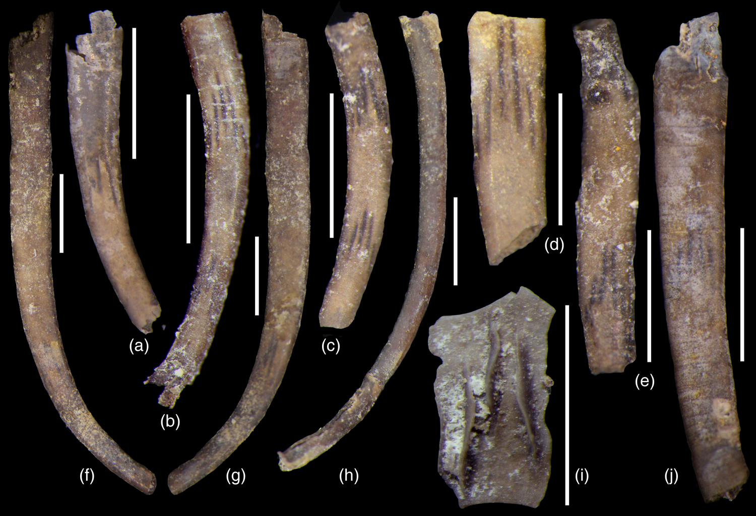
Figure 5 (a–j) Prestephanoscyphus branzovensis sp. nov.; Lochkovian, Lochkov Formation, Kotýs Limestone; locality Bubovice, Branžovy ridge. (a) proximal part of theca showing whorl of apophyses, PCZCU 2297; (b) proximal part of theca showing two whorls of apophyses, PCZCU 2298; (c) part of theca showing two whorls of apophyses, PCZCU 2299; (d) fragment of theca well showing whorl of apophyses, PCZCU 2317; (e) part of theca with constricted diameter, PCZCU 2301; (f, g) holotype, largest known theca, frontal and opposite side, PCZCU 2296; (h) very slender theca, PCZCU 2300; (i) fragment showing three apophyses on internal surface of theca, PCZCU 2383; (j) theca showing annulations of outer surface along with set of apophyses visible from the inner surface, PCZCU 2302. Bar equal to 1 mm. All figures are light photography of uncoated specimens.

Figure 6 (a–kk) Prestephanoscyphus branzovensis sp. nov.; Lochkovian, Lochkov Formation, Kotýs Limestone; locality Bubovice, Branžovy ridge. (a) part of nearly straight theca, PCZCU 2303; (b) part of theca showing annulations, PCZCU 2304; (c, y, z) proximal part of theca and detail of theca showing constriction of theca between external older and narrower younger theca, PCZCU 2305; (d) proximal part of theca with fragment of upper side of basal disc, PCZCU 2306; (e) strongly bent proximal part of theca, PCZCU 2307; (f) inner surface of theca, PCZCU 2308; (g, m) inner surface of theca with whorl of apophyses, PCZCU 2309; (h, i) ripped distal part of theca indicating degree of wall flexibility, PCZCU 2310; (j) chipped theca showing whorl of apophyses, PCZCU 2311; (k, l) parts of thecae showing whorl of apophyses, PCZCU 2312 (k), PCZCU 2313 (l); (n) paratype, proximal part of theca with attachment disc, PCZCU 2355; (o, p, bb) different views to basal part of theca with disc, PCZCU 2314; (q, r) distal parts of thecae showing annulations, PCZCU 2315 (q) and PCZCU 2315 (r); (s, v) cross-sections of thecae at whorl of apophyses, PCZCU 2318 (s) and PCZCU 2321 (v); (t, aa) cross-section of theca at whorl of apophyses and detail of apophyses, PCZCU 2319; (u, w) cross-sections of thecae between whorls of apophyses, PCZCU 2320 (u) and PCZCU 2322 (w); (x, ii) proximal part of theca with attachment disc and its detail showing the constriction between basal and proximal parts of theca (marked by arrow at ii), PCZCU 2323; (cc, jj) proximal part of theca constriction at basal part and detail showing (marked by arrows) the constriction between basal and proximal theca (arrow with *) and borders between annulations in proximal portions of theca, PCZCU 2325; (dd, kk) basal part of theca with disc, PCZCU 2324; (ee) robust disc in upper view, PCZCU 2327; (ff) irregular thin disc in upper view, PCZCU 2328; (gg, hh) basal part of theca with disc adhered to another theca in two views, PCZCU 2326. Bar lengths are in μm. All figures are scanning electron microscope pictures.
Holotype: Theca figured on Fig. 5f, g, specimen PCZCU 2296.
Paratype: Theca figured on Fig. 6n, specimen PCZCU 2355.
Type horizon and locality: Lochkovian; Lochkov Formation, Kotýs Limestone. Bubovice, old section near entry to an abandoned quarry on the north slope of the Branžovy ridge elevation; Central Bohemia, Czech Republic.
Material: More than hundred specimens (mostly fragmented tubular thecae, some with attachment disc or loose attachment discs) preserved by phosphatic matter but few specimens are preserved as phosphatised internal moulds; figured material PCZCU 2296–PCZCU 2318, PCZCU 2320–PCZCU 2328, PCZCU 2355, PCZCU 2364 and PCZCU 2374.
Etymology: After locality name ‘Na Branžovech’.
Diagnosis: Prestephanoscyphus having weakly and regularly expanding bent thecae, changing from basally circular to distally depressed elliptical in cross-section; apophyses blade-like, triangular in tangential view, often having sigmoidally curved crests; apophyses radially arranged, converging adaperturally, ordered in a bilateral symmetry pattern; apophyses discontinuous, forming discrete whorls, first appearing in early theca and with next whorls separated with smooth interior; exterior with broad and uniformly wide, weakly expressed annulations 60–150 μm wide depending on size of theca; thecal wall of medium thickness.
Description: Thecae are slightly to moderately bent since the early growth, mostly becoming less curved in next growth stages (Figs 5a–e, 6n). Theca starts with circular cross-section in early growth stages, becoming moderately oval to depressed elliptical in later growth stages. Theca slightly and regularly widens towards the aperture, with apical angle 4–5° along its entire length. The maximum observed length of the complete theca is 5.75 mm. The estimated length of large theca reaches 10–12 mm but it likely could be even longer. The diameter in basal part of theca is 200–275 μm, increasing to 500 μm in distance 5 mm from the basal disc. The largest observed diameter of theca is 2 mm. Thecal wall is thin and flexible near the aperture but reinforced by more inner layers from the earlier growth stages. Vertical ridges or grooves are not seen in sides of theca. A weak constriction of theca from 425 μm diameter to 300 μm diameter in subsequent growth annulation has been observed (Fig. 5e).
The radial apophyses are arranged in the bilateral symmetry pattern on the inner wall. Apophyses are blade-like in radial view, and low and long triangular in tangential view, some having sigmoidally curved crest. The crests of apophyses are entire and simple, devoid of secondary thickening, rhomb-shaped projections, digitations or similar structures known in other species of the genus. The apophyses are of limited length, arranged in discontinuous sets in whorls externally marked by the darker pigmentation of thecal wall (Fig. 5). Two pairs of longest and largest apophyses are attached to middle part of the inner wall in the planar part of theca. The longest apophyses weakly but distinctly converge toward the aperture. Other apophyses, generally two in each side, are located on the planar inner wall. They are smaller, shorter and more abaperturally placed. The earliest whorl appears shortly after formation of the theca, some 1 mm above the attachment disc. The next whorl appears at 1.6–2.0 mm distance from the first one. Five or more whorls were likely present in the large thecae. There is only a weak variation of the shape, length and arrangement of apophyses in the whorls.
Outer surface of theca bears noticeable annulations, with border lines 100–150 μm apart from each other in the distal part of theca (Fig. 6q) but only 60–80 μm in the proximal part. Annulations are distinct on all parts of theca apart from the earliest basal part near the attachment disc.
The attachment disc is circular to irregularly oval in outline, broadly conical, 500 μm in diameter at its base. The base of the disc may be flat and simple (Fig. 6dd, kk) but there are various modifications of this ideal form. The real shape of the disc depended on the substrate relief and structure. Therefore, small to large indentations and lobes may modify the outer outline of the disc (Fig. 6o, bb, ff). Some discs have a finely to coarsely digitate inner edge and wrinkled or grooved surface, which mimic the superficial structures of the animal body. One specimen (Fig. 6gg, hh) has its basal disc laterally adjoined to the cylindrical tube of another byronid but the lower surface of the basal disc mimics the slightly convex surface of an unknown substrate.
The wall of attachment discs is multilayered. This gives evidence of recurrent deposition of the material which retained only a small open hole at the base of the disc. The thinner cover of the disc floor is often broken off in fossilised specimens.
Remarks: The new species is similar to Prestephanoscyphus devonicus Bischoff, Reference Bischoff1989 from the Lower Devonian (Garra Formation, upper Lochkovian) of New South Wales, Australia. Bischoff (Reference Bischoff1989) noted the presence of narrow, very long blade-like apophyses reaching almost to the centre of the theca in some specimens. It is not uncommon in the Australian species that the number of apophyses is lower than prevalent eight per whorl. Prestephanoscyphus devonicus is represented only by small fragments of thecae, mostly circular in cross-section. The preserved specimens of P. branzovensis are much more complete showing regular increase of diameter and several successive whorls of apophyses in theca. The length of apophyses is uneven in a particular whorl but they are generally shorter that those of P. devonicus. Other species described by Bischoff (Reference Bischoff1989) from Australia differs even more distinctly. Prestephanoscyphus rosemariae Bischoff, Reference Bischoff1989 from the early Llandovery differs by having obviously rhomb-shaped apophyses. The bases of apophyses may grade to equivalently positioned apophysis of the next whorl (Bischoff Reference Bischoff1989). Prestephanoscyphus boreensis Bischoff, Reference Bischoff1989 from the early Wenlock differs by shorter apophyses from P. branzovensis. The same feature distinguishes also Prestephanoscyphus cobcreensis Bischoff, Reference Bischoff1989 of late Llandovery age.
Dzik et al. (Reference Dzik, Baliñski and Sun2017) described and figured a tubular fossil which likely belongs to our new species. The tube referred by Dzik et al. (Reference Dzik, Baliñski and Sun2017) to Sphenothallus ruedemani (Kobayashi, Reference Kobayashi1934)? shows a similar arrangement of internal apophyses and general outline of theca. The specimens come from the Early Devonian Mytkiv Formation of Podolia, Ukraine. This formation is likely coeval with the Lochkov Formation and the brachiopod fauna of the Mytkiv Formation (Baliński Reference Baliński2012) resembles that observed at Bubovice, Bohemia (Mergl Reference Mergl2003). We are not convinced that all Byronia-like tubes and rings figured by Dzik et al. (Reference Dzik, Baliñski and Sun2017) belong to a single long-lasting biological species.
The circular discs of similar shape we describe, occurring usually loose and reported from elsewhere as Phosphannulus universalis Müller et al., Reference Müller, Nogami and Lenz1974 were already referred to byronids (Holmer Reference Holmer1987; Bischoff Reference Bischoff1989; Holmer in Malinky et al. Reference Malinky, Wilson, Holmer, Lardeux, Webby, Paris, Drsoser and Percival2004). However, they also were considered to be the fixation discs of conulariids (Bischoff Reference Bischoff1973) or putative endobiont of crinoids (Welch Reference Welch1976; Werle et al. Reference Werle, Frest and Mapes1984). Attachment discs of Sphenothallus are of a similar outline but their affinity is left open (Ferreti et al. Reference Ferreti, Cardini, Crampton, Serpagli, Sheets and Štorch2013). Some other authors (Dzik et al. Reference Dzik, Baliñski and Sun2017) suggest that byronids are merely the basal portions of sphenothallid shells. We provide here undisputable direct evidence that the basal attachment discs in our material from the Kotýs Limestone belong to this byronid species because several specimens of P. branzovensis display the disc prolonged into the narrow cylindrical basal part of the theca. After the formation of the primordial phosphatic wall of the basal disc, the prolonged growth formed a short thin-walled cylindrical basal part of a subcylindrical theca (Fig. 6bb, ii). This basal part is clearly defined by the growth line and/or weak constriction of the theca (Fig. 6x, ii, jj). A distinct circle is seen on the inner surface of the basal disc (Fig. 6kk) marking the border line between attachment disc and the earliest basal part of a tubular theca.
Occurrence: Only the type (1) locality.
Prestephanoscyphus robustus sp. nov.
Figure 4aa–gg, 9a
Holotype: Fragment of theca with preserved apophyses, figured in Fig. 4cc, dd, specimen PCZCU 2357.
Paratypes: Incomplete theca, figured in Fig. 4bb, specimen PCZCU 2356; incomplete theca figured in Fig. 4gg, specimen PCZCU 2319.
Type horizon and locality: Uppermost Eifelian; Srbsko Formation, ‘upper dark interal’ (=equivalent of the Kačák Member); Nowakia otomari Zone, Polygnathus ensensis Zone. Koněprusy, west-facing low wall of Jirásek's Quarry; Central Bohemia, Czech Republic.
Material: Five fragmental tubes and one disc; figured material: PCZCU 2319, PCZCU 2329, PCZCU 2356, PCZCU 2357 and PCZCU 2379.
Etymology: After unusually thick wall of theca.
Diagnosis: Prestephanoscyphus having regularly expanding, straight to weakly bent theca changing from basally circular to distally depressed elliptical cross-section; apophyses short, lamellose, two in each flatter side of distal theca; annulations weakly defined, 20–25 μm wide; wall of theca very thick relative to the size of theca.
Description: The theca is slender, 300–600 μm in diameter, weakly expanding, thick-walled (70–100 μm), subcircular in cross-section in the proximal part of theca, changing to elliptical in cross-section in the distal part of theca. Weakly defined annulations are observable on the outer surface, with border lines only 20–25 μm apart from each other (Fig. 4ee, ff). Thin blade-like converging apophyses are present on interior of theca, arranged in one pair on each opposite subplanar side of the theca (Fig. 4cc, dd). The associated attachment disc (Fig. 4aa) is sturdy, thick walled, attaining 500 μm diameter and with a small Y-shaped basal opening.
Remarks: Prestephanoscyphus robustus sp. nov. differs from P. branzovensis sp. nov. by the very thick wall of the theca relative to its diameter and by distinctly narrower annulations. This species is the stratigraphically youngest known species of the genus so far observed. The earliest known species of the genus is P. boreensis Bischoff, Reference Bischoff1989 which comes from the lower Llandovery (New South Wales, Australia) while P. devonicus Bischoff, Reference Bischoff1989 comes from the upper Lochkovian of the same area. The Bohemian species P. branzovensis sp. nov. is also of Lochkovian age. Rare discs were found associated with the thecae of P. robustus (Fig. 4aa). Width of a broad circular groove on their upper surface reflects the thickness to thecal wall and clearly supports an assignment of this disc to the same species.
Occurrence: Only the type (8) locality.
Genus Parabyronia gen. nov.
Type species: Parabyronia elegans sp. n.; Emsian; Zlíchov Formation, Chýnice Limestone; Central Bohemia, Czech Republic.
Diagnosis: Byronid with thick-walled theca of spindle-shaped cross-section, externally ornamented by low transverse ridges, sides with acute edge of crowded spines with tips directed towards the aperture of theca; interior with fine thread-like structure arranged with bilateral symmetry pattern; wall structure multi-layered, of alternating compact and granular laminae.
Remarks: The genus differs from Byronia by distinct regular transverse ridges on exterior of the theca. Some species referred to Byronia (Vinn et al. Reference Vinn, Kirsimäe, Parry and Toom2016) also have the transverse ridges but these are less regularly arranged and are of uneven size. The acute edges of theca forming its spindle-shaped cross-section differs from the rounded sides of Byronia and Prestephanoscyphus. Unlike Byronia and Prestephanoscyphus, short spine-like prolongations terminate tips of both side edges at the aperture. Parabyronia has a smooth internal surface unlike the several whorls of internal apophyses present in Prestephanoscyphus. Similarly shaped and sized Sphenothallus lacks regular transverse ridges on surface of theca and wall of its theca is thin and often deformed. The mineralisation of Sphenothallus may be weak (Vinn & Kirsimäe Reference Vinn and Kirsimäe2015) but the shared feature of Parabyronia and Sphenothallus is presence of thickened lateral edges forming resistant rods.
Species assigned: Parabyronia elegans sp. nov.
Parabyronia elegans sp. nov.
Figure 7
Holotype: Fragment of theca, figured in Fig. 7b, i, k, specimen PCZCU 2359.

Figure 7 (a)–(x) Parabyronia elegans sp. nov.; upper Emsian, Zlíchov Formation, Chýnice Limestone; locality Bubovice, hillside of Čeřinka elevation. (a) paratype, proximal part of theca, PCZCU 2358; (b, i, k) holotype, part of theca and its details, PCZCU 2359; (c, d) fragments of thecae, PCZCU 2360 (c) and PCZCU 2361 (d); (e–g) fragment of theca in oblique, lateral and front views, PCZCU 2363; (h) part of theca showing healed walls, PCZCU 2367; (j, n, o) fragment of theca and details of its naturally broken wall showing its lamellar structure, PCZCU 2362; (l, m) sides of thecae in external lateral view, PCZCU 2366 (l), paratype PCZCU 2365 (m); (p) attachment disc which perhaps belongs to the species, PCZCU 2368; (q) inner surface of theca, PCZCU 2369; (r, s, v) inner surface of theca showing thread-like structures, PCZCU 2370; (t) side of theca in oblique view showing interior, PCZCU 2371; (u) side of theca in internal side view, PCZCU 2372; (w, x) inner surface of theca showing transverse thread-like structures and irregular scratches, PCZCU 2373. Bar lengths are in μm. All figures are scanning electron microscope pictures.
Paratypes: Proximal part of incomplete theca, figured in Fig. 7a, specimen PCZCU 2358, fragment of side part of theca figured in Fig. 7m, specimen PCZCU 2365.
Type horizon and locality: Emsian; Zlíchov Formation, rosy Chýnice Limestone; Nowakia barrandei to Nowakia elegans zones. Bubovice, hillside of the Čeřinka elevation; Central Bohemia, Czech Republic.
Material: Four tubular incomplete thecae and 15 fragments of flat sides of thecae and thecal edges, figured material: PCZCU 2258–PCZCU 2263 and PCZCU 2265–PCZCU 2273.
Diagnosis: As for genus.
Description: The theca is flat tubular, gently expanding to almost parallel sided, thick-walled, likely longer than 10 mm (estimated from length/width ratio of fragments and their length). Cross-section is broadly (in proximal part) to narrowly (in distal part) spindle shaped, with acute lateral edge. The proximal width of theca is less than 400 μm judging from the smallest part of theca observed (Fig. 7a), the shape of outline and the larger diameter of associated attachment disc (Fig. 7p). The wall of theca is thick, multilamellar (Fig. 7j, n, o), with lateral edges (Fig. 7m, t) thicker than the weakly bent median part of the theca.
Exterior of theca is covered by prominent transverse wrinkles arranged in regular intervals (Fig. 7b, d, e, m) although denser ridges are sporadically present in some parts of the surface (Fig. 7c, d). General shape of ridges recalls the marine current ripple-marks. Crests of ridges are only 40 μm apart from each other in proximal part (Fig. 7a) to 120–140 μm in distal part of the theca. The courses of ridges are weakly uneven except for the most lateral parts of ridges which are gently bent backwards. Acute edge on each side of the theca is formed by an aligned broad spinose prolongation which shortly projects frontally at the aperture of theca.
Almost the entire inner surface of the theca is covered by thread-like ornament, which was likely imprinted by a superficial striation or wrinkled parts of the byronid animal body. These wrinkles are arranged in a sheaf-like structure along the axial part of the low vaulted part of theca, with tips facing the thecal aperture. The wrinkles run longitudinally and become curved obliquely to almost transversally sidewards. They disappear at thickened lateral sides of the theca where a stepped inner surface is developed. This arrangement of threads repeats once more at the same fragment of theca, indicating a repetitive deposition of phosphatic substance on the inner wall of theca.
Associated discs are elongate oval, with maximum inner diameter 440 μm. Basal opening is oval, surrounded by a thin shelf ring. Outer surface is steeply sloping toward the base.
Remarks: The new species differs from other known byronids from the Devonian of Central Bohemia by its larger size, unique external ornament and acute lateral edges with short spinose projections above the apertural margin. The attachment disc is referred to the new species due to the comparable size of inner diameter and, above all, the shape and character of the transverse outline which follows up with the outline of the proximal part of theca.
One fragment of theca (Fig. 7h) is unique in having two sets of deep incisions which were internally pasted by material devoid of transverse ridges. This indicates the ability of the byronid animal to withstand a strong attack/impact and repair the heavily damaged theca. Another small fragment of theca (Fig. 7w, x) shows a tangle of short, more or less similarly oriented scratches. These scratches look like traces after gnawing of soft tissues from a solid phosphatic wall of the theca.
Occurrence: Only the type (3) locality.
Order Conulariida Miller & Gurley, Reference Miller and Gurley1896 emended Moore & Harrington, Reference Moore, Harrington and Moore1956
Genus Pidiconularia gen. nov.
Type species: Pidiconularia tubulata sp. n.; Emsian; Zlíchov Formation, Chýnice Limestone; Central Bohemia, Czech Republic.
Diagnosis: Minute phosphatic test of conulariid appearance, gently curved to almost straight, very weakly expanding toward the aperture; test roundly quadrate in cross-section; faces gently convex; test wall thin but thicker at corners where broad low internal carina is developed; external transverse ribs of uniform size, thin, gently bent toward aperture; crests of transverse ridges bear small tubercles of uniform size; internal face of test smooth, lacking distinct midline.
Remarks: The test of the type species looks like a very small conulariid. However, evenly sized external ornament and almost cylindrical test justifies an assignment of the type species to the new genus. This form of test hardly represents a juvenile stage of any larger conulariid taxon because: (i) the width of distal part of the test is almost constant, without any distinct expansion toward the aperture indicating a tiny size of the adult specimens; (ii) size and density of transverse ribs is far below the lower size limit of transverse ribs of common conulariid genera; (iii) associated true conulariid fragments have much coarser transverse ribs and much coarser spines or tubercles on external surface; (iv) interiors lack a septum at the midline; and (v) there is no corner groove. We found no apertural structure including rests of lappets. However, the general resemblance to the conulariid test and tetramerous symmetry justify attribution of Pidiconularia to the order Conulariida Miller & Gurley, Reference Miller and Gurley1896.
Among conulariid genera only a few taxa are comparable in test size to our material. Pidiconularia and Microconularia Percival, Reference Percival2009 are very small, have rounded corners, very weakly expanding test at an apical angle of less than 3° and lack a midline. However, Pidiconularia has very fine transverse ridges with a row of small tubercles. Microconularia fragilis Percival, Reference Percival2009 bears coarse, widely and evenly spaced arched transverse ribs without tubercles. Another small-sized genus Climacoconus Sinclair, Reference Sinclair1942 is distinct by alternate transverse ribs. Teresconularia Leme et al., Reference Leme, Heredia, Rodrigues, Simões, Aceñolaza and Milana2003 has considerably widely expanding test and possesses a distinct midline.
Pidiconularia tubulata sp. nov.
Figure 8
Holotype: Partly broken test figured in Fig. 8a, specimen PCZCU 2375.
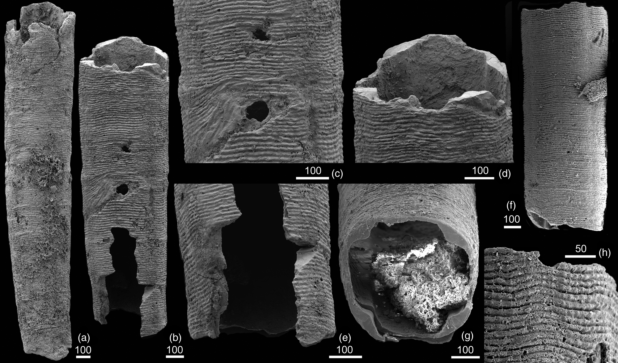
Figure 8 (a–h) Pidiconularia tubularia sp. nov.; upper Emsian, Zlíchov Formation, Chýnice Limestone; locality Bubovice, hillside of Čeřinka elevation. (a) holotype, part of test, PCZCU 2375; (b–e) paratype, part of test showing healed wall and detail of test, PCZCU 2376; (f–h) paratype, part of test, its cross-section and detail of exterior, PCZCU 2377. Bar lengths are in μm. All figures are scanning electron microscope pictures.
Paratypes: Incomplete test with healed injury figured in Fig. 8b–e, specimen PCZCU 2376 and incomplete test figured in Fig. 8f, g, h, specimen PCZCU 2377.
Type horizon and locality: Emsian; Zlíchov Formation, rosy Chýnice Limestone; Nowakia barrandei to Nowakia elegans zones. Bubovice, hillside of the Čeřinka elevation; Central Bohemia, Czech Republic.
Material: Three incomplete tubular tests, figured material: PCZCU 2275–PCZCU 2277.
Diagnosis: As for genus.
Description: The test is tubular, gently to almost imperceptible expanding adaperturally, straight or slightly bent in the proximal part (Fig. 8a, b, f). Estimated length may reach 4 mm or even more. The cross-section of test is roundly quadrate, with more convex corners and less curved faces (Fig. 8g). Corner grooves and midlines are not perceptible externally. A broad and low carina is present on interior of each corner (Fig. 8d, g) indicating the tetrameric symmetry. Median septum is absent. Transverse ridges evenly cover the test exterior. Ridges are of uniform size, 70–90 in number per 1 mm. Ribs are gently bent toward the aperture. Narrow and deep groove separates the individual ribs. Each rib bears some 5 μm sized tubercles aligned along the crest of rib. The test interior is smooth.
Remarks: Affinities to other conulariid taxa are discussed above. The specimen PCZCU 2376 shows a repaired test. Initially, the test was obliquely broken (Fig. 8b, c). The lost part of test was healed by a rapidly formed sheet which bears oblique and irregular wrinkles. The sheet was likely thinner and more fragile. This is evident from a hole penetrating this sheet. The regular growth of test continued after a complete repair. The same growing structures, namely the transverse ribs, before and above the repaired sheet indicate that the tissues producing the test were not seriously damaged.
Occurrence: Only the type (3) locality.
4. Discussion on morphological aspects
Features and function of the byronid thecae are discussed in this section.
4.1. Structure of byronid theca
Unlike other byronids from Central Bohemia, a few thecae of Prestephanoscyphus branzovensis sp. nov. display favourably preserved microstructure of the tube wall. Although the thickness of the tube apparently depends on age of the animal and location on the theca an average thickness is 10–30 μm and is almost constant over circumference at particular parts of the tube. The fossilised tubes consist solely of thin laminae of apatite. The original amount of organic substance is unknown but may be estimated from degree of porosity of thecal layer. The development and thickness of laminae are variable. Laminae are 0.55–2.5 μm thick, with sharp boundaries. Lateral changes through laminae are inconspicuous. The microstructure of laminae is homogeneous. Two types of laminae are distinguished. The first type are the compact laminae of <500 nm long (c-axis) and approximately 50 nm broad (a-axis) columnar crystallites of apatite. Similar elongate crystals are known from the tube wall of the related cnidarians Torellella and Sphenothallus (Vinn Reference Vinn2006, Vinn & Mironenko Reference Vinn and Mironenko2021). Their c-axes are generally subparallel with the external surface of the tube and margin of the aperture; deviations of their c-axes from this subparallel orientation are less than 30° (Fig. 9h, i). The second type is formed by clusters of small apatite crystallites leaving small space and chambers around. Crystallites have short c-axis forming more or less isometric grains approximately 50 nm in size. Granulose laminae regularly alternate with compact laminae. Both types of laminae have a comparable thickness. Observed fracture in the compact lamina (Fig. 9g) which follows the plane between aligned crystallites indicates that compact lamina was more tough but fragile. The granulose laminae originally supported a flexibility of the tube due to assumed higher content of organic substance incorporated between clusters of apatite crystallites. The ‘sandwich’ like structure of wall allowed some flexibility together with toughness necessary for a long thin dwelling tube erected above substrate. The laminated structure also indicates a regular alternation of metabolic activity of the ectoderm.

Figure 9 (a) Prestephanoscyphus robustus sp. nov; upper Eifelian, ‘upper dark interval’ above the Acanthopyge Limestone; locality Koněprusy, east wall of Jirásek Quarry. Part of theca showing the detached and deformed externalmost lamella, arrows indicate borders between annulations, PCZCU 2379. (b–i) Prestephanoscyphus branzovensis sp. nov.; Lochkovian, Lochkov Formation, Kotýs Limestone; locality Bubovice, Branžovy ridge. (b, c) surface of inner lamina of theca; wrinkles and normal fila are preserved on outer surface of one of internal microcrystalline lamella that imprints wrinkled microbial (?) mat; rope-like structures are phosphatic infillings of endoboring of ichnogenus Orthogonum ichsp., PCZCU 2364; (d–i) cross-section of theca (d); arrow indicates part of theca magnified on figure (e and f; e–g) section showing alternation of microcrystalline (mc) and granular (gr) laminae, note the fracture splitting the microcrystalline lamina on g (indicating boundary of potential detachment of layers) and by arrow marking the layer of diagenetic crystallic apatite attached to inner wall of the theca in (f; h, i) details of (f) and (g), showing microcrystalline (mc) and granular (gr) laminae of thecal wall, PCZCU 2378. Bar lengths are in μm. All figures are scanning electron microscope pictures.
Microstructure of apophyses is less distinct. Apophyses seem to be homogeneous, lacking lamination. Their bases are attached with a sharp boundary to the innermost concentric lamina of theca.
The most external layer of the theca was observed to be less than 1 μm thick. One specimen of Prestephanoscyphus robustus (Fig. 9a) has the external layer detached from the underlying layer. The detached layer is ripped and folded and is the only layer where the borders between annulae are preserved (Fig. 9a). The structure of this outer layer is unknown but apparent flexibility indicates a higher content of the organic substance in relation to compact lamina of larger columnar crystallites inwards to the wall.
Inner surfaces of preserved tubes are commonly covered by a thin, about 700 nm thick, sheet of apatite crystallites (Fig. 9e, f). These are much larger than crystallites forming the laminae of thecal wall, having size about 600–800 nm, that is, a-axis (crystallites forming the tube wall are 50 nm sized in average a-axis). These larger crystallites are definitely secondary and therefore of a diagenetic origin. This larger form of apatitic crystallites assembled precise infillings of microboring traces (Fig. 9b, c).
We interpret the microcrystalline structure of lamellae of Prestephanoscyphus as the original or only weakly recrystallised structure of the theca. A similar extremely thin multilamellose structure is known in Ordovician Sphenothallus (Van Iten et al. Reference Van Iten, Cox and Mapes1992; Vinn & Kirsimäe Reference Vinn and Kirsimäe2015), Phosphannulus (Müller et al. Reference Müller, Nogami and Lenz1974) and conulariids (Van Iten Reference Van Iten, Simonetta and Morris1991, Reference Van Iten1992; Ford Reference Ford2011; Ford et al. Reference Ford, Van Iten and Clark2016; Mergl et al. Reference Mergl, Ferrová and Frýda2016; Van Iten et al. Reference Van Iten, Mironenko and Vinn2022). A similar laminated structure is apparent in tubes of Parabyronia elegans sp. nov. All these animal groups strengthen the wall theca from inside and utilise any form of carbonate apatite and organic substance or organic substance alone (Van Iten et al. Reference Van Iten, Cox and Mapes1992; Vinn et al. Reference Vinn, Kirsimäe, Parry and Toom2016; Vinn Reference Vinn2022). The pristine test composition of these groups, origin and relation of mineral and organic laminae have been discussed by several authors (see Muscente & Xiao Reference Muscente and Xiao2015; Chang et al. Reference Chang, Clausen, Zhang, Feng, Steiner, Bottjer, Zhang and Shi2018; Van Iten et al. Reference Van Iten, Leme, Simões and Cournoyer2019) and is not repeated here.
4.2. Growth of byronid theca
Bischoff (Reference Bischoff1989) briefly described the mode of growth of the theca. He noted that it grew in apertural direction increasing in length and more or less noticeably in width. He noted two types of layers (Bischoff Reference Bischoff1989; p. 477) with the inner one composed of several lamellae. Our observation slightly differs from those of Bischoff (Reference Bischoff1989). The specimen figured in Fig. 9d shows two types of layers, with the thin external layer contrasting with several alternating layers inside the wall. Each of the internal layers reinforced the thecal wall and gradually strengthen especially basal and proximal parts of theca. Total number of laminae could be 40 or even more. Their number is perceptible in cross-sectioned naturally corroded attachment discs (Fig. 4t–v).
Although the attachment disc may be conspicuously thick, the floor of the disc stays always open by more or less regular opening. Van Iten (Reference Van Iten1992) suggests that similar holdfast of Sphenothallus was floored by a thin basal membrane. The earliest wall of the disc immediately flanking this central opening is very thin, having the lobate edges (Fig. 6ff). Central opening indicates the permanent contact of soft basal part of byronid body with the surface of substrate. We suggest that this part of the body has a form and function similar to the pedal disc of extant polyps. The apatite lamellae were secreted along the periphery of such a pedal disc and along the adorally growing cylindrical body. The surface of secretory ectodermal cover of the animal body was nearly or entirely smooth leaving the inner surface of theca essentially featureless. Only the lowest part of the body, just above the pedal disc, formed an annulus which is obvious as the circular ring between the disc and the earliest portion of the tube. It is evident that secretion of new lamina covered the entire inner surface of theca, that is, from the attachment disc to the apertural edge of the theca, because no growth lines were observed inside the theca.
4.3. Bilateral symmetry of byronids
No doubt the theca of byronids was flexible and its cross-section changed with growth. The proximal part of the theca was circular in cross-section. It became more and more oval with progressive growth until its long apertural axis could be twice the short one. This flattening is not only post-mortem (cf. Bischoff Reference Bischoff1989). This natural change is also evident from shaping of apophyse whorls which indicates switch to bilateral symmetry from alleged tetramerous symmetry of the earliest growth stages. Unlike Bischoff (Reference Bischoff1989) we interpret the adorally convex-up arrangement of apophyses (Fig. 5a–e) on opposite sides of theca as the result of original bilateral symmetry. We have no direct data about symmetry of the larva because subcircular nature of discs does not exclude either bilateral or radial symmetry.
Suggested bilateral symmetry of byronids is comparable with the absence of tetramerous symmetry in the likely related cnidarian Sphenothallus. A discovery of paired tentacles in Sphenothallus (Fauchald et al. Reference Fauchald, Stürmer and Yochelson1986) does not militate against its cnidarian affinity (Van Iten et al. Reference Van Iten, Cox and Mapes1992). Similar structure of holdfast and thin lamellose mineralised theca of Sphenothallus are very similar to those of byronids. Spindle-shaped or oval cross-section in byronids and sphenothallids support the model of secondary bilateral symmetry of these cnidarians. However, we do not adopt the conclusion of Dzik et al. (Reference Dzik, Baliñski and Sun2017) about identity of Sphenothallus and byronids. Although most cnidarians are characterised by radial symmetry, biradial or bilateral symmetry is not uncommon in modern cnidarian taxa, especially among some anthozoans (Ruppert et al. Reference Ruppert, Fox and Barnes2004).
4.4. Function of byronid apophyses
Bischoff (Reference Bischoff1989; p. 482) suggested that apophyses were not used for attachment of muscles. This contrasts to the assumed function of internal carinae of conulariids and Stephanoscyphus (Van Iten Reference Van Iten, Simonetta and Morris1991). Bischoff (Reference Bischoff1989) assumed that apophyses supported the rigidity of thecal wall. With caution we reject this interpretation. While apophyses of Australian Prestephanoscyphus rosemaria are very long and their whorls seemingly support the rigidity of the wall leaving only short breaks between consecutive ‘supported’ parts, in P. branzovensis the apophyses are short and sparsely arranged (Fig. 5b, c, e, j). Smooth surfaces between whorls are much extensive than part ‘supported’ by a whorl of apophyses. Such supporting structure would not work well.
The supporting function of apophyses for enlargement of the digestive surface of gastric septa is also questionable. Short apophyses provide little support due to their short length discontinuity inside the theca. We argue for a passive defensive function of apophyses. The whorl of apophyses reduces the size of aperture and divides it in to several much smaller interstices. This constriction makes a barrier relevant for protection of the byronian body. The withdrawn body survived behind the nearest whorl of apophyses because a predator cannot enter the theca through the aperture. Several whorls of apophyses make sense when the byronid body got bigger with age. The function of byronid short lamellose apophyses imitates the checked protective function of denticles and lamellae in otherwise unrelated gastropods. For instance, the shell aperture of terrestrial gastropod genera Chondrina, Gastrocopta, Granaria, Vertigo and many others is constricted by numerous denticles and plicae which avert entering of their predators (terrestric planarians, predatory gastropods, larvae of Diptera, etc.) inside the shell.
4.5. Mode of life
Byroniids from the Devonian of Central Bohemia prove that Phosphannulus is, at least in part (for different interpretation see Suttner & Kido Reference Suttner and Kido2020), the attachment structure of byronids. Co-occurrence of attachment discs with more tubular thecate fossils indicates that byronids, sphenothallids and conulariids utilised various substrate for firm and stable attachment (see Van Iten et al. Reference Van Iten, Leme, Simões and Cournoyer2019) although the semi-infaunal mode of life of some tubes not associated with discs cannot be excluded (cf. Thayer Reference Thayer1975; Van Iten et al. Reference Van Iten, Tollerton, Ver Straeten, Leme, Simões and Rodrigues2013). The solid bases could be rocks, blocks, stones, pebbles, shells and also seaweed fronds. The latter two cases signifying epibiontic mode of life of byronids on alive hosts was directly documented by deformed crinoid stems (Welch Reference Welch1976; Werle et al. Reference Werle, Frest and Mapes1984) and organophosphatic brachiopods (Holmer in Malinky et al. Reference Malinky, Wilson, Holmer, Lardeux, Webby, Paris, Drsoser and Percival2004). Inferred from the preservation and abundance of tubes (see below) and a character of associated fauna, abundant associated brachiopods were promising candidates to the hosts of byronids in the Kotýs Limestone at Bubovice, Branžovy ridge. However, direct evidence has not been found (cf. Mergl Reference Mergl2021). Only one observed byronid specimen of Prestephanoscyphus branzovensis attached to another byronid tube proved the epibiontic mode of life (Fig. 6gg, hh) and one byronid specimen was observed inside a malformed crinoid stem with apparent stereomic response of the live host.
5. Discussion on general aspects of distribution
Distributional patterns and influence of taphonomy on phosphatic tubes are subjects of the following discussion resulting in a brief general comment on phosphatic shells supporting the model developed and discussed by Kraft & Mergl (Reference Kraft and Mergl2022).
5.1. Occurrence of phosphatic tubes
The tubes are described from a limited number of Devonian localities in the Prague Basin. Their stratigraphic range is almost throughout the entire Devonian succession (Fig. 10) and various limestone facies along the whole basin (Figs 1, 2). As stated above the described tubes with basal discs, regardless of their taxonomic classification and affinities, needed an appropriate base for their attachment. This mode of life apparently controlled the infrequent, only locally abundant occurrences of these fossils depending on the substrate distribution. In addition, common occurrences of relatively large but still tiny fragments to almost complete tubes of Prestephanoscyphus branzovensis at Bubovice, Branžovy ridge indicate a short transport in association with other fossils that eliminated grinding after their detachment.

Figure 10 Stratigraphic ranges of the studied taxa and their abundances in the order scale.
An absence of pebbles in the sediment and lack of rocky bottom or hard ground nearby point to preference of the above mentioned epibiontic mode of life rather than sessile on the inorganic substrate. Two theoretical models can be considered for this mode of life: (1) attachment to solid shells from inorganic compounds (carbonates or phosphates); and (2) organic substrate that can be decayed. Both cases also provide an explanation for the common occurrences of detached holdfasts with preserved basal plane by their attachment to living organisms rather than to remains of dead individuals.
(1) No remain of byronid from the Devonian of the Barrandian area was observed directly attached to the firm brachiopod, trilobite or other shell. It can likely be due to the presence of organic periostracal layer on the exterior of brachiopod shells. This was a subject of early microbial decay after death of a brachiopod and released the attached byronid thecae. Indeed, no holdfast was also observed on brachiopod shells, alive and dead, at Bubovice (Mergl Reference Mergl2021).
(2) Seaweeds should be considered as alternative substrate for attachment of byronids. Although this suggestion is theoretical, modern cnidarians often utilise algae as a substrate (e.g., Ronowicz et al. Reference Ronowicz, Włodarska-Kowalczuk and Kukliński2013) and similar epiphytic habitat can be suggested for their fossil relatives. In addition, most of the studied specimens occur in sites which may have been located within the photic zone as inferred from associated benthic fauna. However, no remaining traces of seaweed surfaces have been found on ring bases of byronids after their detachment from surface of the decaying fronds. There is also no direct evidence of accompanying seaweeds.
If the byronids were attached to the rocks they could be preserved almost only broken off and transported to short distances in suspension of agitated seawater, typically to concentrations of allochthonous remains; preservation of complete holdfasts would be rather of a low potential in such case. The rocky or stony bottom with its sessile dwellers has been only exceptionally preserved in general. Thus, byronids and other tiny phosphatic tubes are of a low potential to be directly proved in situ in this habitat.
The occurrences of byronids are apparently taphonomically biased even if the resistant phosphatic shells themselves are of a good preservational potential. On the other hand, their globally sparse Devonian record suggests that they belonged to a minor component of the Devonian communities. Compared to their Cambrian and also Ordovician distribution (see references above) a decline of byronids and also other groups producing phospatic tubular shells is obvious. Due to a successful life benthic strategy inside a tube testified by number of other tubaceous benthic animals through the entire Phanerozoic, from the earliest mineralised shells (Murdock Reference Murdock2020) until modern seas (e.g., annelids), this decline should be related to other factors.
5.2 Decline of byronids
The decline of byronids and other phosphate-shelled animals (conulariids, sphenothallids, organophosphatic brachiopods and phyllocarid crustaceans) was likely related to changes of phosphorus cycle in the Devonian (Kraft & Mergl Reference Kraft and Mergl2022). Rapid evolution of the pelagic jawed vertebrates not only increased the predation in marine environments and gave rise to restructuralisation of marine ecosystems (e.g., Bambach Reference Bambach and Valentine1985; Klug et al. Reference Klug, Kröger, Kiessling, Mullins, Servais, Frýda, Korn and Turner2010, Harper et al. Reference Harper, Cascales-Miñana and Servais2020) but concentration of phosphate in bones, teeth and scales of large vertebrates dramatically changed the phosphorus cycle in the Devonian seas (Kraft & Mergl Reference Kraft and Mergl2022). The byronids, together with other benthic suspension-feeding invertebrates were heavily affected by decrease of accessibility of phosphate near a sea bottom. This restriction had several consequences which could be traced in the Mid to Late Palaeozoic phosphate-shelled invertebrates: (i) decreasing shell size (way used by some discinoid brachiopods or conulariids such as dwarf Pidiconularia); (ii) reduction of biomineralisation, with partial or weak mineralisation of shell (way used by some conulariids but also some byronids); and (iii) rapid (byronids, siphonotretid and acrotretid brachiopods) to gradual (conulariids) extinction. Indeed, the byronids were one of the first victims of this change.
6. Conclusions
New material of byronids and similar tubular fossils from the Lower and Middle Devonian of the Barrandian area, Czech Republic, shed new light on understanding phosphatic tubular fossils not only in the Devonian but also in a broader perspective and context.
Byronids are generally rare among phosphatic fossil records in the Barrandian area but their remains are easily identifiable by regular annulations of the most external layer of their theca if it is preserved. Phosphatic circular rings, at least most of their fossils’ records, described by Müller et al. (Reference Müller, Nogami and Lenz1974) and named Phosphannulus are the holdfast structure of byronids. This proves the sessile mode of life similar to that known in Sphenothallus and conulariids. The wall of byronid theca was multilayered with alternation of compact phosphatic laminae of cylindrical crystallites and laminae of isometric crystallites embedded in the suggested organic matrix. The sandwich structure of the theca supports both hardness and flexibility of the test. This structure is essentially identical with the structure of wall observed in Sphenothallus. The strengthening of the byronid theca came exclusively from the inside and new lamina covered the entire inner face of the theca except for the basal central part of the holdfast. This basal opening likely accommodated the soft pedal disc of a scyphozoan polyp character. Whorls of apophyses of Prestephanoscyphus interior likely had a protective function and show a bilaterally symmetrical arrangement. This together with the oval outline of aperture in the distal part of byronid theca refer to bilateral instead of tetramerous symmetry of byronid animal. This feature is shared with Sphenothallus and indicates closer affinity of byronids to sphenothallids rather than to conulariids and coronate scyphozoans.
Diversity of small sessile animals with tubular phosphatic tests was higher in some Devonian environments. This is proved by associated byronids Byronia sp. A, Parabyronia elegans and a dwarf conulariid Pidioconularia tubulata in the Chýnice Limestone of the Emsian age.
The studied tubular organisms were relatively ecologically undemanding. As the sessile and probably microphagous organisms they were apparently not hindered in their potential dispersion and prosperity by a lack of food or substrate as supported by other groups with similar trophic and habitat demands. Therefore, their decline has to be related to other factors. An increasing restriction of phosphate accessibility was probably one of key causes. Deteriorated ability of biomineralisation of phosphatic shells tend toward the extinction or to creation of only organic-walled protection of body, or ultimately, to unprotected naked body. Byronids were victims of a combination of circumstances in a long-lasting trend for phosphorus. The critical decline coincided with the rise of vertebrates and their boom in the Devonian. In that time phosphorus switched from abundant to the limiting biogenic element.
7. Acknowledgement
We thank two anonymous reviewers for their valuable comments and language corrections that improved our manuscript.
8. Financial support
This paper was financially supported by a grant from the Czech Science Foundation 23-05217S. It was also supported by the institutional budget (SGS-2022-11) of the University of West Bohemia (to M. M.) and by Charles University, Prague, through the project Cooperatio GEOL (to P. K.).
9. Competing interests
None.


 Open access