


Introduction
Glaciological studies at t-3 were carried out as part of a research programme of the Arctic Institute of North America under contract to the Geophysics Research Directorate, Air Force Cambridge Research Laboratories. Professor Ukichiro Nakaya, who directed the research project, unfortunately died in 1962. The authors express their heartfelt regret over Professor Nakaya’s death and their sincere thanks for his guidance throughout the work.
The field study was carried out in 1960. After completing the field investigations, the ice samples were packed in dry ice and carried back to Hokkaido University, where supplementary physical investigations were made in the cold room of the Low Temperature Institute.
Core drilling through the entire thickness of the island was accomplished by the authors. The core sample study and surface morphology results are presented in this paper, together with a schematic diagram of the structure of t-3, as inferred from the new data.
Surface Morphology of T-3 Ice
Candle ice
When lake ice begins to melt in the spring it usually displays a candle-like structure. It is, in fact, called candle ice. Candle ice is abundant on t-3, and is very useful when a large amount of ice is needed, because ice with this structure is easy to crush.
The nature of candle ice was studied for 200 samples taken from seven sites at t-3. A candle contains many bubbles and is characterized by internal melting when exposed to solar radiation. This internal melting takes place primarily in a vertical direction and, as a result, many vertical columnar voids are formed. Axis orientation of the samples was determined by observing Tyndall figures. A whole candle was usually one single crystal. Figure 1 is a photograph of a bundle of candle ice taken between crossed polaroids. Each column or candle is a single crystal containing many bubbles. A bundle separates into component candles when exposed to solar radiation, since melting starts chiefly along the crystal boundaries. Inside each individual crystal, melting also occurs, starting from the enclosed bubbles, and forming the many vertical columnar voids mentioned above. In order to show this structure, vertical and horizontal sections were made of single crystals, and prints of the faces were made by applying soft paper and rubbing with black ink. Figure 2 shows prints of the vertical and horizontal faces of two crystals. Sections (a) and (b) are of ice with the c-axis nearly vertical, while (e) and (d) have a nearly horizontal c-axis.

Fig. 1. Vertical cross-section of a bundle of candle ice between crossed polaroids

Fig. 2. Prints of cross-sections of candle ice. Sections (a) and (b) are of ice with c-axis vertical (θ = 0°), while (c) and (d) are of ice with c-axis nearly horizontal (θ = 85°)
The statistical distribution of horizontal and vertical c-axes was studied and it was found that in ice from one site the orientation of the c-axis was mostly vertical, and in ice from another site it was horizontal. Figure 3 shows three examples. At sites cs-4 and cs-6, the c-axes of most candles are horizontal, and at site cs-5 they are vertical. Histograms of the lengths of the candles are also shown in Figure 3. The most frequent value is 12 cm., with more elongated candles when the c-axis is nearly vertical.
Knowledge of the crystallography of lake-ice aggregates is of major importance in determining the most useful age of ice as a landing platform for aeroplanes. Many investigations have been made along this line. The candle structure of the ice of Angiussaq Lake in north-west Greenland has been reported by Reference Taylor and LyonsTaylor and Lyons (1959). In that case, dominantly horizontal c-axes were observed. Small vertical tubules perforating the component candles in a manner similar to that of the t-3 samples were also reported. Many experiments have been reported which give statistics of the orientation of c-axes, as observed in ice made in the laboratory under various conditions. References to some of these studies are given by Reference Lyons and StoiberLyons and Stoiber (1962), who point out that the thickness of the supercooled water layer and more rapid velocity of crystallization in the basal plane of ice are among the factors influencing preferred orientation of the c-axis. Although much has been written about the preferred orientation of the c-axis, its relation to candle structure has not been studied in detail.

Fig. 3. Histograms of candle ice orientation and length
Stratified ice
Near the shore of Colby Bay (Figs. 4 and 5), steeply dipping strata of unsoaked basement ice are frequently observed, as pointed out by Reference Marshall and BushnellMarshall (1960) This peculiar structure is called “banded ice” by Reference SmithSmith (1960) He reports that large banded or foliate structures, spaced 15 to 60cm. apart, dip into the island at angles ranging from 25 to 40 degrees. In 1960 foliate structures 20 to 25 cm. apart were most frequently observed, and the angle of dip varied from 0 to 90 degrees. Close observation of the distribution of strata and the structure of icc was carried out at seven sites (sites I to VII in Figure 5). A photograph of this stratified ice is shown in Figure 6. Samples were cut from sites I–VII with a mechanical chain saw, and the mode of aggregation of crystals and their crystallographic orientations were studied by observing Tyndall figures produced inside component crystals. A very interesting phenomenon was observed in the case of this particular ice. A block of ice taken from one stratum is not a single crystal but is made of many crystals. When this block is exposed to solar radiation, the boundaries are etched and becomes visible. This is shown in Figure 7, which is a sketch of a carbon rubbing of the etched surface. The component crystals are fairly large, some being over 10 cm. in length. The numerous short horizontal bars shown in Figure 7 are the marks of Tyndall figures, which show the orientation of the basal plane of the crystal. The point of interest is that the orientation of the c-axes is almost the same for all grains belonging to an aggregate; the boundaries are small-angle boundaries, and at a glance the whole block looks like a single crystal. Reference SmithSmith (1960) says in his report that this stratified ice consists of extremely large crystals, some as large as 120 cm. in diameter. Our findings indicate that such a block is composed of many crystals with small-angle boundaries. A close examination of the orientation of the Tyndall figures reveals that the c-axes are not exactly perpendicular to the stratified layer. The mean direction of the c-axis is measured for each of the crystals, and the sum of the areas of the domains with the same inclination, θ, is plotted as a function of θ, which is the deviation of the c-axis from the line perpendicular to the stratum, taken in a clockwise direction (Fig. 8). The ordinate in this figure may be taken as the tendency of the crystal to orient in the direction of θ, or the mode of aggregation of crystals with small angle boundaries. For the sample taken from site nt, the average direction of the c-axis tilts 7 degrees from the line perpendicular to the stratum, while for the sample from site 1 the inclination is − 4 degrees. These values, however, are not too significant because they are affected by any error in determining the direction of the strata, which are not clearly defined. The width of the peak is more important because it shows that the mean deviation in the orientation of the c-axis is about ± 1 or 2 degrees, which means that the component crystals are consistently oriented.

Fig. 4. Map of ice island t-3

Fig. 5. Map showing locations where ice samples were obtained

Fig. 6. Stratified ice at site III with strata 20 to 25 cm. thick

Fig. 7. Grain boundaries and Tyndall figures of stratified ice from sites I and III

Fig. 8. Sum of areas of domains with same c-axis inclination, as a function of inclination
The average direction of the c-axes is taken as the direction of the c-axis of the aggregate. The directions of this c-axis were determined for sites I to VII and are shown in Figure 9. It was found that the c-axis was perpendicular to the stratum at all seven sites. At site v, where the stratum is nearly vertical, a fine-layered structure is seen on the surface of the ice (Fig. 10). Here the striae are several centimeters apart. As the c-axis is horizontal at this site, the basal plane is vertically oriented. Since the melting is predominantly in the basal plane, the ice surface becomes foliated, that is, similar to a stack of papers. Figure 10 is the end view of this stack. A regular pattern of this kind is often observed during the melting of the surface of ice or snow.

Fig. 9. Diagram showing direction of c-axis at sites I to VII. Note that c-axis is always perpendicular to the strata

Fig. 10. Striation of ice surface at site V.
Ice of radial structure
A peculiar pattern of ice structure was observed on the cliff of a gorge at a spot designated Q′ in Figure 5. Figure 11 is a photograph of this ice. The pattern is characterized by a narrow tunnel at the centre, radial structure, and nearly concentric lines. In Figure 11 a part of a concentric line is shown by an arrow although most of it is covered by new snow. This ice is easily broken into large blocks along the radial lines. Each block, sometimes more than 50 cm. in the largest dimension, was found to be a single crystal. Most of the radial lines are the boundaries between single crystals, and the concentric line is a row of bubbles usually enclosed in a single crystal. A part of this block photographed between crossed polaroids is shown in Figure 12. In this photograph ab and ac are the radial lines that correspond to crystal boundaries. DE shows a series of rows of bubbles. It can be seen that rows of bubbles are enclosed in the single crystal cab.
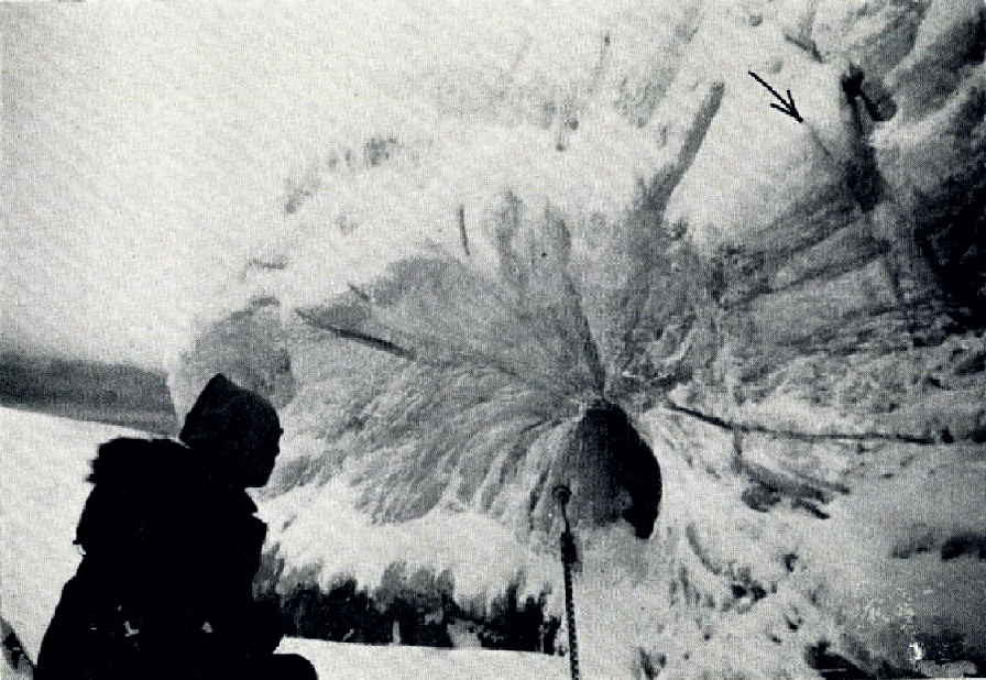
Fig. 11. Ice of radial structure at site Q′

Fig. 12. Cross-section of ice of radial structure photographed between crossed polaroids
The origin of this peculiar pattern of ice is not known, but it is possible that the present form is a result of freezing of water in a tunnel, which is believed to be a remnant of a very deep trough. As the freezing of water near the bottom of a trough must be very slow because of the low heat conductivity of the frozen ice of the surface, it would be reasonable to expect large single crystals of ice to form in the tunnel.
Ice Physics of Core Samples
Core drilling through the ice island
Two holes were drilled through the ice island, one at site p and the other at q (Fig. 5). Site q is near the camp and site p is very close to the edge of the island in the Colby Bay area. The drilling was done using a SIPRE hand core drill. Measurements obtained in the two holes are given in Table I.
Table I. Measurements of ice island t-3 from core drilling
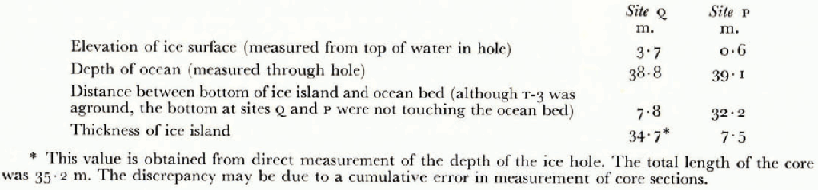
The thickness of the island near the old camp was measured in 1954. by seismic methods (Reference CraryCrary, 1954). From Crary’s values, it is estimated that the thickness near site q was about 45 m. in 1954, a value about 10 m. greater than that measured in 1960. This 10 m. difference is more than the estimated ablation of 5 to 6 m. for the period 1954–60. Since the sites of the 1954 measurements, located about 3.5 km. from site q, are thought to be at higher elevation than site q, it is inferred that the thickness of the island at the old camp site (Fig. 4) is several metres more than at site q. If we assume a difference of about 4 or 5 m., which is reasonable, our findings are consistent.
The first deep drilling on t-3 was made in 1952 to a depth of 15.8 m. (Reference CraryCrary, 1958). Subsequent holes were drilled as shown in Table II.
Table II. Holes Drilled through t-3

Hole No. 10 was the first to be drilled completely through the ice island. Core samples were obtained in a satisfactory manner all through the drilling process. A heavy dirt layer was observed at a depth of 8.4 m., and the character of ice below this dirt layer was quite different from that above this layer. A heavy dirt layer was also found in the previous deep drilling. The depth of this layer was quite different under the ridge from what it was under the trough, being about 25 m. for the former and about 15 m. for the latter. Since site q is in a region of lake ice, it is supposed that the underground structure is similar to that at hole No. 8 (Fig. 13). As the ice surface is smooth in a lake-ice region, the ablation is usually less than that in a ridge region. If we assume that the ablation in the lake region has been 5 m. between the period when hole No. 8 was drilled and 1960, and that the underground structure is similar to that near the old camp, a heavy dirt layer would be expected at around 8 or 9 m. Actually a heavy dirt layer was found at a depth of 8.4 m.

Fig. 13. Hole cored through the ice island in 1960 shown superimposed on profile based on earlier coring (Reference CraryCrary, 1958)
Crystallographic studies of core samples at site q
Samples of the ice core from site q were cut in lengths of about 30 cm. each. Vertical and horizontal thin sections were then cut from each sample by an electric band saw and photographs of the thin sections were taken between crossed polaroids. Figure 14 includes an index of the photographic data. A rectangular bar about 30 cm. long was cut from each sample for visco-elastic measurements. Chlorinity was measured by using the rest of the material of the same sample.

Fig. 14. Profiles of ice core from site q
The crystallographic profile of the core from site q is shown diagrammatically in Figure 14. As defined by Reference CraryCrary (1958) and Reference Marshall and BushnellMarshall (1960), the portion above the heavy dirt layer is called the iced firn stratum, and the main body of the island below this heavy dirt layer is called basement ice. Below this basement ice a thin stratum of ice about 2 m. in thickness is found; this is identified as sea ice by its crystallographic structure and chlorinity content. The iced firn stratum is about 8 m. in thickness and the basement ice is 25 m. thick.
Thin sections of one sample of the iced firn are shown in Figures 15 and 31, the former being a vertical section and the latter a horizontal section. The structure is granular, and the appearance of the vertical and horizontal sections is similar. Most of the grain boundaries are straight and display crystalline facets. This is characteristic of iced firn. The mean size of grains varies with depth, as shown in Figure 14. Many horizontal bubble layers are observed, but the dirt layers seen by Crary and Marshall were not found in our core samples.

Fig. 15. Vertical section of iced firn photographed between crossed polaroids

Figs. 16–19. Vertical sections of basement ice photographed between crossed polaroids

Figs. 20–24. Vertical sections of basement ice photographed belween crossed polaroids

Figs. 25, 26. Vertical sections of basement ice photographed between crossed polaroids

Figs. 27, 28. Vertical sections of sea ice found at bottom of ice island photographed between crossed polaroids

Fig. 29. Heavy dirt layer found between iced firn stratum and basement ice

Fig. 30. Basement ice, stratum IV, with uniformly scattered dirt particles

Figs. 31–42. Horizontal sections of iced firn, basement ice, and sea ice photographed between crossed polaroids
Basement ice may be divided into four strata. Stratum I is composed of granular ice crystals similar in shape to the iced firn. Figure 16 shows the vertical sections of two samples. In general the structure is similar to that of the iced firn stratum (Fig. 15) and some of the boundaries show crystalline facets. This stratum is characterized by inclined bubble layers.
Stratum II is quite different in structure from the stratum described above. The component crystals are large and usually elongated in the vertical direction. The crystals are enmeshed with each other, and no crystalline facets are observed in the boundaries. Figures 17 and 18 show typical views of the boundaries. The crystals in stratum II are classified into four categories depending on length: (1) small crystal, less than 3 cm.; (2) medium crystal, from 3 to 7 cm.; (3) large crystal, from 7 to 15 cm.; (4) very large crystal, more than 15 cm.
Figures 18 and 33 are the vertical and horizontal sections of sample q-8, which was taken from a depth of 12.5 m. ± 0.15 m. These two photographs show that the component crystals are elongated in the vertical direction, and the block has a structure more or less similar to that of candle ice. In the case of sample q-7 the vertical section (Fig. 17) is similar to that of q-8 (Fig. 18), but the horizontal section (Fig. 32) was taken at the upper end of the sample and is a part of a single crystal.
Sample q-12 is an example of a very large single crystal. No texture appears in the vertical section (Fig. 20), thus the whole specimen is a perfect single crystal. The horizontal section (Fig. 35) also shows that most of the specimen belongs to a large single crystal. This is, however, a rather exceptional case; other specimens belonging to the stratum called “very large crystal” in Figure 14 are better called “apparently single crystals”. From the vertical sections shown in Figures 19 and 21 the samples q-10 and q-78 look like single crystals. However, the corresponding horizontal sections, Figures 34 and 36, show definite structures, and on the basis of these photographs they cannot be called single crystals. These specimens are aggregates of many crystals with small-angle boundaries. This statement is understood if the vertical sections are examined carefully; at first glance they look uniform, but slight textures are noticed. These slight textures show the existence of small-angle boundaries. Sample q-10 is a little different from the others; the major portion of this specimen is a large single crystal (Fig. 19), but a few other crystals are observed near the upper end. The orientation of these crystals deviates slightly from that of the main body. This is not apparent in the photograph because the direction of deviation is toward the camera. The horizontal section (Fig. 34) shows the structure near the upper end of q-10, which is something like sheet structure. The vertical section must have been made in one of these sheets. Samples at q-78 are aggregates of crystals with small angle boundaries. These boundaries are almost vertical and they appear in the vertical sections as very faint textures (Fig. 21). In the horizontal sections these small angle boundaries show a pattern (Fig. 36) which is quite different from normal boundaries; the mode of aggregation of crystals is very similar to that of the stratified ice which is shown in Figure 7. All component crystals are oriented almost vertically, but the direction of the c-axis of each crystal deviates a few degrees from the mean direction. This kind of ice is considered to be an advanced stage of development of the stratified ice which is observed near the Colby Bay area.
Stratum III is characterized by its chlorine content. The chlorinity suddenly increases at a depth of 22.5 m. and a stratum of 5.5 m. in thickness exists below this level which is rich in chlorine content. The crystallographic structure is quite different from that in stratum II, compare Figure 21 with Figure 22. Figures 22 and 23 show the structure of this ice with the high chlorine content. The boundaries are very intricate, the general texture being similar to that of sea ice. No crystalline facets are observable.
Stratum IV of the basement ice is characterized by the dirt particles scattered in the ice. Dirt particles are scattered in the stratum more or less uniformly. One example of the scattered dirt is shown in Figure 30. The upper half of stratum IV is rich in dirt particles and the structure of the ice is granular. The texture resembles that of the ice with high chlorine content. Sample q-17 (Figs. 24 and 38) and sample q-18 (Figs. 25 and 39) are examples of the ice in the upper layer. In the lower half of this stratum, dirt particles are observed but they are few in number. Most of the crystals are elongated in the vertical direction, and some of them are quite similar to lake ice in structure. This characteristic is clearly observed in the case of sample q-19 (Figs. 26 and 40) .
The lowest layer, 1.8 m. in thickness, is identified as sea ice from its chlorine content and structure. Many vertically oriented columnar bubbles are observed. Vertical sections of these samples are shown in Figures 27 and 28, the horizontal sections in Figures 41 and 42. Although some large crystals are observed (Figs. 27 and 41), most of the samples are composed of medium and small grains, and the boundaries are intricate. This is characteristic of sea ice.
Visco-elastic nature and chlorinity of core samples at site q
Young’s modulus and viscosity were measured by the sonic method for 27 samples taken from various depths; the method is fully described by Reference Nakaya and MugurumaNakaya and Muguruma (1962). All measurements were made at −20° C. Both Young’s modulus and viscosity were successfully measured for 22 of the 27 samples obtained. The coefficient of viscosity is calculated from the loss factor tan 8 by using the Maxwell model, employing the equation
where ω is the angular frequency.
Profiles of viscosity and elasticity are shown in Figure 14. The values of E and η for the sample taken from the iced firn stratum (q-2) are almost the same as those of tunnel ice in the Greenland Ice Sheet (Reference NakayaNakaya, 1959). Sample q-2 has a structure similar to that of the Greenland sample, which is also iced firn (see figs. 4 and 5 of Reference NakayaNakaya, 1959). The visco-elastic nature thus supports the conclusion based on the structure that the upper layer is iced firm
The visco-elastic nature of stratum II of the basement ice, which is made of large candle-like crystals, is different from that of the iced firn layer. The viscosity is several times greater than that of the ice firn, and Young’s modulus is 10 to 20 per cent greater. Both Young’s modulus and the viscosity are of the same order of magnitude as for ordinary commercial ice. Sample q-78 (Fig. 21), which is almost a single crystal, has a very high value of viscosity and Young’s modulus.
Chlorinity titration was performed by L. W. Winkler’s method. The profile of chlorine content is shown in Figure 14. No trace of chlorine is observed down to a depth of 22.3 m., that is to the bottom of stratum II of basement ice. An unexpected phenomenon was observed with respect to strata III and IV of basement ice. About 0.05‰ chlorine content was found in most of the samples taken from stratum III, but very careful observation showed no chlorine in stratum IV, which is situated below stratum III.
For the core samples obtained by Marshall and Crary (Reference CraryCrary, 1958) the salinity was also measured. Crary reports finding some layers rich in saline content with a maximum of about 1‰. Since chlorine content is equal to about one-half of salinity content, the maximum chlorinity of the earlier core samples is about 0.05‰, while the corresponding value of stratum III is 0.05‰, or one-tenth of the value for the earlier sample. Reference Marshall and BushnellMarshall (1960) suggests that the salinity in certain strata may be the result of soaking by sea-water. The small amount of chlorine content in stratum III and its fluctuation with depth may be due to soaking by sea-water through fine fissures. This point will be confirmed if more core samples are obtained near this site.
Stratum IV is characterized by dirt particles more or less uniformly scattered throughout, and by no trace of chlorinity. Its visco-elastic nature is almost the same as that for the upper half of stratum II, that is the layer of candle-like crystals. The mean chlorinity of the sea-ice layer at the bottom of t-3 is about 0.06‰, which is less than one-tenth of that of old pack ice. The method of formation of this sea-ice layer must have been different from that of ordinary sea ice.
Petrography of the dirts
Detailed petrographic studies of dirt particles of t-3 were carried out by Reference Stoiber and BushnellStoiber and others (1960) on samples obtained by Crary and Marshall. Samples obtained from the surface and from the ice cores in 196 were taken back to Hokkaido University, and Professor M. Minato of the Department of Geology was able to identify the mineral content by an X-ray diffraction method. As identification cannot be made on the basis of the diffraction lines alone, the samples were treated with ethylene glycol, NH4NO3, and heat. The diffraction diagram for surface dirt showed a very strong peak at 3.35 A., which is attributed to quartz. Fairly strong peaks were observed at 7.10 Å., 10.0 Å., and 15.2 Å. The peak at 7.10 Å. could have been chlorite, kaolinite, or vermiculite, but it did not change after the sample was heated at 600° C. for one hour, and is therefore identified as chlorite. The peak at 10.0 Å. did not displace with ethylene glycol treatment, nor did it vanish after heat treatment at 600° C., therefore this must be illite. The 15.2 Å. peak could be attributed to chlorite, montmorillonite or vermiculite on the basis of X-ray diffraction alone. The peak did not displace to 17Å. with ethylene glycol treatment, however, so is not montmorillonite. Heat treatment at 600° C. and NH4NO3 treatment did not affect the line, therefore this peak is considered as the (001) reflection of chlorite. Thus the chief components of the surface dirt are chlorite and ilIite mixed with the primary mineral, quartz.
Diffraction patterns of samples taken from the depths of 28.8 m. and 29.3 m. are almost the same. That obtained for the sample taken at a depth of 28.8 m. is shown in Figure 43. Strong peaks are observed at 3.35 Å., 7.1 Å., and 10.0 Å. The peak at 3.35 Å. is quartz, and that at to 10.0 Å. is proven by ethylene glycol and heat treatment to be illite. The 7.1 Å. peak in this case vanishes by heat treatment at 600° C., and is considered to be kaolinite. The diffraction pattern of the sample of 29.3 m. depth shows chlorite and illite, but not kaolinite.

Fig. 43. X-ray diffraction intensity from the dirt from 28.8 m. depth at site Q
The surface dirts are of silt containing a great many biotite flakes. The chlorite and illite registered in the diffraction diagram are clay material from this mica. Microscopic investigations were carried out by Dr. K. Kizaki in Professor Minato’s laboratory, and the following minerals were identified in the surface dirts: plagioclase, microcline, green hornblende, quartz, biotite, zircon, pyroxene. They are thought to be dusts of Precambrian granite or gneiss (or both), which are the prevailing rocks in Ellesmere Island. The origin of the uniformly scattered dirt particles in stratum IV is also thought to be dust from Ellesmere rocks brought by wind from year to year. These observations confirm the former theory of the origin of t-3 (Reference CraryCrary, 1960).
Core samples at site p
Site p is close to the edge of the island in the Colby Bay area (Fig. 5), where the thickness of the island is only 7.5 m. Stratigraphic data for the core obtained here are shown in Figure 44. The island at this site is made of two layers. The upper layer is 3.3 m. in thickness, and has varying dirt content. The chlorinity is zero. This layer is considered to be the continuation of stratum IV of basement ice at site q. The lower layer, 4.2 m. in thickness, has the same structure as the sea ice at site q. Columnar bubbles oriented vertically were observed in this layer. The vertical and horizontal sections of the samples taken from this layer are shown in Figures 45–51. The chlorinity is between 0.05 and 0.05‰. This is a little higher than that of the sea-ice layer at site q, but is about one-tenth that of old sea ice.

Fig. 44. Profiles of ice core from site p

Fig. 45–51. Vertical and horizontal sections of sea ice samples from site p photographed between crossed polaroids
Structure of T-3
In order to infer the structure of t-3 from the previously gathered data and the new data obtained in 1960, the foIIowing assumptions are made:
a. The ablation between 1954 and 1960 was 6 m. on the ridge and 5 m. in the trough.
b. The aggregate of candle-like crystals with small angle boundaries, stratum II of the basement ice, is the same kind of ice as the stratified ice observed on the island surface near Colby Bay area.
c. The “lake ice” mentioned in Crary’s report is the same as stratum II.
d. The elevation of the ridge after ablation where hole No. 2 was made is the same as that of site q.
These assumptions do not seem to be unreasonable. The structure of t-3 as inferred under the assumptions described above is shown in Figure 52.

Fig. 52. A schematic diagram of the structure of t-3 (vertical exaggeration 6:1)
The thickness of the upper layer of iced firn is almost the same at site q as at hole No. 8. If the “lake ice” of Crary is an aggregate of candle-like crystals with small angle boundaries, three of his cores are consistent with the present core both in thickness and also in depth of “lake ice”. From its crystallographic nature, the stratified ice in the Colby Bay area is considered to be the outcrop of stratum II. In the same way the uniform dirt layer at site p is the outcrop of stratum IV. Although the peculiar nature of stratum III (with its chlorinity) is not clarified, the schematic sketch shown in Figure 52 is felt to illustrate the structure of t-3 to the best of our present knowledge.
A point of interest is the origin of the stratified ice, that is the aggregate of fairly large crystals with small-angle boundaries. This kind of ice has not been observed before. The aggregate of candle-like crystals with small-angle boundaries is also new. It has not been determined whether or not this peculiar kind of ice is the result of the melting of snow and subsequent re-freezing. The so-called “lake ice” may not be lake ice. To discuss the mechanism of formation of t-3, it is necessary to clarify the origin of this peculiar kind of ice, which is the major part of the body of the island.
Acknowledgements
The authors are much obliged to all colleagues at the camp on t-3 for their kind help in our field investigations, specially to the camp commander, Capt. William E. Cohagan. Special thanks are due to Dr. Kou Kusunoki for his collaboration at t-3. They are also indebted to the authorities of the Low Temperature Institute of Hokkaido University for permission to use the cold room and its facilities. They also thank Professor M. Minato and Dr. K. Kizaki of the Department of Geology, Hokkaido University, for their petrographic studies of the dirt of t-3.


