Hostname: page-component-848d4c4894-ttngx Total loading time: 0 Render date: 2024-06-10T12:28:03.103Z Has data issue: false hasContentIssue false

Reversal of Bilateral Sudden Deafness with Thrombectomy

Published online by Cambridge University Press:  28 January 2021

Rosalie E. Morrish
Affiliation:
Clinical Neurosciences, University of Calgary, Calgary, Alberta, Canada
Raed A. Joundi
Affiliation:
Clinical Neurosciences, University of Calgary, Calgary, Alberta, Canada
Christopher D. d’Esterre
Affiliation:
Clinical Neurosciences, University of Calgary, Calgary, Alberta, Canada
Connor C. McDougall
Affiliation:
Clinical Neurosciences, University of Calgary, Calgary, Alberta, Canada
William F. Morrish
Affiliation:
Diagnostic Imaging, University of Calgary, Calgary, Alberta, Canada
Philip A. Barber*
Affiliation:
Clinical Neurosciences, University of Calgary, Calgary, Alberta, Canada
*
Correspondence to: Philip Barber, Foothills Medical Centre, Room 1212, Calgary, AlbertaT2N 4N1, Canada. Email: pabarber@ucalgary.ca
Rights & Permissions [Opens in a new window]

Abstract

Type
Neuroimaging Highlights
Copyright
© The Author(s), 2021. Published by Cambridge University Press on behalf of The Canadian Journal of Neurological Sciences Inc.

A 65-year-old man anticoagulated for atrial fibrillation and previous stroke presented with sudden deafness. Examination revealed normal fluent language, but he was deaf to intense sound. The computed tomography (CT) showed encephalomalacia in the left temporal lobe, and the multiphase CT angiogram revealed hypoperfusion in the superior right temporal lobe (Figure 1A) causing the acute deafness. Thrombectomy successfully recanalized the MCA branch occlusion (Figure 1B) with the return of the patient’s hearing. Magnetic resonance imaging (MRI) confirmed the acute infarct in the right Heschl’s gyrus (Figure 1C/D) and the chronic left transverse temporal gyrus infarct (Figure 1D). Sudden deafness is an uncommon, but treatable presentation of stroke. Reference Leussink, Andermann and Reiners1

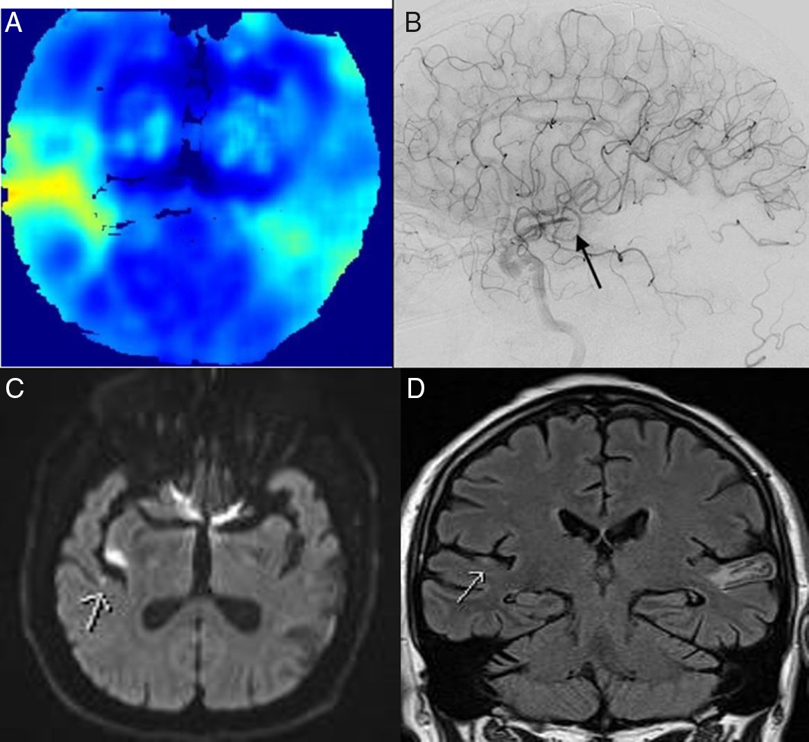
Figure 1: CTA perfusion image (SPIRAL)Reference McDougall, Chan and Sachan2 showing right superior temporal lobe hypoperfusion (A). Right internal carotid catheter angiogram (B) demonstrating right posterior M3 branch occlusion. Diffusion weighted image (C) showing small right transverse temporal gyrus acute infarct following thrombectomy. Coronal FLAIR MRI (D) demonstrating acute right temporal and chronic left contralateral infarcts.

Disclosures

The authors have no conflicts of interest to declare.

Statement of Authorship

REM: drafting and revision of manuscript. RAJ: data collections and interpretation; revision of manuscript. CDdE: data collection and interpretation. CCM: data collection and interpretation. WFM: revision for intellectual content; data collection. PAB: data collection; drafting and revision of manuscript.

References

Leussink, V, Andermann, P, Reiners, K, et al. Sudden deafness from stroke. Neurology. 2005;64(10):1817–18.10.1212/01.WNL.0000162028.82189.E7CrossRefGoogle Scholar
McDougall, C, Chan, L, Sachan, S, et al. Dynamic CT angiography derived perfusion maps predict final infarct volume – the simple perfusion reconstruction algorithm (spiral). Am J Neuroradiol. 2020;41:2034–40.10.3174/ajnr.A6783CrossRefGoogle Scholar
Figure 0

Figure 1: CTA perfusion image (SPIRAL)2 showing right superior temporal lobe hypoperfusion (A). Right internal carotid catheter angiogram (B) demonstrating right posterior M3 branch occlusion. Diffusion weighted image (C) showing small right transverse temporal gyrus acute infarct following thrombectomy. Coronal FLAIR MRI (D) demonstrating acute right temporal and chronic left contralateral infarcts.