
5 results
General intelligence in adult patients with early- and adult-onset schizophrenia
-
- Journal:
- European Psychiatry / Volume 66 / Issue S1 / March 2023
- Published online by Cambridge University Press:
- 19 July 2023, pp. S490-S491
-
- Article
-
- You have access
- Open access
- Export citation
-
Introduction
Early-onset schizophrenia (EOS) is a relatively uncommon disorder with psychotic symptoms emerging before 18 years of age. Although still under debate, EOS may be a more severe disorder relative to adult-onset schizophrenia (AOS), with worse prognosis. Cognitive deficits are a core feature of schizophrenia, accounting for a large part of the detrimental effect of the disorder and may reflect underlying neurodevelopmental disturbances. Some but not all previous studies show that the magnitude of cognitive deficits, including intelligence quotient (IQ), in patients with schizophrenia is dependent on the age of onset.
ObjectivesWe aimed to assess IQ in adult patients with EOS and AOS, and healthy controls. We hypothesized that patients with EOS would show lower IQ than those with AOS, and both patient groups lower IQ than HC.
MethodsWe included 136 adult patients with EOS (mean age: 24.7 (7.7) years, mean duration of illness: 9.3 (8.5) years, 50% women), 382 patients with AOS (mean age: 32.4 (9.5) years, mean duration of illness: 5.7 (6.6) years, 40.1% women) and 896 adult healthy controls (mean age: 33.2 (9.2) years, 47.1% women). We assessed current IQ with the Wechsler Abbreviated Scale of Intelligence (WASI) which yielded verbal (VIQ), performance (PIQ) and full-scale IQ (FIQ) scores. In a post-hoc analysis, we estimated premorbid IQ using the National Adult Reading Test (NART). We applied analyses of covariance (ANCOVAs) to investigate the putative differences in IQ scores and IQ change between patients with EOS, patients with AOS and healthy controls.
ResultsIn sex-, and age-adjusted models, FIQ and PIQ, but not VIQ, were significantly lower in EOS than in AOS (p=0.03, p<0.001 and p=0.428, respectively) (Image). Patients with EOS had fewer years of education than patients with AOS (p<0.001); the PIQ but not the FIQ difference between EOS and AOS remained significant after adjustment for education years (p=0.016 and p=0.333, respectively). Both patient groups had significantly lower IQ scores than healthy controls (Image). Further, patients with EOS and patients with AOS did not significantly differ in estimated premorbid IQ (109 and 110 units, respectively, p=0.092), whereas patients with EOS had a significantly larger estimated IQ decline after the disease onset compared to patients with AOS (12 and 9 units decline, respectively, p=0.015).
Image:
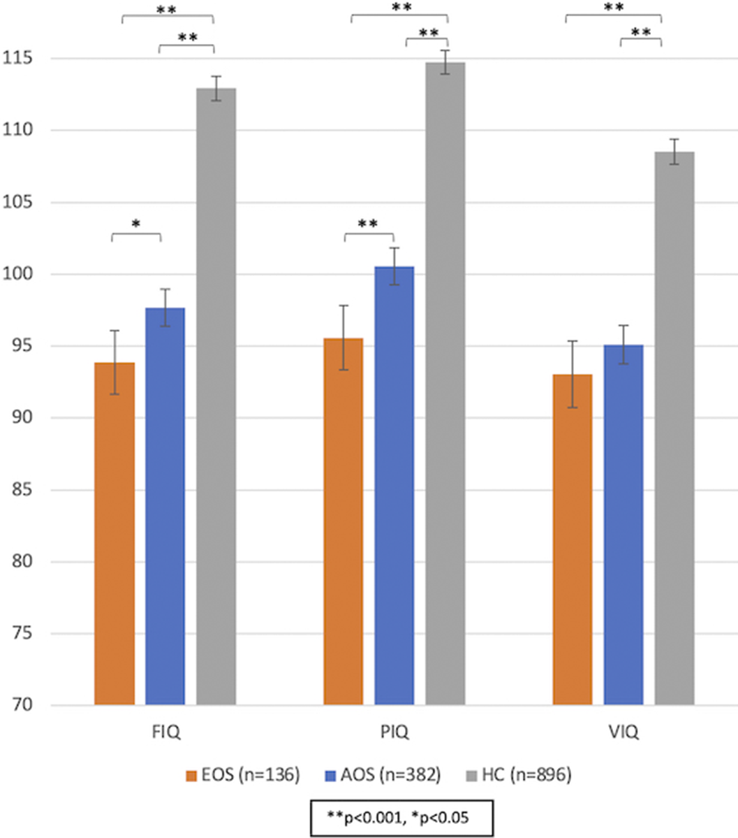 Conclusions
Our findings show that adult patients with EOS have significantly lower PIQ and FIQ scores, and significantly larger IQ decline after the disease onset, but not lower premorbid IQ, compared to patients with AOS. The adolescent onset of psychotic symptoms is linked, as expected, to fewer total years of education, which appears to explain the lower FIQ but only partially the lower PIQ in EOS, which may thereby be linked to the disorder per se.
Disclosure of InterestT. Calkova: None Declared, L. Mørch-Johnsen: None Declared, R. Elle Smelror: None Declared, K. Nordbø Jørgensen: None Declared, S. Cervenka: None Declared, K. Collste: None Declared, A. Vaskinn: None Declared, A. Margrethe Myhre: None Declared, O. A. Andreassen Consultant of: HealthLytix, Speakers bureau of: Lundbeck and Sunovion, T. Ueland: None Declared, I. Agartz: None Declared, D. Andreou: None Declared

Auditory Cortex Characteristics in Early Onset Psychosis and its Associations with Auditory Hallucinations: A Structural MRI Study
-
- Journal:
- European Psychiatry / Volume 41 / Issue S1 / April 2017
- Published online by Cambridge University Press:
- 23 March 2020, p. S59
-
- Article
-
- You have access
- Export citation
Prefrontal cortical thinning links to negative symptoms in schizophrenia via the ENIGMA consortium
-
- Journal:
- Psychological Medicine / Volume 48 / Issue 1 / January 2018
- Published online by Cambridge University Press:
- 26 May 2017, pp. 82-94
-
- Article
- Export citation
Alcohol use is associated with thinner cerebral cortex and larger ventricles in schizophrenia, bipolar disorder and healthy controls
-
- Journal:
- Psychological Medicine / Volume 47 / Issue 4 / March 2017
- Published online by Cambridge University Press:
- 10 November 2016, pp. 655-668
-
- Article
- Export citation
Increased MRI-based cortical grey/white-matter contrast in sensory and motor regions in schizophrenia and bipolar disorder
-
- Journal:
- Psychological Medicine / Volume 46 / Issue 9 / July 2016
- Published online by Cambridge University Press:
- 06 April 2016, pp. 1971-1985
-
- Article
- Export citation
