

Book contents
- Frontmatter
- Dedication
- Contents
- List of Contributors
- Editor’s note on the Foreword to the third edition
- Foreword to the third edition
- Foreword to the second edition
- Foreword to the first edition
- Preface
- Acknowledgments
- List of acronyms
- Introduction
- Section I Skeletal trauma
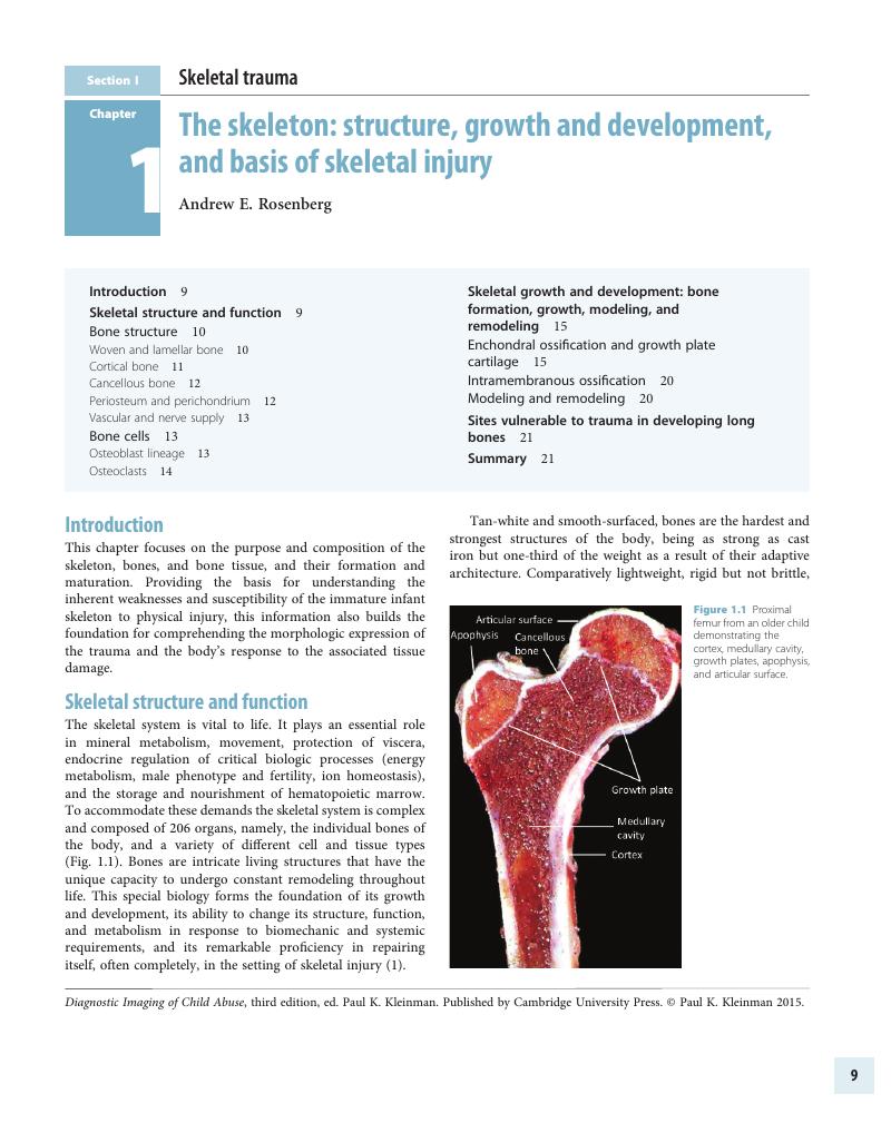
- Chapter 1 The skeleton: structure, growth and development, and basis of skeletal injury
- Chapter 2 Skeletal trauma: general considerations
- Chapter 3 Lower extremity trauma
- Chapter 4 Upper extremity trauma
- Chapter 5 Bony thoracic trauma
- Chapter 6 Dating fractures
- Chapter 7 Differential diagnosis I: diseases, dysplasias, and syndromes
- Chapter 8 Differential diagnosis II: disorders of calcium and phosphorus metabolism
- Chapter 9 Differential diagnosis III: osteogenesis imperfecta
- Chapter 10 Differential diagnosis IV: accidental trauma
- Chapter 11 Differential diagnosis V: obstetric trauma
- Chapter 12 Differential diagnosis VI: normal variants
- Chapter 13 Evidence-based radiology and child abuse
- Chapter 14 Skeletal imaging strategies
- Chapter 15 Postmortem skeletal imaging
- Section II Abusive head and spinal trauma
- Section III Visceral trauma and miscellaneous abuse and neglect
- Section IV Diagnostic imaging of abuse in societal context
- Section V Technical considerations and dosimetry
- Index
- References
Chapter 1 - The skeleton: structure, growth and development, and basis of skeletal injury
from Section I - Skeletal trauma
Published online by Cambridge University Press: 05 September 2015
Edited by

Book contents
- Frontmatter
- Dedication
- Contents
- List of Contributors
- Editor’s note on the Foreword to the third edition
- Foreword to the third edition
- Foreword to the second edition
- Foreword to the first edition
- Preface
- Acknowledgments
- List of acronyms
- Introduction
- Section I Skeletal trauma
- Chapter 1 The skeleton: structure, growth and development, and basis of skeletal injury
- Chapter 2 Skeletal trauma: general considerations
- Chapter 3 Lower extremity trauma
- Chapter 4 Upper extremity trauma
- Chapter 5 Bony thoracic trauma
- Chapter 6 Dating fractures
- Chapter 7 Differential diagnosis I: diseases, dysplasias, and syndromes
- Chapter 8 Differential diagnosis II: disorders of calcium and phosphorus metabolism
- Chapter 9 Differential diagnosis III: osteogenesis imperfecta
- Chapter 10 Differential diagnosis IV: accidental trauma
- Chapter 11 Differential diagnosis V: obstetric trauma
- Chapter 12 Differential diagnosis VI: normal variants
- Chapter 13 Evidence-based radiology and child abuse
- Chapter 14 Skeletal imaging strategies
- Chapter 15 Postmortem skeletal imaging
- Section II Abusive head and spinal trauma
- Section III Visceral trauma and miscellaneous abuse and neglect
- Section IV Diagnostic imaging of abuse in societal context
- Section V Technical considerations and dosimetry
- Index
- References
Summary
A summary is not available for this content so a preview has been provided. Please use the Get access link above for information on how to access this content.
- Type
- Chapter
- Information
- Diagnostic Imaging of Child Abuse , pp. 9 - 22Publisher: Cambridge University PressPrint publication year: 2015
References
, . Bone cell biology: the regulation of development, structure, and function in the skeleton. Am J Anat. 1988;183(1):1–44.CrossRefGoogle ScholarPubMed
, , , , , , et al. Morphology and physiology of the epiphyseal growth plate. Folia Histochem Cytobiol. 2009;47(1):5–16.CrossRefGoogle ScholarPubMed
, , , . Les rapports de la virole perichondrale et du cartilage en croissance normale et pathologique. Ann Radiol. 1968;11:327–35.Google Scholar
, . The periphysis and its effect on the metaphysis. I. Definition and normal radiographic pattern. Skeletal Radiol. 1992;21(5):283–6.CrossRefGoogle ScholarPubMed
, , . Osteoblast physiology in normal and pathological conditions. Cell Tissue Res. 2011;343(2):289–302.CrossRefGoogle ScholarPubMed
, , . The cellular biology and molecular biochemistry of bone formation. In , , eds. Disorders of Bone and Mineral Metabolism. New York, NY: Raven Press; 1992, pp. 241–63.Google Scholar
, , , . Bone organic matrix components: their roles in skeletal physiology. J Endocrinol Invest. 2010;33(7 Suppl.):13–15.Google ScholarPubMed
, , . Signaling pathways governing osteoblast proliferation, differentiation and function. Histol Histopathol. 2009;24(12):1593–606.Google ScholarPubMed
, , , . Signaling networks that control the lineage commitment and differentiation of bone cells. Crit Rev Eukaryot Gene Expr. 2009;19(1):1–46.CrossRefGoogle ScholarPubMed
, , , , , , et al. Regulation of osteogenic differentiation during skeletal development. Front Biosci. 2008;13:2001–21.CrossRefGoogle ScholarPubMed
, . Transcriptional regulatory cascades in Runx2-dependent bone development. Tissue Eng Part B Rev. 2013;19(3):254–63.CrossRefGoogle ScholarPubMed
. Building strong bones: molecular regulation of the osteoblast lineage. Nat Rev Mol Cell Biol. 2012;13(1):27–38.CrossRefGoogle Scholar
, . Integration of BMP, Wnt, and notch signaling pathways in osteoblast differentiation. J Cell Biochem. 2011;112(12):3491–501.CrossRefGoogle ScholarPubMed
, , , , . Skeletal (stromal) stem cells: an update on intracellular signaling pathways controlling osteoblast differentiation. Bone. 2015;70:28–36.CrossRef
. Osteocytes. In , ed. Primer on the Metabolic Bone Diseases and Disorders of Mineral Metabolism. Washington, DC: American Society for Bone and Mineral Research; 2009, pp. 22–7.Google Scholar
, , . The osteocyte: an endocrine cell and more. Endocr Rev. 2013;34(5):658–90.CrossRefGoogle ScholarPubMed
, . Osteocyte signaling in bone. Curr Osteoporos Rep. 2012;10(2):118–25.CrossRefGoogle ScholarPubMed
. Osteocyte-driven bone remodeling. Calcif Tissue Int. 2014;94(1):25–34.CrossRefGoogle ScholarPubMed
, . Molecular mechanisms of triggering, amplifying and targeting RANK signaling in osteoclasts. World J Orthoped. 2012;3(11):167–74.CrossRefGoogle ScholarPubMed
, . Osteoclasts: malefactors of disease and targets for treatment. Discov Med. 2012;13(70):201–10.Google Scholar
, , , , . Regulation of osteoclast function. Mod Rheumatol. 2012;22(2):167–77.CrossRefGoogle ScholarPubMed
, , , . The skeleton: a multi-functional complex organ: the role of key signalling pathways in osteoclast differentiation and in bone resorption. J Endocrinol. 2011;211(2):131–43.CrossRefGoogle ScholarPubMed
, . Osteoclasts: more than bone eaters. Trends Mol Med. 2014;20(8):449–59.CrossRefGoogle ScholarPubMed
, , , , . Endochondral ossification: how cartilage is converted into bone in the developing skeleton. Int J Biochem Cell Biol. 2008;40(1):46–62.CrossRefGoogle ScholarPubMed
, . Development of endochondral skeleton. Cold Spring Harb Perspect Biol. 2013;5(1):1–20.CrossRefGoogle ScholarPubMed
. Origin of the perichondrial osseous ring. First example of a phenomenon of induction in skeletal development. In , ed. The Organization of Bones. Philadelphia, PA: Blakiston Co.; 1951, pp. 90–7.Google Scholar
, , , , . High-resolution CT with histopathologic correlates of the classic metaphyseal lesion of infant abuse. Pediatr Radiol. 2014;44(2):124–40.CrossRefGoogle Scholar
, . Relationship of the subperiosteal bone collar to metaphyseal lesions in abused infants. J Bone Joint Surg. 1995;77(10):1471–6.CrossRefGoogle ScholarPubMed
, , , . Normal metaphyseal radiologic variants not to be confused with findings of infant abuse. AJR. 1991;156(4):781–3.CrossRefGoogle Scholar
, , . Genetic regulation of the growth plate. Front Endocrinol (Lausanne). 2011;2:113.Google ScholarPubMed
, , , , . Mechanisms of growth plate maturation and epiphyseal fusion. Horm Res Paediatr. 2011;75(6):383–91.CrossRefGoogle ScholarPubMed
, , . The skeleton: a multi-functional complex organ: the growth plate chondrocyte and endochondral ossification. J Endocrinol. 2011;211(2):109–21.CrossRefGoogle ScholarPubMed
. Regulatory mechanisms for the development of growth plate cartilage. Cell Mol Life Sci. 2013;70(22):4213–21.CrossRefGoogle ScholarPubMed
. Current understanding on the molecular basis of chondrogenesis. Clin Pediatr Endocrinol. 2014;23(1):1–8.CrossRefGoogle ScholarPubMed
, , , . Quantitative analysis of trabecular morphogenesis in the human costochondral junction during the postnatal period in normal subjects. Anat Rec. 1997;248(1):1–12.3.0.CO;2-Z>CrossRefGoogle ScholarPubMed
, . Skeletal system. In , , eds. Comparative Anatomy and Histology. Waltham, MA: Academic Press; 2012, pp. 53–70.CrossRefGoogle Scholar
, , , , . Discordant radiologic and histological dimensions of the zone of provisional calcification in fetal piglets. Pediatr Radiol. 2013;43:1606–14.CrossRefGoogle ScholarPubMed
, . Determination of proliferative characteristics of growth plate chondrocytes by labeling with bromodeoxyuridine. Calcif Tissue Int. 1993;52(2):110–19.CrossRefGoogle ScholarPubMed
, , . Quantitation of chondrocyte performance in growth-plate cartilage during longitudinal bone growth. J Bone Joint Surg Am. 1987;69-A:162–73.CrossRefGoogle ScholarPubMed
, . Quantitative histology of cartilage cell columns in the human costochondral junction: findings in newborn and pediatric subjects. Pediatr Res. 1989;25(2):202–4.CrossRefGoogle ScholarPubMed
. The imprinting of nutritional disturbances on the growing bone. Pediatrics. 1964;33(Suppl.):815–62.Google ScholarPubMed
, , . Fine structural aspects of vascular invasion of the tibial epiphyseal plate of growing rats. Acta Anat (Basel). 1968;69(1):1–17.CrossRefGoogle ScholarPubMed
, . Studies on experimental rickets in rats. I. Structural modifications of the ephiphyseal cartilages in the tibia and other bones. Am J Anat. 1934;55:135–65.CrossRefGoogle Scholar
. The acrophysis: a unifying concept for enchondral bone growth and its disorders. I. Normal growth. Skeletal Radiol. 2003;32(3):121–7.CrossRefGoogle ScholarPubMed
. Cellular mechanisms of bone remodeling. Rev Endocr Metab Disord. 2010;11(4):219–27.CrossRefGoogle ScholarPubMed
, , , . Bone remodeling: new aspects of a key process that controls skeletal maintenance and repair. Osteoporos Int. 2005;16(Suppl. 2):S18–24.CrossRefGoogle ScholarPubMed
, , . Control of bone remodeling by the peripheral sympathetic nervous system. Calcif Tissue Int. 2014;94(1):140–51.CrossRefGoogle ScholarPubMed
. The reversal phase of the bone-remodeling cycle: cellular prerequisites for coupling resorption and formation. Bonkey Rep. 2014;6:1–8.Google Scholar
, . Injuries involving the epiphyseal plate. J Bone Joint Surg. 1963;45(3):587–622.CrossRefGoogle Scholar
. Some traumatic lesions in growing bones other than fractures and dislocations: clinical and radiological features. BrJ Radiol. 1957;30:225–38.CrossRefGoogle ScholarPubMed
, , . The metaphyseal lesion in abused infants: a radiologic–histopathologic study. AJR. 1986;146(5):895–905.CrossRefGoogle ScholarPubMed
- 1
- Cited by