


Folic acid (FA) supplementation or food fortification began in the USA on 1 January 1998 (Food & Drug Administration, 1996), because of its effect on the prevention of neural tube defects (MRC Vitamin Study Research Group, 1991; Czeizel & Dudas, Reference Czeizel and Dudas1992) and the potential association with reduced risk for vascular disease (Homocysteine Lowering Trialists' Collaboration, 2005) and cancer (Giovanucci et al. Reference Giovanucci, Stampfer, Colditz, Hunter, Fuchs, Rosner, Speizer and Willet1998). Canada, Chile and Israel have more recently started universal mandatory FA fortification (Giovanucci et al. Reference Giovanucci, Stampfer, Colditz, Hunter, Fuchs, Rosner, Speizer and Willet1998; Bailey et al. Reference Bailey, Rampersaud and Kauwell2003). Public health policies promoting daily intake of FA supplements by women of childbearing age have not resulted in increased supplement use. In contrast, food fortification has been associated with a reduction in neural tube defect rates in the USA of approximately 26 % (Centers for Disease Control & Prevention, 2004) and in Canada up to 54 % (Persad et al. Reference Persad, Van den Hof, Dubé and Zimmer2002; Ray, Reference Ray2004). However, there is some limited evidence to suggest that additional FA in the diets of population groups (e.g. children and elderly) not initially targeted for fortification may suffer adverse effects. Such concern is mainly focused on the potential masking of vitamin B12 deficiency, a condition that affects 10–15 % of the population over age 60 (Bailey et al. Reference Bailey, Rampersaud and Kauwell2003). Except for this concern, FA is mainly considered a non-toxic vitamin (Bailey & Berry, Reference Bailey and Berry2005). However, in man, although the currently available data are not sufficient, it has been hypothesized that excess folate may enhance the development and progression of already existing, undiagnosed premalignant and malignant lesions (Sohn et al. Reference Sohn, Stempak, Reid, Shirwadkar, Mason and Kim2003; Kim, Reference Kim2004). It has also been recently reported that unmetabolized FA in plasma is associated with decreased natural killer cytotoxicity among postmenopausal women (Troen et al. Reference Troen, Mitchell and Sorensen2006). In addition, few data exist on the potential effect of long-term high FA intake in children and teenagers. In rats, diets containing 40 mg FA/kg diet (20 times the recommended dietary concentration for rats) accelerated cancer progression in rodent models of cancer (Kim et al. Reference Kim, Salomon, Graeme-Cook, Choi, Smith, Dallal and Mason1996; Song et al. Reference Song, Medline, Mason, Gallinger and Kim2000; Kim, Reference Kim2004). Recent data from our group showed also a negative effect of high-dose FA supplementation on dietary metabolic protein utilization in pregnant and virgin rats as well as in aged rats, suggesting that the vitamin may act under these conditions as a xenobiotic more than as a nutrient (Achón et al. Reference Achón, Alonso-Aperte, Reyes, Úbeda and Varela-Moreiras1999, Reference Achón, Alonso-Aperte, Reyes, Úbeda and Varela-Moreiras2000, Reference Achón, Alonso-Aperte and Varela-Moreiras2002). Folate coenzymes are involved in many processes including DNA synthesis, normal cell division, purine synthesis and amino acid interconversions, especially in growing individuals (Institute of Medicine, 1998). FA plays a critical role in the nutritional regulation in methionine metabolism (Fig. 1). By donation of a methyl group, 5-methyltetrahydrofolate participates in the synthesis of methionine in the reaction catalysed by the vitamin B12-dependent methionine synthase (MS). Methionine acts as a substrate for the synthesis of S-adenosylmethionine (SAM), a molecule which serves as a methyl donor in a wide range of transmethylation reactions involved in different aspects of cell life. By donating its methyl group, SAM is converted to S-adenosylhomocysteine (SAH), which in turn is hydrolysed to homocysteine and adenosine. Homocysteine can then be remethylated to methionine (via MS or additionally in liver by betaine homocysteine methyltransferase (BHMT)), thus completing the cycle, or be degraded via the transsulphuration pathway (Finkelstein, Reference Finkelstein1990).

Fig. 1 Methionine cycle.
On this basis, the present study was conducted to examine the effects of supranormal FA dietary supplementation on several biomarkers involved in the methionine cycle in weanling rats.
Materials and methods
Animals and diets
Thirty weanling male Wistar rats (initial weight approximately 50 g; Animal Service, Universidad San Pablo-CEU, Madrid, Spain), were classified into two groups on the basis of the experimental diet administered, FA supplemented (40 mg FA/kg diet, n 15) v. control (1 mg FA/kg diet, n 15). The rats were given free access to the diets for 4 weeks, after a 1-week adaptation period. Rats were individually housed in metabolic cages and were maintained in a room with a 12 h light/dark cycle, 20–23°C, and with an appropriate ventilation system. Both diets were adjusted to rat requirements (National Research Council, Reference Overton1995), and were based on the pure amino acid diet (17 % amino acid; Dyets, Bethlehem, PA, USA) (Walzem & Clifford, Reference Walzem and Clifford1988), differentiating in succinylsulphathiazole, which was not included. This is one of the most reliable systems to study the exclusive effect of dietary FA, without confounding factors, as has been demonstrated in several studies (Varela-Moreiras & Selhub, Reference Varela-Moreiras and Selhub1992; Varela-Moreiras et al. Reference Varela-Moreiras, Ragel and Pérez de Miguelsanz1995; Alonso-Aperte & Varela-Moreiras, Reference Alonso-Aperte and Varela-Moreiras1996; Achón et al. Reference Achón, Alonso-Aperte, Reyes, Úbeda and Varela-Moreiras1999, Reference Achón, Alonso-Aperte, Reyes, Úbeda and Varela-Moreiras2000, Reference Achón, Alonso-Aperte and Varela-Moreiras2002; Roncalés et al. Reference Roncalés, Achón, Manzarbeitia, Maestro de las Casas, Ramírez, Varela-Moreiras and Pérez-Miguelsanz2004). All rats were also given free access to water.
On day 29, rats were anaesthetized with CO2 and killed by decapitation.
The present study was approved by the Ethical Committee of Universidad San Pablo-CEU, according to the present law for laboratory animals.
S-Adenosylmethionine and S-adenosylhomocysteine
Hepatic SAM and SAH levels were determined by HPLC according to the method described by Fell et al. (Reference Fell, Benjamin and Steele1985), with some modifications (Miller et al. Reference Miller, Nadeau, Smith, Smith and Selhub1994). Aliquots of frozen liver (approximately 100 mg) were homogenized in four volumes of 0·4 m-perchloric acid, and then centrifuged at 10 000 g, 4°C, for 10 min. The clear supernatants were removed, filtered, and appropriate aliquots were analysed for SAM and SAH.
DNA methylation assay
The capacity of hepatic DNA preparations to serve as methyl group acceptors was determined using the method of Christman et al. (Reference Christman, Weich, Schoenbrun, Schneideman and Asc1980) which was modified by replacing the DNA methylase from Friend erythroleukaemia cells with SssI methylase from Escherichia coli (New England Biolabs, Beverly, MA, USA). Briefly, DNA (2 μg), SssI (4 U) and [3H-methyl]SAM (5 μCi) in 20 μl buffer containing 50 mm-NaCl, 10 mm-Tris-HCl, pH 8·0, and 10 mm-EDTA were incubated for 3 h at 37°C. The reaction was stopped by heating the mixture for 20 min at 65°C. The mixture was then applied on to a disk of Whatman DE-81 paper and soaked in 50 ml 5 % NaH2PO4 for 45 min. Radioactivity retained on the disk was determined by scintillation counting using a non-aqueous scintillation fluor. The amount of radioactivity bound to a filter from an incubation mixture lacking only DNA was used as background and was subtracted from the values obtained with mixtures containing DNA. Because this is an inverse assay, a higher incorporation of 3H-methyl groups into DNA in the in vitro assay indicates a diminished in vivo methylation of DNA.
Homocysteine
Serum homocysteine levels were determined using a Chromsystems Reagent Kit for HPLC analysis of homocysteine in serum (Chromsystems, Munich, Germany), which uses a simple isocratic HPLC system with an attached fluorescence detector (λ Ex 385 nm; λ Em 515 nm).
Folate and vitamin B12
Serum folate and vitamin B12 levels were measured by using the Abbot Imx folate and vitamin B12 assays, respectively (Abbot Laboratories, Abbot Park, IL, USA), as previously reported (Roncalés et al. Reference Roncalés, Achón, Manzarbeitia, Maestro de las Casas, Ramírez, Varela-Moreiras and Pérez-Miguelsanz2004).
Vitamin B6
The determination of vitamin B6 serum levels was done using a Chromsystems Reagent Kit for HPLC analysis of vitamin B6 in serum, which uses a simple isocratic HPLC system with an attached fluorescence detector (λ Ex 300 nm; λ Em 400 nm).
Enzymes
Methionine adenosyltransferase (MAT), BHMT and MS activities were measured in liver extracts using radioenzymatic assays as described by Martín-Duce et al. (Reference Martín-Duce, Ortiz, Cabrero and Mato1988), Finkelstein & Mudd (Reference Finkelstein and Mudd1967) and Keating et al. (Reference Keating, Weir and Scott1985), respectively. The results were expressed as mol product synthesized/time per mg protein. In all three cases, total protein content in liver extracts was measured by the method of Bradford (1976).
Serum biochemical markers
Whole blood was collected from all rats and serum and plasma were separated by centrifugation, and kept at − 20°C until analysed. Aspartate aminotransferase, alanine aminotransferase, urea, glucose oxidase, creatinine, total bilirrubine and uric acid seric levels were determined with a Coulter chemistry profile analyser (Kemia Científica S.A., Madrid, Spain), as previously reported (Roncalés et al. Reference Roncalés, Achón, Manzarbeitia, Maestro de las Casas, Ramírez, Varela-Moreiras and Pérez-Miguelsanz2004).
Statistics
Results are expressed as means and their standard errors. Differences in means were studied by one-way ANOVA. When ANOVA resulted in significant differences, multiple comparisons between means were studied by the Tukey test. Differences were considered significant at P < 0·05 (SYSTAT Version 11; Systat, Chicago, IL, USA).
Results
General nutritional status
All the rats appeared to be healthy, and FA supplementation did not alter dietary intake (16·6 (sem0·79) g/d supplemented rats v. 17·1 (sem 0·78) g/d control rats, NS) or body weight gain (6·1 (sem 0·25) g/d supplemented v. 6·2 (sem 0·20) g/d control, NS). Consistent with previous studies (Achón et al. Reference Achón, Alonso-Aperte, Reyes, Úbeda and Varela-Moreiras1999, Reference Achón, Alonso-Aperte, Reyes, Úbeda and Varela-Moreiras2000, Reference Achón, Alonso-Aperte and Varela-Moreiras2002), the folate-supplemented rats did not show a different pattern of growth after 4 weeks of the dietary intervention.
Vitamins and homocysteine concentrations
Levels of vitamins involved in the nutritional regulation of the methylation cycle were also examined (Table 1). As expected, supplementation with FA led to a significant increase in serum folate concentrations (P < 0·001). In fact, supplemented animals had folate concentrations three times higher than that found in the control group. FA administration, conversely, did not affect serum vitamin B12 or B6 concentrations significantly.
Table 1 Serum folate, vitamin B12 and vitamin B6 levels in weanling male Wistar rats fed folic acid-supplemented (40 mg/kg diet) or control (1 mg/kg) diets for 29 d (Mean values with their standard errors)

Mean value was significantly different from that of the control group: ***P < 0·001.
On the other hand, plasma homocysteine concentrations were significantly lower in weanling rats receiving extra folate (P < 0·01) when compared with control animals (Table 2).
Table 2 Hepatic S-adenosylmethionine (SAM) and S-adenosylhomocysteine (SAH) concentrations, values for methylation ratio (SAM:SAH) and plasma homocysteine concentrations in weanling male Wistar rats fed on folic acid-supplemented (40 mg/kg diet) or control (1 mg/kg diet) diets for 29 d (Mean values with their standard errors)

Mean value was significantly different from that of the control group: **P < 0·01.
Hepatic S-adenosylmethionine and S-adenosylhomocysteine concentrations, global DNA methylation and enzymatic activities
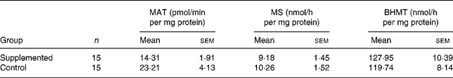
Regarding the main biomarkers involved in the functioning of the methionine methylation cycle (Table 2), hepatic SAM concentrations were similar in both groups, supplemented and control. Hepatic SAH concentrations were not affected by folate supplementation. In consequence, the SAM:SAH concentration ratio (‘methylation ratio’) remained unchanged in the supplemented group compared to the control group. Accordingly, hepatic DNA global methylation did not show significant intergroup differences (Fig. 2). With respect to the enzymatic activities implicated in the methionine cycle, hepatic MAT activity was slightly reduced in the group of animals receiving extra folate, although differences were not significant (P = 0·06; Table 3). Folate supplementation did not affect either hepatic MS or BHMT activities.

Fig. 2 Global DNA methylation in liver from weanling male Wistar rats fed on folic acid-supplemented (40 mg/kg diet; n 15) or control (1 mg/kg diet; n 15) diets for 29 d. Values are means with their standard errors depicted by vertical bars. Results are expressed as methyl group (dpm) incorporation into DNA (2 μg) isolated from rat liver. As an inverse assay, a greater in vitro incorporation of methyl groups indicates a lower degree of in vivo DNA methylation, and vice versa.
Table 3 Hepatic methionine adenosyltransferase (MAT), methionine synthase (MS) and betaine homocysteine methyltransferase (BHMT) activities in weanling male Wistar rats fed folic acid-supplemented (40 mg/kg diet) or control (1 mg/kg) diets for 29 d (Mean values with their standard errors)
Serum biochemical markers
FA supplementation did not alter serum aspartate aminotransferase, alanine aminotransferase, urea, glucose oxidase, total bilirrubine and uric acid. However, serum creatinine concentrations were significantly lower (P < 0·05) in supplemented rats (12·44 (sem 1·25) μmol/l) compared to control rats (16·71 (sem 1·36) μmol/l; Table 4).
Table 4 Serum alanine aminotransferase (ALT), aspartate aminotransferase (AST), total bilirrubine, creatinine, urea, uric acid and glucose oxidase in weanling male Wistar rats fed on folic acid-supplemented (40 mg/kg diet) or control (1 mg/kg diet) diets for 29 d (Mean values with their standard errors)

Mean value was significantly different from that of the control group: *P < 0·05.
Discussion
Dietary surveys regarding folate generally deal with adults. In consequence, there is an urgent need to study the effects of long-term exposure to the presence of unmetabolized FA in growing individuals. On the basis of this lack of information, we studied FA long-term effects on the methionine cycle in an animal model as weanling rats.
When general nutrition status was studied, we observed that FA supplementation did not affect normal dietary intake or growth in rats. This is in accordance with other studies which did not show an improved growth response when animals were fed with FA-enriched diets (Clifford et al. Reference Clifford, Wilson and Bills1989; Achón et al. Reference Achón, Alonso-Aperte, Reyes, Úbeda and Varela-Moreiras1999, Reference Achón, Alonso-Aperte, Reyes, Úbeda and Varela-Moreiras2000, Reference Achón, Alonso-Aperte and Varela-Moreiras2002). The present results, therefore, indicate an adequate growth with both FA levels in the diet. To our knowledge, there are no other comparable studies at present using our dietary FA level and experimental design.
Folate requirements are not specifically established in weanling rats. Still, the normal growth pattern in rats is well known to progress from early growth, that is predominantly by cellular hyperplasia and later by a combination of both hyperplasia and hypertrophy, eventually to a stage in which growth in most organs is primarily due to cellular hypertrophy (Winick & Noble, Reference Winick and Noble1965). According to this pattern, McNulty et al. (Reference McNulty, McPartlin, Weir and Scott1993, Reference McNulty, McPartlin, Weir and Scott1995) reported an association between folate catabolism and hyperplasic growth situations as weaning and pregnancy. It has been proposed that this association is possibly related to vitamin utilization in cell replication. In the present study, serum FA was elevated by three-fold in supplemented animals compared to control and, according to the animals' growth, both diets were adequate to cover the rat requirements in a physiological situation of intense growth such as weaning.
SAM is essential in many transmethylation reactions and, therefore, in development and growth (Finkelstein, Reference Finkelstein1990). Folate deficiency has been shown to modulate SAM and SAH in the brain (Ordóñez & Wutman, Reference Ordóñez and Wutman1994; Varela-Moreiras et al. Reference Varela-Moreiras, Pérez-Olleros, García-Cuevas and Ruiz-Roso1994), pancreas (Balaghi et al. Reference Balaghi, Horne and Wagner1992) and liver (Balaghi et al. Reference Balaghi, Horne and Wagner1993; Miller et al. 1994) in rats. Nevertheless, in the rodent model, a definite effect on SAM:SAH concentration ratio, also known as methylation ratio, after FA supplementation has not been established yet. In the present study, both groups of animals had similar hepatic SAM concentrations, despite the diet. SAH values also remained unchanged. In consequence, the value for the SAM:SAH concentration ratio was unaffected by the diet. In addition, hepatic MAT activity, which leads to SAM synthesis, did not change because of the dietary treatments. A reduction in MAT activity has been described in classic studies of hepatic injury in man (Cabrero et al. Reference Cabrero, Martín-Duce, Ortiz, Alemany and Mato1988) as well as in rats (Corrales et al. Reference Corrales, Giménez and Alvarez1992). However, there are no published studies reporting the effects of supranormal amounts of folate on this critical enzymatic activity and in different physiological situations. We have also reported no effect on hepatic SAM and SAH concentrations after feeding aged rats with FA-enriched diets (Achón et al. Reference Achón, Alonso-Aperte and Varela-Moreiras2002), although it has been frequently hypothesized that a greater methyl group supply could lead to more SAM synthesis. Additionally, in accord with the present findings, Sohn et al. (Reference Sohn, Stempak, Reid, Shirwadkar, Mason and Kim2003) have shown that moderate folate supplementation with 8 mg/kg diet did not modify colonic mucosal concentrations of SAM and SAH, and SAM:SAH ratios at any point after 5 weeks of dietary treatment. The present results, therefore, could indicate that very high levels of folate supplementation do not affect this stable SAM:SAH ratio and raises the speculation of whether or not available FA excess does really increase the efficiency of methylation in the liver. Moreover, it opens the question of the priority destination of the nutrient whether in deficiency or supranormal state.
Hepatic genomic DNA global methylation was not significantly affected by the diet. The study by Sohn et al. (Reference Sohn, Stempak, Reid, Shirwadkar, Mason and Kim2003) also reported that folate supplementation did not modulate genomic DNA methylation at any time. We have previously observed this same lack of effect of FA dietary supplementation in other physiological situations also related to growth, such as pregnancy (Achón et al. Reference Achón, Alonso-Aperte, Reyes, Úbeda and Varela-Moreiras2000). There is also a very interesting recent study by Choi et al. (Reference Choi, Friso, Keyes and Mason2005) that showed increased hepatic DNA methylation in folate-supplemented aged rats (8 mg FA/kg diet) compared with a folate-depleted group (0 mg FA/kg diet) after 20 weeks of dietary treatment. In their study it is concluded that the ability to detect this effect of folate supplementation on DNA methylation is probably related to the age of the animals and/or the heightened accuracy of the method utilized to measure methylation. It is also very important to emphasize that in that same study differences in DNA methylation between supplemented animals and control (2 mg FA/kg diet) were not detected at any point. Their results thus support our present results, since they provide evidence that both moderate and high folate supplementation do not involve an increase in DNA methylation. It seems then that dietary folate appears to be a determinant of DNA methylation under deficiency circumstances and not in supplementation or supranormal status.
FA supplementation resulted in a reduction for homocysteine concentration compared to control diets. The capacity of FA/folates to reduce homocysteine concentrations has been widely described in man (Selhub et al. Reference Selhub, Jacques, Wilson, Rush and Rosenberg1993; Homocysteine Lowering Trialists' Collaboration, 2005) as well as in animal models. Indeed, the main concern at present is to ascertain the lowest dose of FA associated with the maximum reduction in homocysteine concentrations and to determine the additional relevance of vitamins B12 and B6 as well as B2. It is interesting to remember that the present results reflect this homocysteine-lowering effect of the FA supplement even when comparing to data from animals fed with FA-sufficient diets. We have reported this same effect in pregnancy (Achón et al. Reference Achón, Alonso-Aperte, Reyes, Úbeda and Varela-Moreiras2000). The ability of FA to reduce even homocysteine reference values has been also described in man (Santhos-Kumar et al. Reference Santhos-Kumar, Deutsch, Ryder and Kolhouse1997; Jacques et al. Reference Jacques, Selhub, Bostom, Wilson and Rosenberg1999). One of the main arguments used to support mandatory fortification is that elevated blood concentrations of homocysteine have been suggested as a modifiable risk factor for coronary artery disease, stroke and dementia (Clarke et al. Reference Clarke, Smith, Jobst, Refsum, Sutton and Ueland1998; Malinow et al. Reference Malinow, Duell, Irvin-Jones, Upson and Graf2000; Homocysteine Studies Collaboration, 2002).
Homocysteine can be remethylated to methionine via MS (main pathway) or additionally in liver by BHMT. Dietary FA did not affect MS activity or BHMT activity, despite folate indeed being involved in this reaction. Vitamin B12, a cofactor involved in MS activity, also was unaffected. A reduction in MS activity in animals exposed to valproate, a well-known antifolate, has been reported (Alonso-Aperte et al. Reference Alonso-Aperte, Úbeda, Achón, Pérez-Miguelsanz and Varela-Moreiras1999). The activity of MS can be also reduced under circumstances that lead to deficiency of vitamin B12 (Scott, Reference Scott1992). However, the effect of high FA dietary supplementation has not been evaluated in other studies. Vitamin B6, which is involved in the final conversion of homocysteine to glutatione through the transsulphuration pathway, also remained unchanged. This same effect has been described under moderate FA supplementation (Alonso-Aperte, Reference Alonso-Aperte1997) in rats. Creatinine is a waste product in muscle metabolism and is a well-known biomarker for renal function. It is one of the numerous compounds requiring a methyl group from SAM for its synthesis. Additionally, serum creatinine has been positively related to homocysteine concentrations in healthy 28–82-year-old women and men (Jacques et al. Reference Jacques, Bostom, Wilson, Rich, Rosenberg and Selhub2001) as well as dialysed patients (Bostom, Reference Bostom2000). Consistent with this pattern and together with the homocysteine-lowering effect, we observed a reduction in serum creatinine as a result of FA supplementation. In this sense, the present findings could confirm the ability of creatinine to be a good marker for homocysteine levels.
Since aspartate aminotransferase, alanine aminotransferase and billirubine were unchanged, we can conclude that FA supplementation did not induce adverse effects on these hepatic markers in the studied conditions. There are no other animal studies evaluating FA effects on these biomarkers. However, our group has actually described that FA supplementation not only did not damage but improved liver morphology in aged rats (Roncalés et al. Reference Roncalés, Achón, Manzarbeitia, Maestro de las Casas, Ramírez, Varela-Moreiras and Pérez-Miguelsanz2004), which even had the same number of hepatocytes when compared to young animals. In the present study, we have also evaluated how folate supplementation may alter some biomarkers related to glycaemic metabolism and renal function. Specifically, either glucose, uric acid or urea seem to be unaffected by the dietary treatment.
On the basis of the observed results, we can conclude that there were no apparent adverse effects on one-carbon metabolism after FA supplementation in the studied conditions. More studies need to be undertaken in order to confirm the present results and to ascertain the possible mechanisms involved.
Acknowledgements
The study has been supported by a grant from Ministerio de Ciencia y Tecnología, Madrid, Spain (Ref. BFI2003-09 538) and Universidad San Pablo-CEU, Madrid, Spain (Ref. USP-CEU 02/15). Part of this work was presented at the VIII Meeting of the Spanish Nutrition Society, Murcia, 24–27 October 2001 [Achón M, Alonso-Aperte E & Varela-Moreiras G (2001) Dietary folate supplementation in weanling and aged rats. Relationship with methionine metabolism. Nutrition 17, 1018].


