


The risk of transmission of tuberculosis (TB) depends on multiple factors, including the extent of disease in the index patient, the duration and proximity of contacts, the presence of medical conditions that impair immune competence and increase the susceptibility to infection. The likelihood of TB transmission is also influenced by the age of the index case: although transmission from children aged <10 years is unusual, children should be regarded as infectious if they have smear-positive pulmonary TB or cavitary TB on chest X-ray (CXR) [1–Reference Curtis3]. Nosocomial transmission of TB has been described in almost every type of healthcare setting, and its prevention is based on early diagnosis of active TB cases, adoption of isolation measures and initiation of proper treatment [Reference Jensen4]. However, there are incidents where patients with active TB are not appropriately isolated, and considerable resources are used in contact investigation activities aimed at preventing future cases of infection.
In this report, we describe the epidemiological investigation undertaken in response to a case of prolonged in-hospital exposure to an infant with active TB.
The patient was a 4-month-old girl born in Italy from Romanian parents, who presented with respiratory distress to the emergency department of a paediatric hospital. As the Italian national immunization programme does not include TB vaccination, she had not previously received BCG. About 1 month earlier, she was hospitalized for 10 days in another hospital for an acute episode of fever and cough; Mantoux tuberculin skin testing (TST) and CXR performed during that hospitalization were reported as negative. Past history did not reveal failure to thrive, or persistent fever. Diagnosis at admission was bronchiolitis and otitis externa; she had no fever, and her weight and length were at the 25th percentile for age and gender (5·9 kg, 60 cm). She was hospitalized in ward A, treated with aerosol bronchodilators, corticosteroids, and oral antimicrobials. During the first week of hospitalization, cytomegalovirus infection was confirmed by real-time PCR on blood. After 2 weeks, she had no clinical improvement and presented persistent cough; CXR performed on day 17 of hospitalization showed millet-sized nodules distributed throughout the parenchyma, suggestive of miliary pulmonary TB. TST with 5 tuberculin units of purified protein derivative was found to be positive (15 mm induration), and fasting gastric aspirate specimens collected on three consecutive days showed abundant acid-fast bacilli (AFB). When a culture on liquid media [BBL™ MGIT™ (mycobacteria growth indicator tube)] tested positive, Mycobacterium tuberculosis complex identification was performed using sequencing analysis of 500-bp 16s rDNA gene (MicroSeq; Applied Biosystems Inc., CA). Final identification as M. tuberculosis subsp. tuberculosis was obtained on the basis of analysis of gyrase B gene polymorphism (GenoType® MTBC, Hain Lifescience, Germany). As rapid detection of drug resistance to rifampicin (RMP) and/or isoniazid (INH), rpoB, katG and inhA gene mutations were evaluated by using a molecular genetic assay (GenoType® MTBDRplus, Hain Lifescience).
The complete antimicrobial susceptibility of M. tuberculosis [including RMP, INH, streptomycin, pyrazinamide (PZA), ethambutol (EMB)] was tested on MGIT 960 (BD BACTEC™ SIRE and PZA susceptibility testing; USA), showing susceptiblity to all drugs.
The infant was transferred to a negative-pressure isolation room, placed on airborne transmission precautions and treated with RMP, INH, PZA, and EMB. Serology for HIV was negative. Three weeks after commencement of anti-TB therapy, three gastric aspirate samples were collected on consecutive days and tested negative. The patient was discharged after 60 days of starting anti-TB treatment. Therapy was prescribed for a total of 9 months. Compliance to treatment was verified in three outpatient visits, respectively, conducted 13 days, 21 days and 2 months after discharge, thereafter the child was lost to follow-up.
Immediately after diagnosis, information about the patient was transmitted to the local health authority (LHA) and in-hospital contact tracing was started. The mother of the infant had a positive TST result (20-mm induration) and a negative CXR. The infant's household members were investigated by the LHA responsible for contact tracing outside the hospital. None of the household members was found to have active TB. History collected from relatives indicated that a friend from Romania visited the family about 2 months prior to the infant's hospitalization. The friend was reported to have cough, but it was not possible to test him since he was no longer in Italy when contact tracing was conducted; therefore the infant's source of infection remains unknown.
Patients who were hospitalized in ward A during the same period of time as the infant with TB were identified and classified in: (a) patients who shared the room with the infant; (b) patients hospitalized in the same ward, but in a different room.
The parents of the exposed patients were contacted by telephone and mail, advising that the patients and any potentially exposed relatives and visitors should be evaluated for TB. The contact investigation included interviews, screening for symptoms of TB, TST, and CXRs for those with positive TSTs (⩾5 mm induration). TST was performed at baseline and at least 12 weeks after the last exposure. Children aged <5 years were prescribed preventive therapy with 10 mg/kg INH per day and preventive treatment was discontinued after 12 weeks if TST remained negative.
Healthcare workers (HCWs) and volunteers who assisted children and families in the ward were referred to the hospital occupational health service for evaluation; interferon-gamma release assay (IGRA; QuantiFeron®-TB-Gold-IT; Cellestis, Australia) was used to confirm positive TST results.
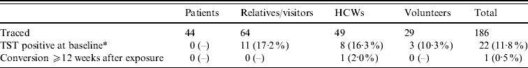
A total of 186 exposed individuals were traced, including 44 patients, 64 relatives and visitors, 49 HCWs and 29 volunteer workers (Table 1).
Table 1. Distribution of contact tracing by category of contacts
HCW, Healthcare worker; TST, tuberculin skin test.
* For HCWs prior to exposure.
The mean age of patients was 5·8 years (range 0–18 years), 45% were female and their mean duration of hospitalization in ward A was 3·7 days (range 1–14 days); none had been previously vaccinated with BCG and 12 had shared a room with the index case for at least one day. At the first visit, none of 44 exposed patients was TST positive; all 23 children aged ⩽5 years started preventive therapy with INH. Forty-three of the 44 patients (97·7%) repeated TST at 12 weeks from the last exposure, and all were negative.
The mean age of the 64 relatives/visitors was 41 years (range 23–82 years); 71% were female. Eleven (17·1%) had a positive TST at the first visit; none reported previous BCG vaccination, while one reported a history of past TB. None was diagnosed with active TB. Fifty-seven were retested (89·1%), and none of the TST negative individuals converted to positive.
Out of 49 HCWs, eight (16·3%) had a positive TST result previously. One of the 41 remaining HCWs had a positive IGRA test at 13 weeks (IFN-γ 0·70 IU/ml, cut-off value 0·35 IU/ml). This individual did not report direct contact with the infant; CXR was negative. Two additional IGRA tests were available for this HCW; the first one was performed 4 months prior to exposure as a periodic screening, and was negative (IFN-γ 0·18 IU/ml). The second one was performed at baseline along with TST (i.e. 3 weeks after exposure), and resulted in a slightly above threshold value (IFN-γ 0·40 IU/ml).
Of the 29 volunteer workers, three (10·3%) were TST positive at baseline; none converted at follow-up.
Nosocomial transmission of M. tuberculosis in paediatric wards has been rarely described, since children aged <10 years with pulmonary TB are rarely contagious. Four previous studies have reported nosocomial transmission where the case age at initial diagnosis was <1 year [Reference Lee5–Reference Matlow8]; in one of these reports, TB transmission was attributed to an infectious adult visitor accompanying the infant [Reference Lee5]. In our experience, neither the infant's mother, who remained with her child during the whole hospital stay, nor other household members who visited the child during hospitalization, showed signs of active infection. Although it was not possible to ascertain where the infant contracted the disease, the patient represented the only probable source of infection in the ward.
We followed a total of 186 exposed patients, relatives/visitors, HCWs and volunteer workers, and we detected only one possibly related conversion in a HCW (0·5%); this is a lower overall conversion rate than those reported in other contact-tracings conducted in paediatric hospitals (1·2–7·1%) [Reference Lee5–Reference George9].
Although we are unable to describe the type and proximity of contact of patients and visitors with the index case, it is likely that they had none or very limited interactions with the infant. None of the patients who shared the room with the infant were infected, confirming that M. tuberculosis transmission from young children is rare. The HCW who converted had no direct interaction with the patient, and although we cannot rule out transmission, it is possible that he was exposed to a different source of infection.
Early diagnosis is a critical issue for TB control [Reference Lee5]. In this episode the delay in diagnosis led to hospitalization of an infant with a positive AFB smear in a normal ward for almost 3 weeks. TB should always be suspected in children with a history of contact with TB or with chronic symptoms suggestive of TB, i.e. persistent cough, fever, and weight loss or failure to thrive [Reference Lee5, Reference Ozuah10]. However, sensitivity of fever to diagnose pulmonary TB in HIV-uninfected children aged <3 years was shown to be 43·6%, while sensitivity of the combination of persistent cough >2 weeks, objective weight loss and reported fatigue was 51·8% [Reference Marais11]. In our case, the initial absence of fever, the normal growth and the absence of a known exposure to TB probably obfuscated the diagnosis. Moreover, the misdiagnosis of bronchiolitis, and the consequent diagnostic approach which does not include a routinely performed CXR [12], can contribute to diagnosis delay.
The symptom-based diagnostic approach may be difficult in small children, in particular in non-endemic areas [Reference Marais11]. However, for proper control of TB it is essential that paediatricians working in low-incidence countries maintain a high level of alert, and include TST in the investigations to be routinely conducted in high-risk groups or in the presence of suggestive symptoms. The use of risk assessment tools evaluated in low-incidence countries could be useful in identifying children who need TST [Reference Ozuah10].
DECLARATION OF INTEREST
None.

