Hostname: page-component-8448b6f56d-cfpbc Total loading time: 0 Render date: 2024-04-24T17:47:50.902Z Has data issue: false hasContentIssue false

Correction of Secondary Fluorescence Across Phase Boundaries in Electron Probe Microanalysis of Mineral Inclusions

Published online by Cambridge University Press:  03 September 2020

Xavier Llovet*
Affiliation:
Centres Científics i Tecnològics, Universitat de Barcelona, Lluís Solé i Sabarís, 1-3, Barcelona08028, Spain
Joaquín A. Proenza
Affiliation:
Departament de Mineralogia, Petrologia i Geologia Aplicada, Universitat de Barcelona, Martí i Franqués s/n, Barcelona08028, Spain
Núria Pujol-Solà
Affiliation:
Departament de Mineralogia, Petrologia i Geologia Aplicada, Universitat de Barcelona, Martí i Franqués s/n, Barcelona08028, Spain
Júlia Farré-de-Pablo
Affiliation:
Departament de Mineralogia, Petrologia i Geologia Aplicada, Universitat de Barcelona, Martí i Franqués s/n, Barcelona08028, Spain
Marc Campeny
Affiliation:
Departament de Mineralogia, Museu de Ciències Naturals de Barcelona, Passeig Picasso s/n, Barcelona08003, Spain
*
*Author for correspondence: Xavier Llovet, E-mail: xavier@ccit.ub.edu
Get access

Abstract

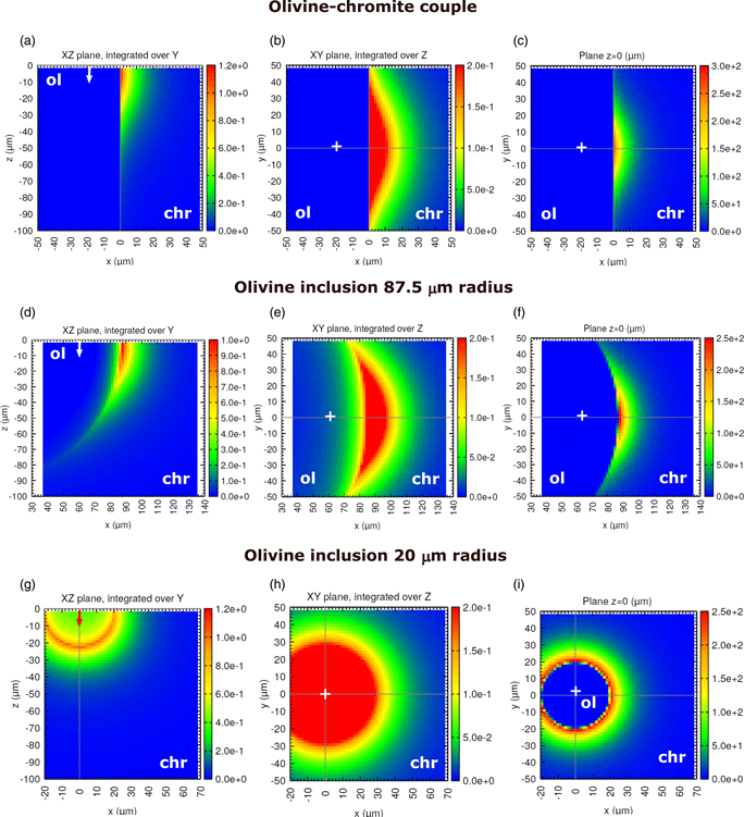
One of the limiting factors for the analysis of minor elements in multiphase materials by electron probe microanalysis is the effect of secondary fluorescence (SF), which is not accounted for by matrix corrections. Although the apparent concentration due to SF can be calculated numerically or measured experimentally, detailed investigations of this effect for fine-grained materials are scarce. In this work, we use the Monte Carlo simulation program PENEPMA to examine and correct the effect of SF affecting micron-sized mineral inclusions hosted by other minerals. A concentration profile across an olivine [(Mg,Fe)2SiO4] inclusion in chromite (Fe2+Cr2O4) is measured and used to assess the reliability of calculations, where different boundary geometries are examined. Three application examples are presented, which include the determination of Cr in olivine and serpentine [Mg3Si2O5(OH)4] inclusions hosted by chromite and of Fe in quartz (SiO2) inclusions hosted by almandine garnet (Fe3Al2Si3O12). Our results show that neglecting SF leads to concentrations that are overestimated by ~0.1–0.8 wt%, depending on inclusion size. In addition, assuming a straight boundary yields to an underestimation of SF effects by a factor of ~2–4. Because its long-range nature, SF severely compromises trace element analyses even for phases as large as 1 mm in size.

Type
Materials Science Applications
Copyright
Copyright © Microscopy Society of America 2020

Access options

Get access to the full version of this content by using one of the access options below. (Log in options will check for institutional or personal access. Content may require purchase if you do not have access.)

References

Ackerson, MR, Watson, EB, Tailby, ND & Spear, FS (2017). Experimental investigation into the substitution mechanisms and solubility of Ti in garnet. Am Mineral 102, 158172.CrossRefGoogle Scholar
Acosta, E, Llovet, X & Salvat, F (2002). Monte Carlo simulation of bremsstrahlung emission by electrons. Appl Phys Lett 80, 32283230.CrossRefGoogle Scholar
Adams, GE & Bishop, FC (1986). The olivine-clinopyroxene geobarometer: Experimental results in the CaO-FeO-MgO-SiO2 system. Contrib Mineral Petrol 94, 230237.CrossRefGoogle Scholar
Almwark, C & Schmitz, B (2009). Relict silicate inclusions in extraterrestrial chromite and their use in the classification of fossil chondritic material. Geochim Cosmochim Acta 73, 14721486.CrossRefGoogle Scholar
Bastin, GF, van Loo, FJJ, Vosters, PJC & Vrolijk, JWGA (1983). A correction procedure for characteristic fluorescence encountered in microprobe analysis near phase boundaries. Scanning 5, 172183.CrossRefGoogle Scholar
Batanova, VG, Sobolev, AV & Kuzmin, DV (2015). Trace element analysis of olivine: High precision analytical method for JEOL JXA-8230 electron probe microanalyser. Chem Geol 419, 149157.CrossRefGoogle Scholar
Borisova, AY, Zagrtdenov, NR, Toplis, MJ, Donovan, JJ, Llovet, X, Asimow, PD, Parseval, P & Gouy, S (2018). Secondary fluorescence effects in microbeam analysis and their impacts on geospeedometry and geothermometry. Chem Geol 490, 2229.CrossRefGoogle Scholar
Brenan, JM (2003). Effects of fO2, fS2, temperature, and melt composition on Fe-Ni exchange between olivine and sulfide liquid: Implications for natural olivine–sulfide assemblages. Geochim Cosmochim Acta 67, 26632681.CrossRefGoogle Scholar
Buse, B, Wade, J, Llovet, X, Kearns, S & Donovan, JJ (2018). Secondary fluorescence in WDS: The role of spectrometer positioning. Microsc Microanal 24, 604611.CrossRefGoogle ScholarPubMed
Capobianco, CJ & Amelin, AA (1994). Metal silicate partitioning of nickel and cobalt: The influence of temperature and oxygen fugacity. Geochim. Cosmochim Acta 58, 125140.CrossRefGoogle Scholar
Cui, JQ, Yang, SY, Jiang, SY & Xie, J (2019). Improved accuracy for trace element analysis of Al and Ti in quartz by electron probe microanalysis. Microsc Microanal 25, 4757.CrossRefGoogle ScholarPubMed
Dalton, JA & Lane, SJ (1996). Electron microprobe analysis of Ca in olivine close to grain boundaries: The problem of secondary X-ray fluorescence. Am Mineral 81, 194201.CrossRefGoogle Scholar
D'Souza, RJ, Canil, D & Coogan, LA (2020). Geobarometry for spinel peridotites using Ca and Al in olivine. Contrib Mineral Petrol 175, 5.CrossRefGoogle Scholar
Elardo, SM, McCubbin, FM & Shearer, CK Jr (2012). The origin of chromite symplectites in lunar troctolite:A new look at an old rock. 43rd Lunar and Planetary Science Conference, Houston, Texas. Abstract #1028.Google Scholar
Endo, S (2014). Cr-rich olivine in deserpentinized peridotite and its implication. Japan Geoscience Union Meeting 2014. Kanagawa, Japan. Abstract SIT04-P06.Google Scholar
Evans, BW, Hattori, K & Baronnet, A (2013). Serpentinite: What, why, where? Elements 9, 99106.CrossRefGoogle Scholar
Feenstra, A & Engi, M (1998). An experimental study of the Fe-Mn exchange between garnet and ilmenite. Contrib Mineral Petrol 131, 379392.CrossRefGoogle Scholar
Fournelle, JH, Kim, S & Perepezko, JH (2005). Monte Carlo simulation of Nb Ka secondary fluorescence in EPMA: Comparison of PENELOPE simulations with experimental results. Surf Interface Anal 37, 10121016.CrossRefGoogle Scholar
Gómez-Pugnaire, MT & Franz, G (1988). Metamorphic evolution of the Palaeozoic series of the Betic Cordilleras (Nevado-Fila´bride complex, SE Spain) and its relationship with the Alpine orogeny. Geol Rundsch 77, 619640.CrossRefGoogle Scholar
Goodrich, CA, Harlow, GE, Van Orman, JA, Sutton, SR, Jercinovic, MJ & Mikouchi, T (2014). Petrology of chromite in ureilites: Deconvolution of primary oxidation states and secondary reduction processes. Geochim Cosmochim Acta 135, 126169.CrossRefGoogle Scholar
Hénoc, J, Maurice, F & Zemskoff, A (1969). Phenomenes de fluorescence aux limites de phases. In 5th International Congress on X-ray Optics and Microanalysis. Mollenstedt G and Gaukler KH (Eds.), pp. 187–192. Berlin: Springer-Verlag.Google Scholar
Hermann, J, O'Neil, HSC & Berry, AJ (2005). Titanium solubility in olivine in the system TiO2–MgO–SiO2: No evidence for an ultra-deep origin of Ti-bearing olivine. Contrib Mineral Petrol 148, 746760.CrossRefGoogle Scholar
Jennings, ES, Wade, J, Laurenz, V & Petitgirard, S (2019 a). Diamond anvil cell partitioning experiments for accretion and core formation: Testing the limitations of electron microprobe analysis. Microsc Microanal 25, 110.CrossRefGoogle ScholarPubMed
Jennings, J, Wade, J & Llovet, X (2019 b). Comment on: “Investigating earth's formation history through copper and sulfur metal-silicate partitioning during core-mantle differentiation” by Mahan et al. (2018). J Geophys Res-Earth 124, 1283712844.CrossRefGoogle Scholar
Jercinovic, MJ, Williams, ML & Lane, ED (2008). In-situ trace element analysis of monazite and other fine-grained accessory minerals by EPMA. Chem Geol 254, 197215.CrossRefGoogle Scholar
Jurewicz, AJG & Watson, EB (1988). Cations in olivine, part 1: Calcium partitioning and calcium-magnesium distribution between olivines and coexisting melts, with petrologic applications. Contrib Mineral Petrol 99, 76185.CrossRefGoogle Scholar
Köhler, TP & Brey, GP (1990). Calcium exchange between olivine and clinopyroxenne calibrated as a geothermobarometer for natural peridotites from 2 to 60 kb with applications. Geochim Cosmochim Acta 54, 23752388.CrossRefGoogle Scholar
Korolyuk, VN & Pokhilenko, LN (2014). Electron probe determination of trace elements in olivine. X-ray Spectrom 43, 353358.CrossRefGoogle Scholar
Kronz, A, Van den Kerkhof, AF & Müller, A (2012). Analysis of low element concentrations in quartz by electron microprobe. In Quartz: Deposits, Mineralogy and Analytics, Götze, J & Möckel, R (Eds.), pp. 191217. Berlin: Springer-Verlag.CrossRefGoogle Scholar
Lanari, P, Vho, A, Bovay, T, Airaghi, L & Centrella, S (2018). Quantitative compositional mapping of mineral phases by electron probe micro-analyser. In: Metamorphic Geology: Microscale to Mountain Belts, vol. 478, Ferrero, S, Lanari, P, Gonçalves, P & Grosch, EG (Eds.), pp. 3963. London: Geological Society, Special Publications.Google Scholar
Li, JP, O'Neil, HSC & Seifert, F (1995). Subsolidus phase relations in the system MgO–SiO2–Cr–O in equilibrium with metallic Cr, and their significance for the petrochemistry of chromium. J Petrol 36, 107132.CrossRefGoogle Scholar
Llovet, X & Galán, G (2003). Correction of secondary X-ray fluorescence near grain boundaries in electron microprobe analysis: Application to thermobarometry of spinel lherzolites. Am Mineral 88, 121130.CrossRefGoogle Scholar
Llovet, X, Pinard, PT, Donovan, JJ & Salvat, F (2012). Secondary fluorescence in electron probe microanalysis of material couples. J Phys D: Appl Phys 45, 225301.CrossRefGoogle Scholar
Llovet, X & Salvat, F (2017). PENEPMA: A Monte Carlo program for the simulation of X-ray emission in electron probe microanalysis. Microsc Microanal 23, 634646.CrossRefGoogle ScholarPubMed
Llovet, X & Salvat, F (2018). Influence of simulation parameters on the speed and accuracy of Monte Carlo calculations using PENEPMA. IOP Conf Series: Mat Sci Eng 304, 012009.CrossRefGoogle Scholar
Llovet, X, Valovirta, E & Heikinheimo, E (2000). Monte Carlo simulation of secondary fluorescence in small particles and at phase boundaries. Microchim Acta 132, 205212.CrossRefGoogle Scholar
Longhi, J, Walker, D & Hays, FJ (1976). Fe and Mg in plagioclase. Proc Lunar Sci Conf 7, 12811300.Google Scholar
McKay, GA (1986). Crystal/liquid partitioning of REE in basaltic systems: Extreme fractionation of REE in olivine. Geochim Cosmochim Acta 50, 6979.CrossRefGoogle Scholar
Melcher, F, Grum, W, Grigore, W, Thalhammer, VT & Stumpfl, EF (1997). Petrogenesis of the ophiolitic giant chromite deposits of Kempirsai, Kazakhstan: A study of solid and fluid inclusions in chromite. J Petrol 38, 14191458.CrossRefGoogle Scholar
Moreno, T, Gibbons, W, Prichard, HM & Lunar, R (2001). Platiniferous chromitite and the tectonic setting of ultramafic rocks in Cabo Ortegal, NW Spain. J Geol Soc 158, 601614.CrossRefGoogle Scholar
Müller, A, Wanvik, JE & Ihlen, PM (2012). Petrological and chemical characterisation of high-purity quartz deposits with examples from Norway. In Quartz: Deposits, Mineralogy and Analytics, Götze, J & Möckel, R (Eds.), pp. 71118. Berlin: Springer-Verlag.CrossRefGoogle Scholar
Myklebust, RL & Newbury, DE (1995). Monte Carlo modeling of secondary X-ray fluorescence across phase boundaries in electron probe microanalysis. Scanning 17, 235242.CrossRefGoogle Scholar
Phillips, D, Harris, JW & Viljoen, KS (2004). Mineral chemistry and thermobarometry of inclusions from De Beers Pool diamonds, Kimberley, South Africa. Lithos 77, 155179.CrossRefGoogle Scholar
Plumper, O, Piazolo, S & Austrheim, H (2012). Olivine pseudomorphs after serpentinized orthopyroxene record transient oceanic lithospheric mantle dehydration (Leka Ophiolite Complex, Norway). J Petrol 53, 19431968.CrossRefGoogle Scholar
Pouchou, JL & Pichoir, F (1991). Quantitative analysis of homogeneous or stratified microvolumes applying the model “PAP”. In Electron Probe Quantitation, Heinrich, KFJ & Newbury, DE (Eds.), pp. 31–75. New York: Plenum Press.Google Scholar
Ramírez, C, Weber, M, Tobón, M, Proenza, JA, Beltrán-Triviño, A, Pujol-Solà, N, Betancur, S, Duque, J & von Quadt, A (2019). Evolución tectónica desde el Jurásico superior al Mioceno en Planeta Rica, Córdoba – Aportes a la historia geológica del Caribe. Memorias del XVII Congreso Colombiano de Geología, pp. 304–305. Available at http://sociedadcolombianadegeologia.org/Memorias/Google Scholar
Reed, SJB (2005). Electron microprobe analysis and scanning electron microscopy in geology. Cambridge: Cambridge University Press.CrossRefGoogle Scholar
Reed, SJB & Long, JVP (1963). Electron-probe measurements near phase boundaries. In: X-ray Optics and X-ray Microanalysis. Pattee, HH, Cosslett, VE & Engström, A (Eds.), pp. 317327. New York: Academic Press.CrossRefGoogle Scholar
Ritchie, NWM (2017). Efficient simulation of secondary fluorescence via NIST DTSA-II Monte Carlo. Microsc Microanal 23, 618633.CrossRefGoogle ScholarPubMed
Robinson, BW, Ware, NG & Smith, DGW.1998). Modern electron-microprobe trace-element analysis in mineralogy. In: Modern Approaches to Ore and Environmental Mineralogy. Short Course Series, vol. 27. Cabri, LJ & Vaughan, DJ (Eds.). Otawa: Mineralogical Association of Canada.Google Scholar
Rui, H, Jiao, J, Xia, M, Yanng, J & Xia, Z (2019). Origin of chromitites in the Songshugou peridotite massif, Qinling Orogen (Central China): Mineralogical and geochemical evidence. J Earth Sci 30, 476493.CrossRefGoogle Scholar
Salvat, F (2015). PENELOPE-2014: A code system for Monte Carlo simulation of electron and photon transport. OECD/NEA Data Bank (NEA/NSC/DOC (2015)3). Retrieved from https://www.oecd-nea.org/science/docs/2015/nsc-doc2015-3.pdf (last access May 31, 2020).Google Scholar
Sato, K & Santosh, M (2007). Titanium in quartz as a record of ultrahigh-temperature metamorphism: The granulites of Karur, southern India. Mineral Magaz 71, 143154.CrossRefGoogle Scholar
Singerling, SA & Brearley, AJ (2018). Primary iron sulfides in CM and CR carbonaceous chondrites: Insights into nebular processes. Meteorit Planet Sci 53, 20782106.CrossRefGoogle Scholar
Smith, JV (1966). X-ray emission microanalysis of rock-forming minerals II. Olivines. J Geol 74, 116.CrossRefGoogle Scholar
Sugawara, T (2001). Effect of secondary fluorescence on microprobe analysis of Fa in plagioclase. Jpn Mag Mineral Petrol Sci 30, 159163.Google Scholar
Vera, JA (1988). Evolucio´n de los sistemas de depo´sito en el margen ibe´rico de las Cordilleras Be´ticas. Rev Soc Geol Esp 1, 373391.Google Scholar
Wade, J & Wood, BJ (2012). Metal–silicate partitioning experiments in the diamond anvil cell: A comment on potential analytical errors. Phys Earth Planet Inter 192–193, 5458.CrossRefGoogle Scholar
Watson, EB (1979). Calcium content of forsterite coexisting with silicate liquid in the system Na2O-CaO-MgO-ALO3-SiO2. Am Mineral 64, 824829.Google Scholar
Yuan, Y, Demers, H, Rudinsky, S & Gauvin, R (2019). Secondary fluorescence correction for characteristic and bremsstrahlung X-rays using Monte Carlo X-ray depth distributions applied to bulk and multilayer materials. Microsc Microanal 25, 92104.CrossRefGoogle ScholarPubMed
Zaccarini, F, Pushkarev, E, Garuti, G & Kazakov, I (2016). Platinum-group minerals and other accessory phases in chromite deposits of the Alapaevsk Ophiolite, Central Urals, Russia. Minerals 6, 108.CrossRefGoogle Scholar
Zhang, D, Chen, Y, Mao, Q, Su, B, Jia, LH & Guo, S (2019). Progress and challenge of electron probe microanalysis technique. Acta Petrol Sin 35, 261274.Google Scholar
Zhang, Z, von de Handt, A & Hirschmann, MM (2018). An experimental study of Fe–Ni exchange between sulfide melt and olivine at upper mantle conditions: Implications for mantle sulfide compositions and phase equilibria. Contrib Mineral Petrol 173, 119.CrossRefGoogle Scholar
Zhao, D, Zhang, Y & Essene, EJ (2015). Electron probe microanalysis and microscopy: Principles and applications in characterization of mineral inclusions in chromite from diamond deposit. Ore Geol Rev 65, 733748.CrossRefGoogle Scholar