


Crossref Citations
This article has been cited by the following publications. This list is generated based on data provided by Crossref.
Ishii, Noriyuki
2023.
C‐shaped dipper: A novel useful auxiliary tool for preparation of specimen grids for transmission electron microscopy.
Microscopy Research and Technique,
Vol. 86,
Issue. 4,
p.
431.
Ishii, Noriyuki
and
Odahara, Takayuki
2023.
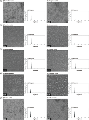
Investigation of the Efficacy of Lanthanoid Heavy Metal Acetates as Electron Staining Reagents for Biomembrane Vesicles.
Microscopy and Microanalysis,
Vol. 29,
Issue. 6,
p.
2080.
Pope, Iestyn
Tanner, Hugh
Masia, Francesco
Payne, Lukas
Arkill, Kenton Paul
Mantell, Judith
Langbein, Wolfgang
Borri, Paola
and
Verkade, Paul
2023.
Correlative light-electron microscopy using small gold nanoparticles as single probes.
Light: Science & Applications,
Vol. 12,
Issue. 1,