


No CrossRef data available.
Article contents
Ultrastructure and Morphometric Analysis of Interstitial Cells of Cajal in the Gastric Wall of the Bullfrog (Rana catesbeiana)
Part of:
Micrographia Collection
Published online by Cambridge University Press: 06 January 2022
Abstract
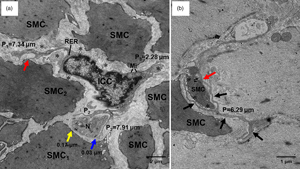
Interstitial cells of Cajal (ICC) play a vital role in the gastrointestinal motility. However, information on ICC in lower vertebrates is rare. Here, ICC and ICC-like features of the gastric wall in the bullfrog (Rana catesbeiana) were observed by light microscopy and transmission electron microscopy. The lengths and distances of the ICC/ICC-like features were measured by morphometric analysis. The gastric wall contained mucosa, submucosa, tunica muscularis, and serosa. The gastric glands contained mucous cells and oxynticopeptic cells. The ICC with 1–3 processes were located among smooth muscle cells (SMC) of the tunica muscularis. Moreover, the ICC-like features were observed among oxynticopeptic cells of the mucosa. The processes of ICC established direct contacts with SMC. Also, the gap junctions were observed between the processes of ICC and nerve fiber bundles in the tunica muscularis. The multivesicular bodies, including shedding exosomes, were frequently observed between ICC and SMC. In addition, ICC-like features and their processes were observed in close proximity to oxynticopeptic cells and blood vessels. Our findings illustrated that ICC are present in the gastric tunica muscularis, and ICC-like features were in the mucosal lamina propria of the gastric wall of R. catesbeiana. These histological evidences supported the notion that ICC are implicated in gastric motility.
- Type
- Micrographia
- Information
- Copyright
- Copyright © The Author(s), 2022. Published by Cambridge University Press on behalf of the Microscopy Society of America
References
Ball, ER, Matsuda, MM, Dye, L, Hoffmann, V, Zerfas, PM, Szarek, E, Rich, A, Chitnis, AB & Stratakis, CA (2012). Ultra-structural identification of interstitial cells of Cajal in the zebrafish Danio rerio. Cell Tissue Res 349, 483–491.CrossRefGoogle ScholarPubMed
De Ceulaer, KM, Van Ginneken, CJ, Philips, WA & Weyns, A (2007). Interstitial cells of Cajal and their role in veterinary gastrointestinal pathologies. Anat Histol Embryol 36, 300–310.CrossRefGoogle ScholarPubMed
Fintl, C, Lindberg, R & McL Press, C (2020). Myenteric networks of interstitial cells of Cajal are reduced in horses with inflammatory bowel disease. Equine Vet J 52, 298–304.CrossRefGoogle ScholarPubMed
Foong, D, Zhou, J, Zarrouk, A, Ho, V & O'Connor, MD (2020). Understanding the biology of human interstitial cells of Cajal in gastrointestinal motility. Int J Mol Sci 21, 4540.CrossRefGoogle ScholarPubMed
Garcia-Lopez, P, Garcia-Marin, V, Martínez-Murillo, R & Freire, M (2009). Updating old ideas and recent advances regarding the interstitial cells of Cajal. Brain Res Rev 61, 154–169.CrossRefGoogle ScholarPubMed
Ghose, D, Jose, L, Manjunatha, S, Rao, MS & Rao, JP (2008). Inherent rhythmicity and interstitial cells of Cajal in a frog vein. J Biosci 33, 755–759.CrossRefGoogle Scholar
Junquera, C, Martinez-Ciriano, C, Castiella, T, Serrano, P, Aisa, J, Calvo, E & Lahoz, M (2001). Enteric plexus and interstitial cells of Cajal: Interrelationship in the stomach of the lizard podarcis hispanica (reptilia). An ultrastructural study. Histol Histopathol 16, 869–881.Google Scholar
Komuro, T (1999). Comparative morphology of interstitial cells of Cajal: Ultrastructural characterization. Microsc Res Tech 47, 267–285.3.0.CO;2-O>CrossRefGoogle ScholarPubMed
Lino, S & Horiguchi, K (2006). Interstitial cells of Cajal are involved in neurotransmission in the gastrointestinal tract. Acta Histochem Cytochem 39, 145–153.Google Scholar
Mandache, E, Popescu, LM & Gherghiceanu, M (2007). Myocardial interstitial Cajal-like cells (ICLC) and their nanostructural relationships with intercalated discs: Shed multivesicular bodies as intermediates. J Cell Mol Med 11, 1175–1184.CrossRefGoogle Scholar
Mitsui, R & Komuro, T (2002). Direct and indirect innervation of smooth muscle cells of rat stomach, with special reference to the interstitial cells of Cajal. Cell Tissue Res 309, 219–227.CrossRefGoogle Scholar
Miyamoto-Kikuta, S & Komuro, T (2007). Ultrastructural observations of the tunica muscularis in the small intestine of Xenopus laevis, with special reference to the interstitial cells of Cajal. Cell Tissue Res 328, 271–279.CrossRefGoogle Scholar
Musara, C & Vaillant, C (2013). Immunohistochemical studies of the enteric nervous system and interstitial cells of Cajal in the canine stomach. Onderstepoort J Vet Res 80, 518.CrossRefGoogle ScholarPubMed
Sanders, KM, Kito, Y, Hwang, SJ & Ward, SM (2016). Regulation of gastrointestinal smooth muscle function by interstitial cells. Physiology 31, 316–326.CrossRefGoogle ScholarPubMed
Sanders, KM, Koh, SD & Ward, SM (2006). Interstitial cells of cajal as pacemakers in the gastrointestinal tract. Annu Rev Physiol 68, 307–343.CrossRefGoogle ScholarPubMed
Sperelakis, N & McConnell, K (2002). Electric field interactions between closely abutting excitable cells. IEEE Eng Med Biol Mag 21, 77–89.CrossRefGoogle ScholarPubMed
Sukhotnik, I, Ben-Shahar, Y, Pollak, Y, Cohen, S, Moran-Lev, H, Koppelmann, T & Gorenberg, M (2020). Intestinal dysmotility after bowel resection in rats is associated with decreased ghrelin and vimentin expression and loss of intestinal cells of Cajal. Am J Physiol Gastrointest Liver Physiol. doi:10.1152/ajpgi.00223.2020Google ScholarPubMed
Wang, Q, Huang, ZP, Zhu, Y, Fu, F & Tian, L (2021). Contribution of interstitial cells of Cajal to gastrointestinal stromal tumor risk. Med Sci Monit 27, e929575.Google ScholarPubMed
Won, KJ, Sanders, KM & Ward, SM (2005). Interstitial cells of Cajal mediate mechanosensitive responses in the stomach. Proc Natl Acad Sci U S A 102, 14913–14918.CrossRefGoogle ScholarPubMed
Yu, H, Liu, Y, Chu, M, Si, Y, Ye, Y, Ge, T, Zhao, H & Zhang, H (2020). Structural relationships between interstitial cells of Cajal and smooth muscle cells/nerve fibers in the gastric muscularis mucosae of Chinese giant salamander. Microsc Microanal 23, 1–9.Google Scholar