

The second Microscopy Today Micrograph Awards competition was again a great success. The premise of these competitions is that scientific micrographs can be interesting in their own right as images with visual impact. Submissions came from 20 US states and 21 other countries. The judges could not see the names of the submitters or their affiliations. The 25 finalist micrographs (Microscopy Today July cover and in the contest gallery) came from the US and 15 other countries.
In this article we show the three prize winners in each category: Published category, for micrographs published in the previous year; Open category, for unpublished micrographs; and the Video category, for clips of movies taken through a microscope and animations of reconstructed images. The following images are the first, second, and third prize winners in each category, as well as the winner of the People's Choice Award and a special award.
Finalists and prize winners were selected by a panel of judges led by Robert Simmons, and the People's Choice Award was selected via public voting at the competition gallery on the MSA website. The judging panel for the 2020 competition was comprised of five judges, all of whom bring their own special expertise. This year Robert Simmons (Chief Judge), Charles Lyman (Senior Editor), and Bob Price (Editor-in-Chief) were joined by Andree Kraker and Jeanette Killius. Andree brings expertise from materials science and is well known as a photographer outside of the scientific realm. Jeanette comes from the biological side of microscopy, is well established as a photographer, and is a Past President of MSA. This group of scientists and artists provides the broad knowledge base needed to address the scientific, technical, and esthetic aspects of our competition.
The original idea for the Microscopy Today Micrograph Awards competition was proposed by Robert and Camille Simmons. They suggested in 2017 that Microscopy Today sponsor a micrograph contest emphasizing both the scientific and artistic merit of micrographs. This concept was developed during 2018, producing a prospectus and a set of instructions for use of the micrograph submission software designed by Nestor Zaluzec.
When submitting a micrograph it is very important to read and follow the instructions for image submission (https://www.microscopy.org/awards/micrograph_submission_instructions.cfm). Following these guidelines makes it much easier for the judges to assess submitted images. The judging process has two steps: once the scientific relevance of an image has been established, the judges evaluate visual impact. Can the micrograph stand on its own as a captivating image without requiring knowledge of the subject or the type of microscopy employed? In other words, would the image look good on a living room or museum wall?
Another goal of our competition is to honor images that may not be eligible or competitive in other micrograph contests. First, all types of micrographs are welcome in this competition, whether they were acquired with a light microscope, electron microscope, X-ray microscope, scanning probe microscope, or some other microanalytical tool. Second, some worthy micrographs are published in journals or magazines without a thought of entering them in a competition. By honoring published images in a separate category we hope to encourage microscopists to think about image composition and visual impact in experiment planning and during image acquisition. Third, the understanding of mechanisms and processes often requires dynamic imaging acquired by in situ microscopy of all types. Thus, we have established a separate category for video micrographs. This category also includes digital animations of reconstructed three-dimensional datasets, for example from cryo-electron microscopy, that provide new insights into cellular and even molecular structures.
Our competition is also driven by image quality from a technical standpoint. Sharpness of image details is important. Imaging of three-dimensional objects with a large depth of field was once the exclusive domain of the scanning electron microscope, but with focus-stacking software, light micrographs now can be in sharp focus over a considerable depth of field. Our judges evaluated submitted micrographs on large high-resolution monitors that can reveal lack of sharpness, as well as other image defects. We request that submitted images have both inherent sharpness and sufficient pixel density to be presented in an 11” × 14” format suitable for hanging in an exhibition. Often image sharpness can be maintained by acquiring the micrograph at a lower magnification than might be required for research purposes. Acquisition at high pixel density is now available to most microscopists since the cost of suitable cameras has decreased dramatically over the last decade. An excellent micrograph with only a modest pixel density is not necessarily eliminated from the competition, but justification may be required to allow the image to be competitive.
So, what makes a winning micrograph? Microscopes reveal interesting features and patterns in objects that are not visible to the naked eye. Some microscopists encounter these images accidentally during the pursuit of other goals, and other microscopists actively seek specific subjects, environmental conditions, and special specimen preparation methods that make acquisition of a great image more likely. Whereas the composition of the image is always important, there are other considerations. Micrographs acquired by electron microscopy or scanning probe microscopy are usually monochrome in nature, which does not prevent an image from winning. However, it is now common to “improve” images with post-acquisition processing to add color to features or to change the background color behind the subject. While such manipulation is acceptable, to be successful the microscopist must make good decisions along the way. Choosing to use artificial color in an image is an artistic choice which should be made carefully. While color may enhance a good image, it will not make up for lack of quality in the original. Color choices are critically important and discordant colors can reduce the appeal of an image as easily as a lack of sharpness. Post-processing should be undertaken carefully with an eye to improving the appeal or clarity of an image.
The editors and judges of Microscopy Today thank all entrants to this year's competition and welcome their submissions in the next contest. The submission site will re-open on October 1, 2020, and close on February 22, 2021.
Published Category

Published 1st Prize.Triceratium morlandii, a diatom from Otago, New Zealand, from a prepared slide by H.J. Baker. This fossil species has no living examples. Phase contrast light microscopy with the background replaced in Photoshop. Similar to an image published in the 2020 Nikon Small World Calendar. Image by Larry Gouliard, Jr., independent microscopist, Pekin, IL.

Published 2nd Prize. Native vanadium dendritic inclusions in the mineral hibonite revealed by X-ray micro-computed tomography (μCT). This image reveals the growth mechanism and has implications for dendritic crystallization in low-oxygen fugacity systems. Specimen was scanned at 50 kV, yielding a tomogram. Published in Microscopy and Microanalysis 25(S2) (2019) 2486. Image by Sarah Gain, Centre for Microscopy, Characterisation, and Analysis of the University of Western Australia, Perth, Australia.
Published 3rd Prize. Gold nanocubes with a monodispersed size distribution (edge = 55 nm) deposited on a silicon wafer. These tiny uniform cubes were produced as reference nanoparticles. SEM image acquired at 10 kV with an in-lens secondary electron detector. Image colored in ImageJ using LUT “Orange hot.” Published in Microscopy and Microanalysis 25(S2) (2019) 2328. Image by Vasile-Dan Hondoroaba, Federal Institute for Materials Research and Testing, Berlin, Germany.
Open Category

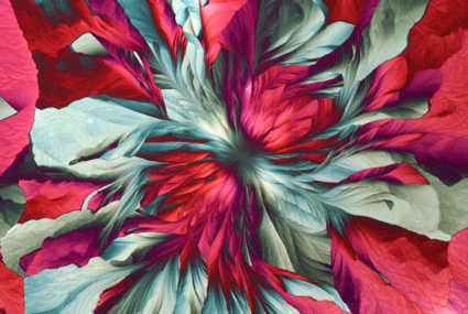
Open 1st Prize. Thin film crystals formed by evaporation of a solution smeared on a glass slide in which the solute was a mixture of iron, aluminum, phosphorus, and ammonium sulfates. The nucleation site in the center was likely a dust particle. Polarized light microscopy with color introduced by a waveplate. Image by Karl Gaff, K Gaff Microscopy, Dublin, Ireland.

Open 2nd Prize. This discharged nematocyst of a Portuguese Man O'War (Physalia physalis) was collected from the Atlantic Ocean off the coast of Florida. The spherical nematocyst capsule (blue), ejected from its nematocyte, is still covered by a microtubular basket (orange). SEM secondary electron image acquired at 4 kV. Image by Connon Thomas, Max Planck Florida Institute for Neuroscience, Jupiter, FL.
Open 3rd Prize. Rat endothelial cells fixed on a glass slide and marked with fluorescent dyes. Nuclei were stained with DAPI (blue), actin filaments with phalloidin (green), and mitochondria with MitoTracker™. Sample from Anja Kraemer of Boehringer Ingelheim Pharma GmbH. Image acquired with a confocal Raman microscope. Image by Damon Strom, WITec GmbH, Ulm, Germany.
Video Category

Video 1st Prize. Soap film colors resulting from interference of reflected light beams: some incident light reflects off the top surface while the remaining light travels to the back of the film and is reflected. Color variations are produced as the thickness of the film varies across the field. Bright-field light microscopy. Image by Gerd Günther, independent microscopist, Düsseldorf, Germany.

Video 2nd Prize. Damselfly larva (nymph) during the hunt showing the operation of a special organ, called a mask, used for capturing and eating prey (ceriodaphnia). For this video the larva was placed on glass, and the ceriodaphnia was added prior to imaging by dark-field light microscopy. Image by Andrei Savitsky, independent microscopist, Cherkasy, Ukraine.
Video 3rd Prize. Video shows a ciliate (Nassula sp.) dying. If local conditions become unfavorable (for example, too dry, high osmotic pressure, bad chemicals in water), the single-cell ciliate stops moving around, and the cell eventually bursts and expels everything it has eaten as well as its own organelles. Bright-field light microscopy. Image by Julia Van Etten, Rutgers University, New Brunswick, NJ.
People’s Choice Award
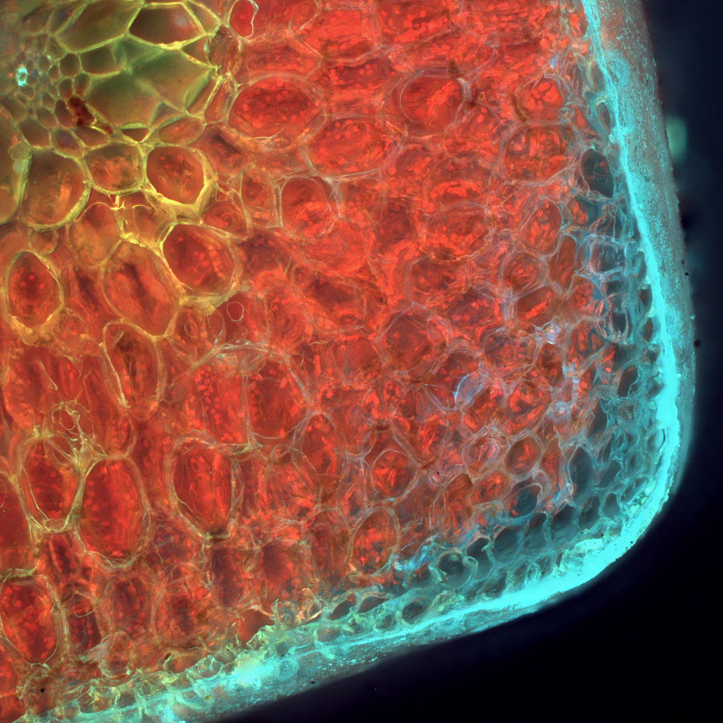
People's Choice Award. Aloe vera leaf imaged with a halogen lamp exciting fluorescence of internal aspects, especially chlorophyll that fluoresces in red. Leaf was cut with a knife to a thickness of 5 mm and imaged directly. Z-stack acquisition of 51 slides in a wide-field fluorescence microscope. Image by Jose Martinez-Lopez, Química Tech Microscopy and Microanalysis, Juarez, Mexico.
Special Honors

Special Award for TEM. Cytoskeletal microarchitecture in Zosterograptus sp. showing microtubules used for feeding. The cytopharyngeal feeding basket (left) is a circular arrangement of wedge-shaped nematodesmata composed of about 25,000 hexagonally packed microtubules (right). Cross section imaged by TEM at Georgetown University. Image by Andrea Brothers, A.B. Brothers Microscopy, Manassas, VA.
