


It is with a heavy heart that I undertake to write this remembrance of an extraordinary member of the microscopy and microanalysis community, Professor David C. Joy (Figure 1), who passed away on August 10, 2022, at the age of 79. However, I am uplifted in this task by the joy of recalling the many wonderful interactions that I had with David over the 53 years of our friendship, and the realization that David helped, inspired, and promoted the work of so many others who could equally well write this tribute.

Figure 1: Dr. David Joy
I first met David in 1969 upon the day of my arrival at the Department of Metallurgy and Materials Science of the University of Oxford at a celebration for the successful defense of his Doctor of Philosophy dissertation, “Investigation of Properties of Magnetic Materials by Scanning Electron Microscopy,” one of the earliest doctoral theses to apply the recently commercialized SEM to a materials science problem. David had come to Oxford following his Master of Arts degree, with First Class Honors, in the Natural Sciences Tripos from Trinity College, Cambridge in 1966, where he worked as a graduate student at Oxford with Dr. John Jakubovics on the nature of SEM contrast of magnetic domains. Upon completing his Doctor of Philosophy, David was awarded a Research Fellowship at Linacre College, an ICI-Unilever Research Fellowship, and a Royal Society Warren Research Fellowship, enabling him to undertake post-doctoral research at Oxford to design and build (in conjunction with Vacuum Generators, Ltd.) the first field emission scanning transmission electron microscope (FE-STEM) outside of the USA (“Project 6”). David's continued presence at Oxford was of great importance to me personally. He somehow found time in his extraordinarily busy schedule to help me in my Doctor of Philosophy research to apply the SEM to study the deformation of materials in situ, including applying electron channeling patterns/contrast, another SEM topic to which David had already made fundamental contributions. Throughout his career, David's extraordinary generosity to anyone who asked for his help was legendary.
David worked seemingly around the clock Monday to Friday on an amazing range of projects, but while at Oxford he made time every weekend to travel to Southampton to play the organ for the Sunday services at his father's church. His musical interests were wide-ranging indeed, from medieval and Renaissance music to the operettas of Gilbert and Sullivan, of which he amassed a huge collection while seeking out every available recording by perusing local record shops (an ancient gathering place for those younger readers unfamiliar with the concept) during his extensive professional travel throughout his long career.
In 1974 David permanently relocated to the United States and became a Member of Technical Staff at AT&T Bell Laboratories, Murray Hill, NJ. At Bell Labs he designed and built the first practical imaging electron energy loss spectrometer for the transmission electron microscope and continued his development of FE-STEM instrumentation (Figure 2). He carried out extensive work applying the SEM to semiconductor device processing, performance of finished devices, and failure mechanisms, as well as investigating fundamentals of voltage contrast and electron beam-induced conductivity. He developed the first practical Monte Carlo electron trajectory simulations to aid SEM image interpretation in support of semiconductor device metrology. While at Bell Labs, David was recruited by Professor Joe Goldstein to lecture on various SEM topics (including electron optics, detectors, electron-specimen interactions, and semiconductor applications) at the annual Lehigh University Summer Microscopy School (LMS), in which he participated for more than 35 years, instructing well over 5,000 attendees. He became an enthusiastic member of the author team of the LMS course textbook, Scanning Electron Microscopy and X-ray Microanalysis by Goldstein et al., (Springer, New York), which is now in its 4th edition (and had over 3.5 million free chapter downloads in 2020 as part of Springer's support of the world scientific community response during the COVID-19 pandemic). He also taught short courses in association with the Microscopy & Microanalysis annual conferences, as well as at numerous microscopy conferences held around the world, including the United Kingdom, Australia, France, Germany, Sweden, Japan, Korea, Thailand, Singapore, South Africa, India, Peru, and Argentina.

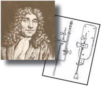
Figure 2: Dr. David Joy working on a STEM system.
In 1987, David was appointed to joint positions as a Distinguished Professor at the University of Tennessee and as a Distinguished Scientist at Oak Ridge National Laboratory of the Department of Energy (DOE). Starting in 2005, he was responsible for the Electron Microscopy Facility at the Center for Nano Materials Science at Oak Ridge, and in 2009 he became Theme Leader for the DOE thrust in “Multiscale Functionality for Nano Materials.”
David was elected Fellow of The Royal Microscopical Society (London, UK), Fellow of The Microscopy Society of America (MSA), and Fellow of the Microanalysis Society (MAS). He received many technical awards including the Semiconductor Research Consortium (SRC) Researcher of the Year Award (1999), National Winner of the Battelle Institute Nanoscience Competition (2001), and the MAS Duncumb Medal (2010). He served as president of MAS (1982) and of MSA (1999). He served as editor of the Journal of Microscopy (1981–1991) and Editor-in-Chief of SCANNING (1992–2011).
David was author or co-author of nine books, nine patents, and more than 425 scientific journal articles. Perusing the titles of these articles reveals the vast range of his interests and the large number of people and institutions with whom he collaborated: aberration corrected SEM; low beam energy SEM; electron beam lithography; helium ion beam microscopy; ion beam nano-fabrication; secondary electron emission under electron or ion bombardment; Monte Carlo electron trajectory simulation and propagation of secondary electrons; energy dispersive X-ray spectrometry (EDS); microcalorimetry EDS; liquid cell visualization of cells in TEM/STEM; nanotip electron guns; variable pressure SEM; voltage contrast, including visualization and quantitative measurement of specimen charging; electron beam-induced conductivity contrast; compilation of fundamental constants critical to electron and ion beam microscopy and microanalysis; length calibration standards for nano-manufacturing; applications of SEM/STEM to semiconductors, magnetic materials, carbon nanofibers, and graphene; serial block facing of biological materials; engineered nanoparticles; oxidation of nanoparticles; fuel cell electrode materials; and many others.
All those who have had the privilege of hearing him speak know that David was a masterful presenter, explaining complex topics clearly and instinctively adjusting his material to the level appropriate to his audience. He was also unflappable when confronted with the unexpected. In 1974 David was scheduled to give several talks at the annual Chicago SEM Conference, where the conference organizer was notorious for making abrupt, last-minute adjustments to the speaker list. I was talking with David at the end of the morning break on the first day when the conference organizer ran up to tell David that he was now the next speaker after the break. David gave his slides for that talk to the conference organizer to take to the second-floor projection booth. These were old-style projector slides consisting of heavy glass plates that sandwiched the 35 mm film in an aluminum frame. As the conference organizer went up the escalator carrying the stack of slides, we saw him suddenly lose his grip and begin to juggle the slides, several of which crashed to the metal treads where they shattered, with the fragments entering the escalator mechanism and bringing it to a grinding halt. (It remained off for the rest of the week!) The conference organizer continued on with the surviving slides to the projection booth. Without any obvious discomfort, David then gave a wonderful presentation despite having to deal with a limited set of randomly arranged slides, some of which were projected upside down or reversed!
David's legacy to us is remarkable: a vast treasure of publications that will continue to enrich the microscopy and microanalysis field for many years. For all of us privileged to have known him personally, pleasant memories will remain of a generous friend always ready and willing to provide technical advice and help. In closing, I'd like to remember David quoting one of his favorite bits from Gilbert and Sullivan:


