The Microscopy Society of America, the Microanalysis Society, and, for the first time, the International Field Emission Society invite you to Microscopy & Microanalysis 2017 in beautiful St. Louis, Missouri. This meeting celebrates the 75th anniversary of MSA and the 50th anniversary of MAS. This year M&M will feature two plenary lectures, more than 36 symposia, and a special series of anniversary lectures, not to mention many educational opportunities in the form of courses and tutorials. And, of course, the meeting will again feature the largest exhibition of microscopy technology in the world.
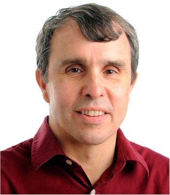
We are honored to have Prof. Erik Betzig from the Janelia Farms Research Campus, Howard Hughes Medical Institute, present a plenary talk titled “Imaging Life at High Spatiotemporal Resolution.” Prof. Betzig obtained a BS in Physics from Caltech and a Ph.D. in Applied Physics at Cornell. In 1988 he became a Principal Investigator at AT&T Bell Labs where he extended his thesis work on near-field optical microscopy, the first method to break the diffraction barrier. By 1993, he held a world record for data storage density. Later he was the co-inventor of the super-resolution technique photoactivated localization microscopy (PALM) with Bell Labs colleague Harald Hess. For this work, he was co-recipient of the 2014 Nobel Prize for Chemistry. Since 2005 he has been a Group Leader at the Janelia Research Campus, developing new optical imaging technologies for biology.
Prof. Erik Betzig

Our other plenary lecturer, Prof. Keith Riles from the University of Michigan, will show us that microscopy and microanalysis can lead to amazing discoveries when applied to the question of whether or not gravitational waves truly exist. Prof. Riles received his B.A from the University of California Berkeley in Physics in 1982 and his Ph.D. in Physics from Stanford in 1989. He began his career at Lawrence Berkeley Laboratory, then moved to the Stanford Linear Accelerator Center, and finally to the University of Michigan in 1982. In his plenary talk, “Detecting Massive Black Holes via Attometry - Gravitational Wave Astronomy Begins,” Professor Riles will explain how the two detectors of the Advanced Laser Interferometer Gravitational-Wave Observatory (Advanced LIGO) simultaneously observed transient gravitational-wave signals. Descriptions will be presented of the first gravitational-wave discoveries and the instruments that made them possible. Dr. Riles’s plenary lecture topic will be expanded in the symposium “Geological Sample Characterization Using Various Imaging Modalities”
High-resolution imaging in the life sciences will be continued with a symposium honoring the memory of Gina Sosinsky who passed away in the Fall of 2015: “Gina Sosinsky Memorial Symposium on Imaging of Cellular Communications.” Gina served as the assistant director of the National Center for Microscopy and Imaging Research at University of California at San Diego and was an advocate for women in science and engineering, spending seven years as co-chair of the UCSD Women in Science Committee. This symposium is intended to interest young scientists in some fast-developing areas in electron microscopy.
Prof. Keith Riles

The Advances in Instrumentation Symposia will cover, in addition to the above techniques, such popular topics as electron and atom probe tomography, electron diffraction, data processing, in situ techniques, and real-world microscopy and microanalysis. Methods bridging the physical and biological sciences will be highlighted. In addition, as part of our celebration of 50 years of atom probe technology, multiple sessions will cover various aspects of this powerful method. The Technologists’ Forum this year will focus on energy-dispersive x-ray spectroscopy methods and will provide insights into applications in life science and materials research. Instrument manufacturers and vendors will have the opportunity to showcase new developments in the “Vendor Symposium: Latest Developments in Tools for Life and Materials Sciences.”
Featured among the life science symposia are those devoted to understanding basic concepts in cellular, molecular, and structural biology: “3D Structures of Macromolecular Assemblies and Cellular Organelles and Whole Cells” and “Microstructure Characterization of Food Systems.” Symposia titled “Utilizing Microscopy for Research and Diagnosis of Diseases in Humans, Plants, and Animals” and “Pharmaceuticals and Medical Science” will cover aspects of pathology and pharmacology. Two novel symposia explore the role of correlative light microscopy and microanalysis and 3D and intravital imaging in development and disease.
Prof. Gina Sosinsky (1955–2015)

Physical science symposia will showcase energy and storage materials with such topics as: The characterization of semiconductor devices, advanced characterization of energy-related materials, and materials characterization methods used in nuclear power systems. Another materials-related symposium will be “Nanoparticles: Synthesis, Characterization, and Applications.” A special symposium celebrating the MSA 75th (diamond) anniversary is titled “Diamonds: From the Origins of the Universe to Quantum Sensing in Materials and Biological Science Applications.”
Other special 75th anniversary lectures at this special M&M meeting will be given by pioneering figures in microscopy and microanalysis: Prof. Robert Glaeser will speak on “Development of High-Resolution TEM for Imaging Native, Radiation-Sensitive Biomolecules,” and Dr. Ondrej Krivanek will give the talk “Smarter than an iPhone: The Emergence of the Modern Microscope.” The Microanalysis Society 50th anniversary lecture in analytical sciences will be given by Dr. Dale Newbury who will talk on “Microanalysis: What is it, where did it come from, and where is it going?” And finally the International Field Emission Society will present a special lecture marking the 50th anniversary of the invention of the atom probe, “Microscopes Without Lenses,” to be given by Dr. John Panitz.
The successful learning opportunities offered at previous M&M meetings will continue with pre-meeting and in-meeting courses taught by specialists in their fields. The topics of the six pre-meeting short courses range from specimen prep for biological EM to 3D reconstruction. Three Technologist Forum Sessions will cover the following topics: “Cryo-Tomography of Macromolecular Complexes in Whole Cells,” “Atomic Force Microscopy for Imaging Materials and Biomaterials,” and “Developing and Applying Light Sheet Imaging Technology.” Short one-hour tutorials will provide insights into CryoEM with phase plates, practical strategies for cryo-CLEM and freeze fracture, large data set acquisition, and entrepreneurship in the microscopy community. We will also showcase our educational opportunities for broader audiences: Project Micro, Microscopy in the Classroom, and It’s a Family Affair.
This year we feature four Pre-Meeting Congresses to be held on Saturday and Sunday. The first titled “Focused Ion Beam Applications and Equipment Developments” is organized by the FIB Focused Interest Group. The second titled “Smaller, Faster, Better: New Instrumentation for Electron Microscopy” is organized by the Aberration-Corrected Electron Microscopy (ACEM) Focused Interest Group. A third congress on “Understanding Radiation Beam-Damage during Cryo-, ETEM, Gas-cell and Liquid-cell Electron Microscopy” will focus on enhancing understanding of the high-energy electron beam effects on various specimens. Finally, an inaugural Pre-Meeting Congress for Students and Early-Career Scientists in Microscopy and Microanaylsis will be held on Saturday.
As usual M&M 2017 will have the world’s largest microscopy and microanalysis instrument exhibition. Over 100 companies will display their latest equipment and services. Don’t miss this exciting annual event. Also in the exhibition hall will be the popular daily poster sessions. The now traditional “Poster Happy Hour” accompanying each day’s poster session will occur again, as well as the presentation of student poster awards.
Descriptions of all the symposia, contributed sessions, educational opportunities, and the multiple award possibilities from the three organizing societies may be found in the Call for Papers (distributed with the November Microscopy Today and at the following website: http://www.microscopy.org/MandM/2017).
The Executive Program Committee suggests that you budget some time to explore St. Louis. Recognized by its world-famous Gateway Arch, St. Louis offers a variety of attractions. The free St. Louis Zoo and Missouri Botanical Gardens offer an excellent retreat for the day. You may want to catch a St. Louis Cardinals baseball game or tour the historic Anheuser Busch Brewery. There is truly something for everyone. See you in St. Louis!


