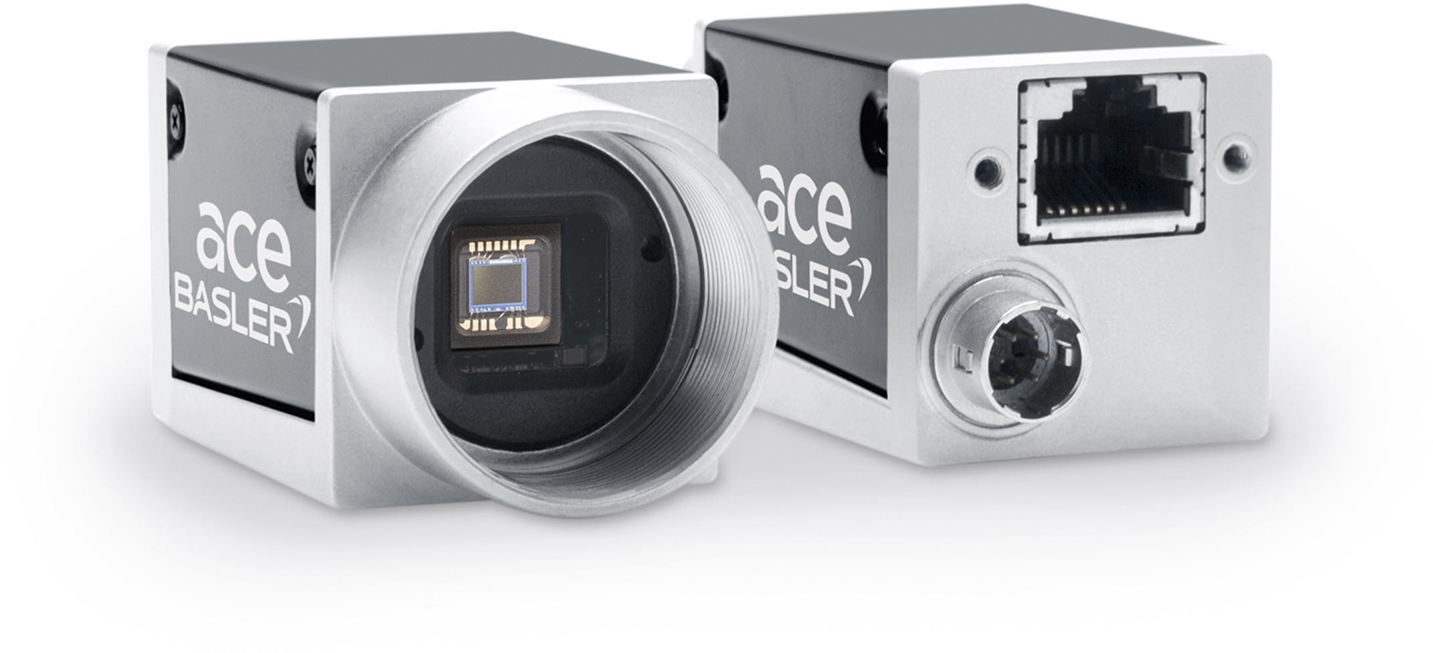
Series Production Start: Four New ace U Models with the IMX287 and IMX273 Sensors from Sony
The four new ace U GigE models from camera manufacturer Basler feature resolutions of VGA and 1.6 megapixels and have now moved into series production. They are equipped with the IMX287 and IMX273 sensors from Sony's Pregius series and deliver up to 291 frames per second. Both sensors feature the Ultra Short Exposure Time Mode, which enables extremely short exposure times to one microsecond.
Baslar AG
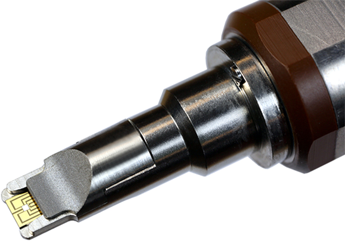
Hummingbird Scientific Introduces a new MEMS Heating + Biasing Platform for the Transmission Electron Microscope
Hummingbird Scientific’s new in situ TEM MEMS Heating + Biasing Specimen Holder allows users to heat and/or electrically bias samples inside the TEM. Heating can be performed to temperatures > 1000°C. MEMS chips are directly inserted into the holder via a proprietary 9-pin connector. The holder is available in both single and double-tilt configurations.
Hummingbird Scientific
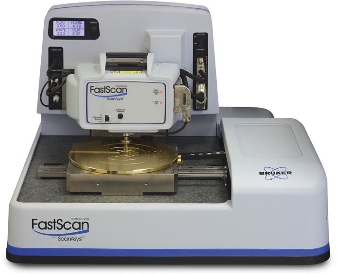
Bruker’s NanoElectrical Lab™ for Dimension AFMs
Bruker’s NanoElectrical Lab™ for Dimension FastScan AFMs is an expansion from conventional contact-based electrical modes to innovative capabilities, such as PeakForce TUNA and PeakForce KPFM, for correlative electrical and mechanical data. Now, DataCube modes further expand these capabilities by enabling the acquisition of multidimensional data cubes. For materials scientists and engineers, this enables simultaneous capture of nanoscale electrical and mechanical characteristics in high-density data cubes, previously impossible to attain in a single measurement.
Bruker Nano Surfaces Division
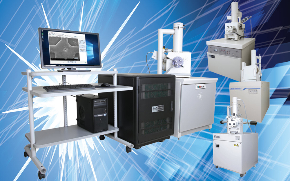
SEMTech Solutions Introduces SEMView8000, a Universal Win10TM Operator’s Console for Scanning Electron Microscopes
SEMTech Solutions is offering a new hardware and software package that will upgrade virtually any SEM to operate on a Windows10™ platform. SEMView8000 replaces existing SEM Operator's Consoles with new electronics to control all electron optics, gun power supply, and electron detectors. In addition, SEMView8000 increases imaging capabilities with a 64-MegaPixel frame grabber. For microscopes lacking factory support, or software that is obsolete, upgrading to SEMView8000 is an option.
SEMTech Solutions, Inc.
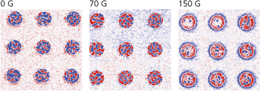
AFM Accessory Enables Advanced Magnetics Research
The new Variable Field Module (VFM4) accessory for Asylum Research MFP-3D atomic force microscopes enables measurements under applied in-plane and out-of-plane magnetic fields in order to better understand the effects on nanoscale magnetic domain structure. The VFM4 can apply either an adjustable in-plane (±8000 G) or out-of-plane (±1200 G) magnetic field to a sample and offers ~1 G field resolution. Learn more about the VFM4 and see recent results at the website below.
Oxford Instruments Asylum Research
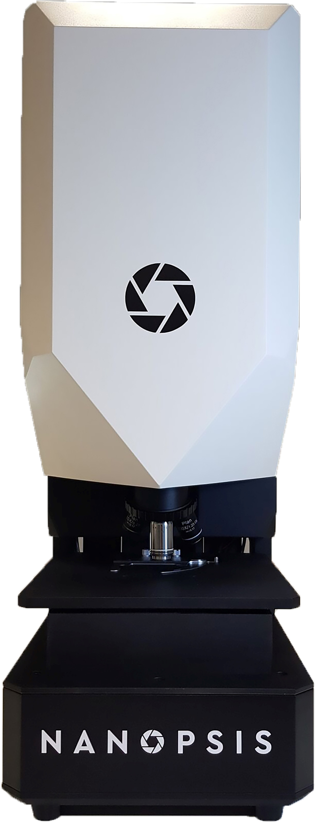
Angstrom Scientific Introduces Nanopsis Super Resolution Microscopy
Angstrom Scientific introduces Nanopsis Super Resolution Microscopy, an exciting optical microscopy technology that allows for imaging below the theoretical limit of light microscopy to provide color images down to the 50 nm level. For biologists, this means there is the potential to see live samples without protein fixation. For semiconductors, this allows imaging of features and lines of different colors due to different materials. The technique bridges the gap between SEM and optical microscopy.
Angstrom Scientific, Inc.
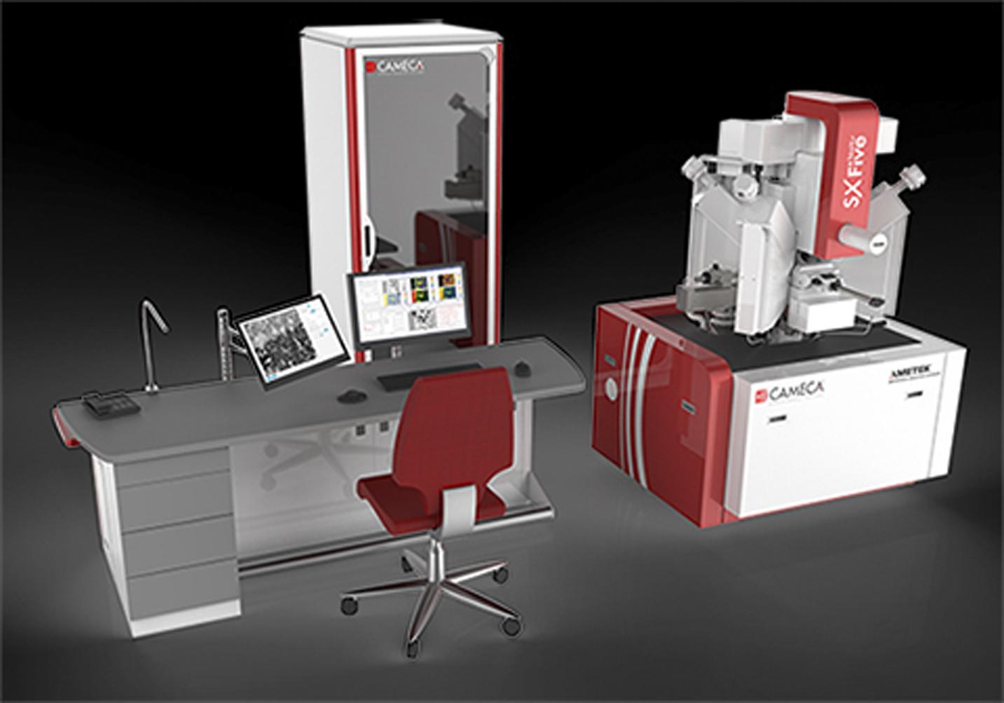
Cameca Launches First Electron Probe Microanalyzer with Touch-Screen Interface
CAMECA announced the launch of SXFive-TACTIS, the newest addition to their CAMECA line of microanalytical instruments, and the only electron probe microanalyzer (EPMA) with a touch-screen interface. SXFive-TACTIS brings together all the best features of CAMECA’s earlier electron probe microanalyzers and innovates with a revolutionary dual interface. It is designed to meet a growing demand from multiuser research facilities for instrumentation that combines highly sophisticated analytical options with extreme ease of use.
Cameca
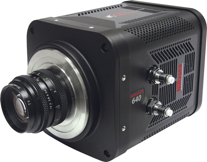
Princeton Instruments Improves NIRvana® SWIR Camera Performance
Princeton Instruments announced an improvement in the performance of its NIRvana:640 short-wave infrared (SWIR) cameras. To meet demanding requirements of scientific research, Princeton Instruments NIRvana:640 cameras are now capable of delivering 40% lower read noise and 10% lower dark current. These improvements address the most critical requirements for quantitative measurements in low-light-level NIR/SWIR imaging and spectroscopy applications.
Princeton Instruments
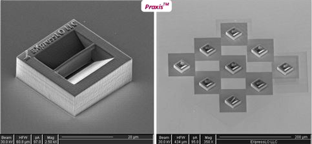
EXpressLO LLC Introduces Praxis™ 3D Printed Specimens
EXpressLO LLC introduced Praxis™ 3D Printed Specimens, patent-pending 3D-printed samples that mimic the size, shape, and geometry of FIB specimens. Praxis™ can be used for practice, training, execution, education, and teaching of either the ex situ or in situ lift-out method. One can easily manipulate numerous specimens with a reduction in FIB time and practice technique and throughput without wasting FIB time. One can also work on final FIB thinning techniques after lift-out.
EXpressLO LLC
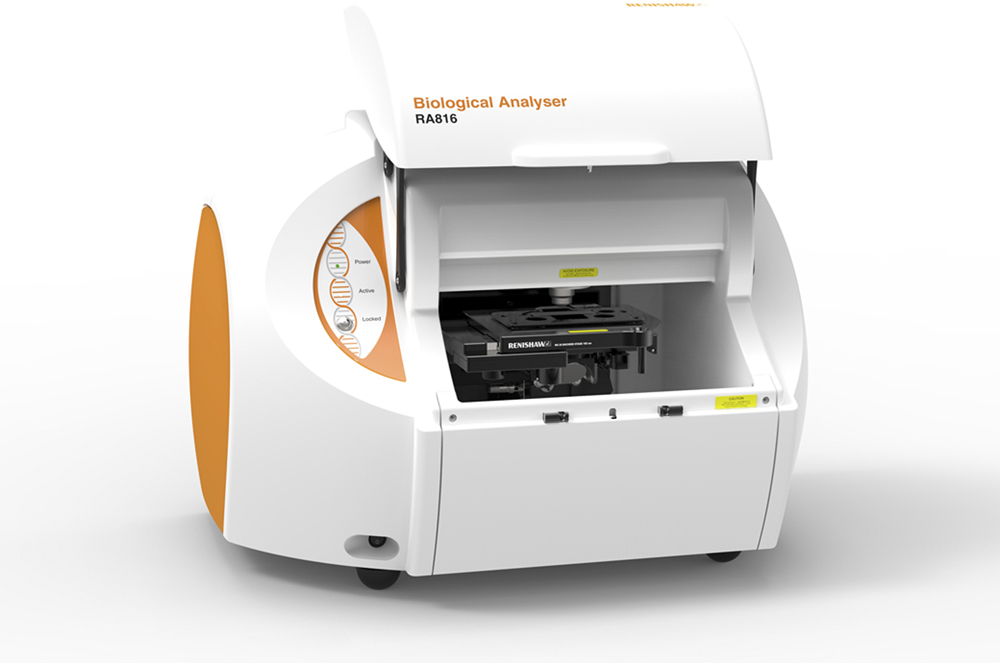
Renishaw’s RA816 Biological Analyser
Imaging and molecular medical diagnostic techniques can be sensitive and specific to information related to the initiation and progression of disease and pathology. These techniques require stains and labels or molecular tags that can be costly in time and money. The Renishaw Biological Analyser identifies and assesses biochemical changes associated with disease formation and progression. There is little to no sample preparation, and no contrast agents or tags are needed.
Renishaw
www.renishaw.com/en/the-renishaw-biological-analyser--43607
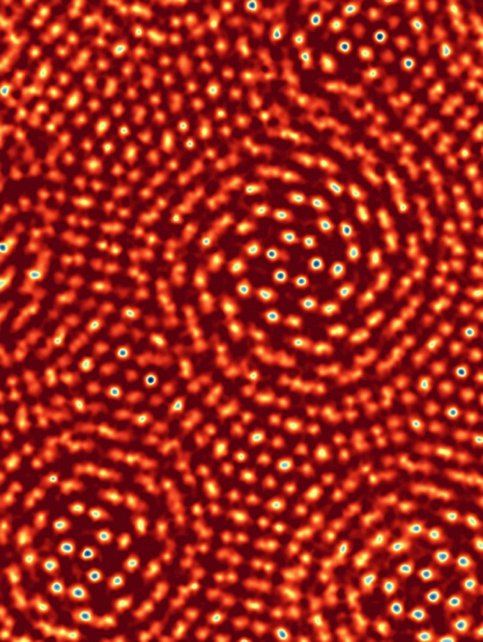
Achieve Equivalent Image Resolution to the Cornell Team's World Record Using Phasefocus πbox Reconstruction Engine
Ptychography is a computational imaging technique that uses algorithms to reconstruct an image from overlapping diffraction patterns. Phasefocus™ has commercialized ptychography as its proprietary platform technology, creating a portfolio of products to accommodate imaging applications in life science, healthcare, engineering, metrology, and more. The Phasefocus πbox reconstruction engine, suitable for electron microscopy, is a processing platform that can be connected to a microscope to deliver reconstructed ptychographic images.
Phase Focus Limited
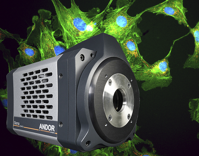
Andor Launches Sona for Fluorescence Microscopy
Andor announced the launch of the new ultrasensitive Sona back-illuminated camera platform for fluorescence microscopy. Featuring 95% quantum efficiency and market-leading vacuum cooling down to -45 °C, Sona has high sCMOS sensitivity, meaning signal to noise can be optimized under reduced illumination conditions, thus preserving living cells during extended measurement periods. Sona also presents an exclusive solution for capturing extremely large fields of cells or whole embryos with exceptional clarity.
Andor, a part of Oxford Instruments Group
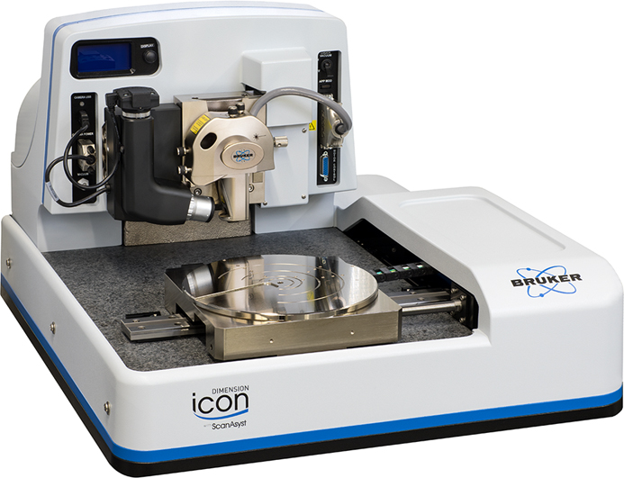
Bruker’s NanoElectrical Lab™ for Dimension AFMs
Bruker’s NanoElectrical Lab™ for Dimension AFMs is an expansion from conventional contact-based electrical modes to innovative capabilities, such as PeakForce TUNA and PeakForce KPFM, for correlative electrical and mechanical data. Now, DataCube modes further expand these capabilities by enabling the acquisition of multidimensional data cubes. For materials scientists and engineers, this enables simultaneous capture of nanoscale electrical and mechanical characteristics in high-density data cubes, previously impossible to attain in a single measurement.
Bruker Nano Surfaces Division
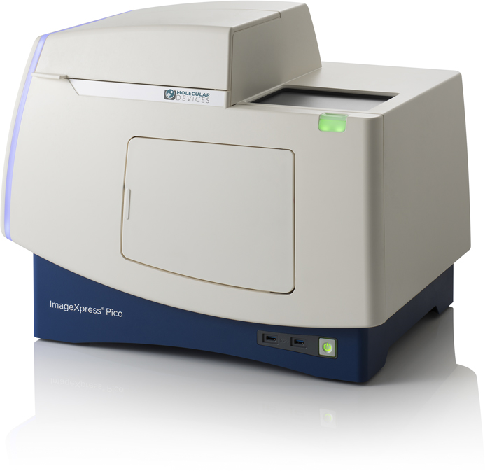
The ImageXpress™ Pico Automated Cell Imaging System
The ImageXpress™ Pico Automated Cell Imaging System is more than a digital microscope, combining high-resolution imaging with powerful analysis. The system comes with CellReporterXpress Image Acquisition and Analysis Software, which features integrated data visualization tools and a comprehensive portfolio of preconfigured protocols that shorten the learning curve, so you can start running experiments quickly. The system’s lab-friendly price affords the convenience of automated digital microscopy in every lab.
Molecular Devices, LLC
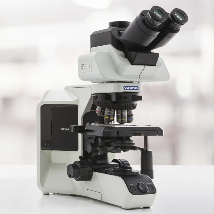
Olympus BX53 Microscope with High-Luminosity LED Provides Bright, Sharp Images
With an LED illuminator equivalent to a 100-watt halogen lamp, the Olympus BX53 microscope delivers outstanding brightness and true-to-life images. The BX53’s ergonomic design and ease of use make it well-suited for clinical laboratories, while the LED illuminator’s brightness makes the system an good solution for multi-head teaching systems. Its consistent color temperature also helps speed up the observation workflow because users don’t have to waste time adjusting a color filter.
Olympus Corporation
www.olympus-lifescience.com/en/microscopes/upright/bx53f2/#!
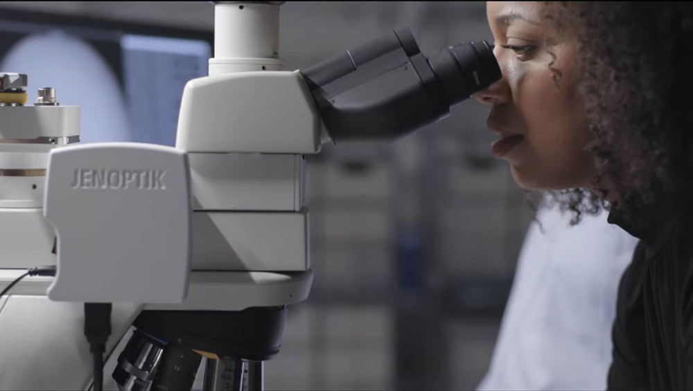
JENOPTIK Demonstrates an Augmented Microscopy Platform for Pathology Applications
JENOPTIK announced that they have developed, in collaboration with Google researchers, an innovative technology to bring AI to conventional microscopy through overlay of digital content directly into a commercial off-the-shelf microscope’s field of view. Google has demonstrated modeling for tumor detection in histopathology slides for prostate cancer and lymph nodes from breast cancer using the platform and presented their results at the annual meeting for the American Association for Cancer Research.
JENOPTIK Optical Systems