Skip to main content Accessibility help
×
Hostname: page-component-76fb5796d-skm99 Total loading time: 0 Render date: 2024-04-25T11:46:48.264Z Has data issue: false hasContentIssue false

Chapter 10 - Brain Imaging in Transient Ischemic Attack and Minor Stroke

from Section 2 - Clinical Features, Diagnosis, and Investigation

Published online by Cambridge University Press:  01 August 2018

Gary K. K. Lau
Affiliation:
University of Oxford
Sarah T. Pendlebury
Affiliation:
University of Oxford
Peter M. Rothwell
Affiliation:
University of Oxford
Get access

Summary

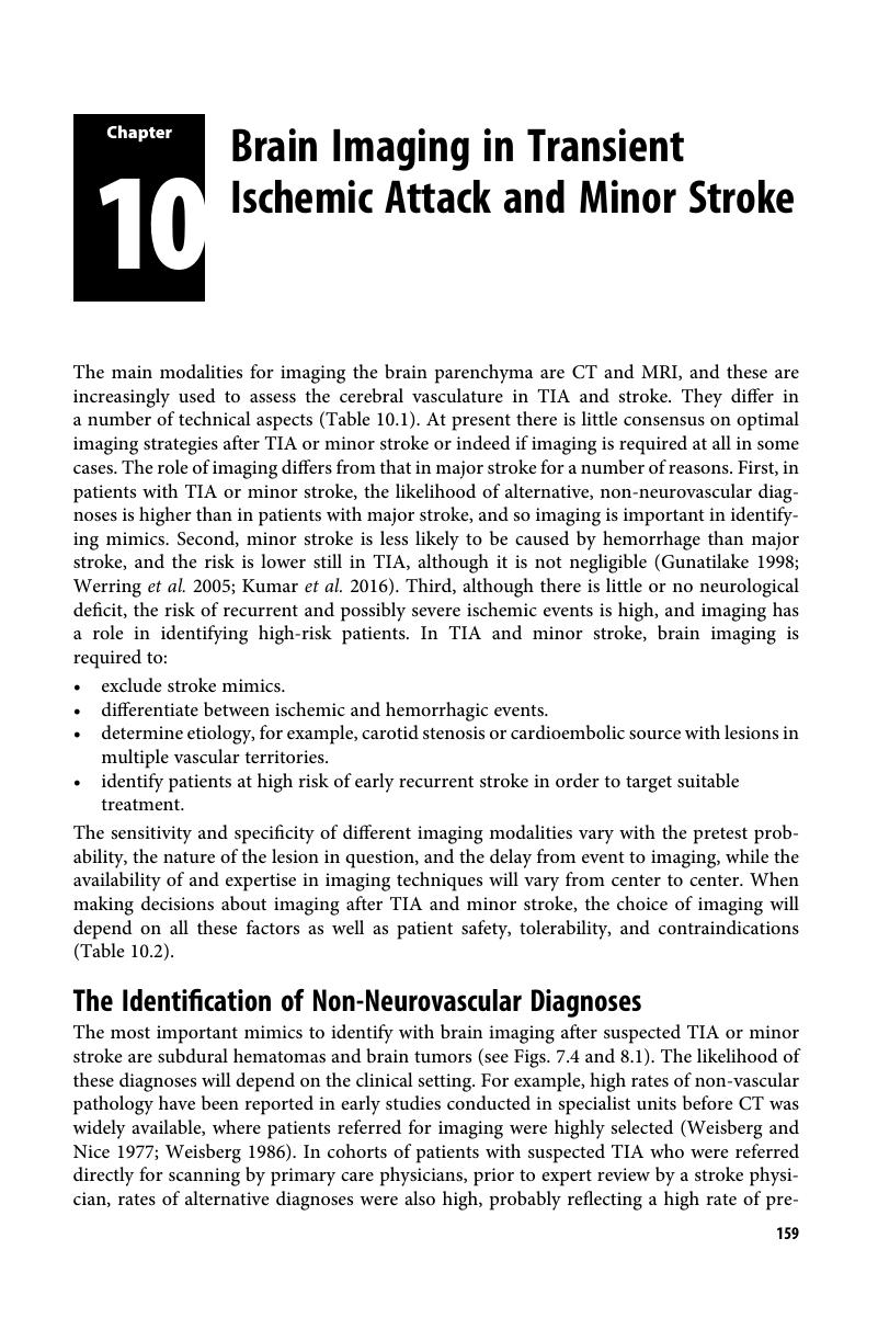
Image of the first page of this content. For PDF version, please use the ‘Save PDF’ preceeding this image.'
Type
Chapter
Information
Transient Ischemic Attack and Stroke
Diagnosis, Investigation and Treatment
, pp. 159 - 175
Publisher: Cambridge University Press
Print publication year: 2018

Access options

Get access to the full version of this content by using one of the access options below. (Log in options will check for institutional or personal access. Content may require purchase if you do not have access.)

References

Albers, GW, Lansberg, MG, Norbash, AM et al. (2000). Yield of diffusion-weighted MRI for detection of potentially relevant findings in stroke patients. Neurology 54:15621567CrossRefGoogle ScholarPubMed
Allen, LM, Hasso, AN, Handwerker, J et al. (2012). Sequence-specific MR imaging findings that are useful in dating ischemic stroke. Radiographics 32:12851297Google Scholar
Ay, H, Oliveira-Filho, J, Buonanno, FS et al. (2002). “Footprints” of transient ischemic attacks: A diffusion-weighted MRI study. Cerebrovascular Digest 14:177186Google Scholar
Bang, OY, Lee, PH, Heo, KG et al. (2005). Specific DWI lesion patterns predict prognosis after acute ischaemic stroke within the MCA territory. Journal of Neurology, Neurosurgery and Psychiatry 76:12221228CrossRefGoogle ScholarPubMed
Chalela, JA, Kidwell, CS, Nentwich, L et al. (2007). Magnetic resonance imaging and computed tomography in emergency assessment of patients with suspected acute stroke: A prospective study. Lancet 369:293298Google Scholar
Chan, S, Kartha, K, Yoon, SS et al. (1996). Multifocal hypointense cerebral lesions on gradient-echo MR are associated with chronic hypertension. American Journal of Neuroradiology 17:18211827Google ScholarPubMed
Cordonnier, C, Al-Shahi Salman, R, Wardlaw, J (2007). Spontaneous brain microbleeds: Systematic review, subgroup analyses and standards for study design and reporting. Brain 130:19882003Google Scholar
Coutts, SB, Simon, JE, Eliasziw, M et al. (2005). Triaging transient ischemic attack and minor stroke patients using acute magnetic resonance imaging. Annals of Neurology 57:848854Google Scholar
Dennis, MS, Bamford, JM, Molyneux, AJ et al. (1987). Rapid resolution of signs of primary intracerebral hemorrhage in computed tomograms of the brain. British Medical Journal 295:379381Google Scholar
Douglas, VC, Johnston, CM, Elkins, J et al. (2003). Head computed tomography findings predict short-term stroke risk after transient ischemic attack. Stroke 34:28942898Google Scholar
Dutch TIA Trial Study Group (1993). Predictors of major vascular events in patients with a transient ischemic attack or nondisabling stroke. Stroke 24:527531CrossRefGoogle Scholar
Fazekas, F, Kleinert, R, Roob, G et al. (1999). Histopathologic analysis of foci of signal loss on gradient-echo T2-weighted MR images in patients with spontaneous intracerebral hemorrhage: Evidence of microangiopathy-related microbleeds. American Journal of Neuroradiology 20:637642Google Scholar
Fiebach, JB, Schellinger, PD, Gass, A et al. (2004). Stroke magnetic resonance imaging is accurate in hyperacute intracerebral hemorrhage: A multicenter study on the validity of stroke imaging. Stroke 35:502506CrossRefGoogle ScholarPubMed
Fisher, M (2014). Cerebral microbleeds: Where are we now? Neurology 83:13041305Google Scholar
Gass, A, Ay, H, Szabo, K et al. (2004). Diffusion-weighted MRI for the “small stuff”: The details of acute cerebral ischaemia. Lancet Neurology 3:3945Google Scholar
Greenberg, SM, Finklestein, SP, Schaefer, PW (1996). Petechial hemorrhages accompanying lobar hemorrhage: Detection by gradient-echo MRI. Neurology 46:17511754CrossRefGoogle ScholarPubMed
Gunatilake, SB (1998). Rapid resolution of symptoms and signs of intracerebral hemorrhage: Case reports. British Medical Journal 316:14951496CrossRefGoogle Scholar
Kidwell, CS, Alger, JR, Di Salle, F et al. (1999). Diffusion MRI in patients with transient ischemic attacks. Stroke 30:11741180Google Scholar
Kidwell, CS, Chalela, JA, Saver, JL et al. (2004). Comparison of MRI and CT for detection of acute intracerebral hemorrhage. JAMA 292:18231830Google Scholar
Knudsen, KA, Rosand, J, Karluk, D et al. (2001). Clinical diagnosis of cerebral amyloid angiopathy: Validation of the Boston Criteria. Neurology 56:537539CrossRefGoogle ScholarPubMed
Kumar, S, Selim, M, Marchina, S et al. (2016). Transient neurological symptoms in patients with intracerebral hemorrhage. JAMA Neurology 73:316320CrossRefGoogle ScholarPubMed
Jauch, EC, Saver, JL, JrAdams, HP et al. (2013). Guidelines for the early management of patients with acute ischemic stroke: A guideline for healthcare professionals from the American Heart Association/American Stroke Association. Stroke 44:870947Google Scholar
Lau, KK, Wong, YK, Teo, KC et al. (2017). Long-term prognostic implications of cerebral microbleeds in Chinese with ischemic stroke. Journal of the American Heart Association 6(12)Google Scholar
Lemesle, M, Madinier, G, Menassa, M et al. (1998). Incidence of transient ischemic attacks in Dijon, France. A 5-year community-based study. Neuroepidemiology 17:7479Google Scholar
National Institute for Health and Clinical Excellence (2017). Stroke and Transient Ischemic Attack in Over 16s: Diagnosis and Initial Management. London: NICEGoogle Scholar
Offenbacher, H, Fazekas, F, Schmidt, R et al. (1996). MR of cerebral abnormalities concomitant with primary intracerebral hematomas. American Journal of Neuroradiology 17:573578Google ScholarPubMed
Purroy, F, Montaner, J, Rovira, A et al. (2004). Higher risk of further vascular events among transient ischaemic attack patients with diffusion-weighted imaging acute lesions. Stroke 35:23132319Google Scholar
Redgrave, JN, Coutts, SB, Schulz, UG et al. (2007). Systematic review of associations between the presence of acute ischemic lesions on diffusion-weighted imaging and clinical predictors of early stroke risk after transient ischemic attack. Stroke 38:14821488Google Scholar
Rolak, LA, Gilmer, W, Strittmatter, WJ (1990). Low yield in the diagnostic evaluation of transient ischemic attacks. Neurology 40:747748Google Scholar
Schaefer, PW, Copen, WA, Lev, MH et al. (2005). Diffusion-weighted imaging in acute stroke. Neuroimaging Clinics of North American 15:503530Google Scholar
Schellinger, PD, Jansen, O, Fiebach, JB et al. (1999). A standardized MRI stroke protocol: Comparison with CT in hyperacute intracerebral hemorrhage. Stroke 30:765768Google Scholar
Schellinger, PD, Thomalla, G, Fiehler, J et al. (2007). MRI-based and CT-based thrombolytic therapy in acute stroke within and beyond established time windows: An analysis of 1210 patients. Stroke 38:26402645Google Scholar
Schulz, UGR, Briley, D, Meagher, T et al. (2003). Abnormalities on diffusion weighted magnetic resonance imaging performed several weeks after a minor stroke or transient ischaemic attack. Journal of Neurology, Neurosurgery and Psychiatry 74:734738Google Scholar
Schulz, UG, Flossman, E, Rothwell, PM (2004). Heritability of ischemic stroke in relation to age, vascular risk factors and subtypes of incident stroke in population-based studies. Stroke 35:819824CrossRefGoogle ScholarPubMed
Shoamanesh, A, Kwok, CS, Benavente, O (2011). Cerebral microbleeds: Histopathological correlation of neuroimaging. Cerebrovascular diseases 32:528534Google Scholar
Sylaja, PN, Coutts, SB, Subramaniam, S et al. (2007). Acute ischemic lesions of varying ages predict risk of ischemic events in stroke/TIA patients. Neurology 68:415419Google Scholar
UK TIA Study Group (1993). Intracranial tumours that mimic transient cerebral ischaemia: Lessons from a large multicentre trial. Journal of Neurology, Neurosurgery and Psychiatry 56:563566CrossRefGoogle Scholar
van Veluw, SJ, Biessels, GJ, Klijn, CJM et al. (2016). Heterogeneous histopathology of cortical microbleeds in cerebral amyloid angiopathy. Neurology 86:867871Google Scholar
Wardlaw, JM, Keir, SL, Dennis, MS (2003). The impact of delays in computed tomography of the brain on the accuracy of diagnosis and subsequent management in patients with minor stroke. Journal of Neurology, Neurosurgery and Psychiatry 74:7781CrossRefGoogle ScholarPubMed
Weisberg, LA (1986). Computerized tomographic abnormalities in patients with hemispheric transient ischemic attacks. South Medical Journal 79:804807Google Scholar
Weisberg, LA, Nice, CN (1977). Intracranial tumors simulating the presentation of cerebrovascular syndromes. Early detection with cerebral computed tomography (CCT). American Journal of Medicine 63:517524Google Scholar
Wen, HM, Lam, WW, Rainer, T et al. (2004). Multiple acute cerebral infarcts on diffusion-weighted imaging and risk of recurrent stroke. Neurology 63:13171319Google Scholar
Werring, DJ, Coward, LJ, Losseff, NA et al. (2005). Cerebral microbleeds are common in ischemic stroke but rare in TIA. Neurology 65:19141918CrossRefGoogle ScholarPubMed
Wilson, D, Charidimou, A, Ambler, G et al. (2016). Recurrent stroke risk and cerebral microbleed burden in ischemic stroke and TIA: A meta-analysis. Neurology 87:110Google Scholar
Wityk, RJ, Pessin, MS, Kaplan, RF et al. (1994). Serial assessment of acute stroke using the NIH Stroke Scale. Stroke 25:362365Google Scholar

Save book to Kindle

To save this book to your Kindle, first ensure coreplatform@cambridge.org is added to your Approved Personal Document E-mail List under your Personal Document Settings on the Manage Your Content and Devices page of your Amazon account. Then enter the ‘name’ part of your Kindle email address below. Find out more about saving to your Kindle.

Note you can select to save to either the @free.kindle.com or @kindle.com variations. ‘@free.kindle.com’ emails are free but can only be saved to your device when it is connected to wi-fi. ‘@kindle.com’ emails can be delivered even when you are not connected to wi-fi, but note that service fees apply.

Find out more about the Kindle Personal Document Service.

Available formats
×

Save book to Dropbox

To save content items to your account, please confirm that you agree to abide by our usage policies. If this is the first time you use this feature, you will be asked to authorise Cambridge Core to connect with your account. Find out more about saving content to Dropbox.

Available formats
×

Save book to Google Drive

To save content items to your account, please confirm that you agree to abide by our usage policies. If this is the first time you use this feature, you will be asked to authorise Cambridge Core to connect with your account. Find out more about saving content to Google Drive.

Available formats
×