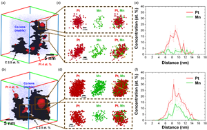
Carbon-supported nanoparticles have been used widely as efficient catalysts due to their enhanced surface-to-volume ratio. To investigate their structure–property relationships, acquiring 3D elemental distribution is required. Here, carbon-supported Pt, PtMn alloy, and ordered Pt3Mn nanoparticles are synthesized and analyzed with atom probe tomography as model systems. A significant difference of Mn distribution after the heat-treatment was found. Finally, the field evaporation behavior of the carbon support was discussed and each acquired reconstruction was compared with computational results from an evaporation simulation. This paper provides a guideline for studies using atom probe tomography on the heterogeneous carbon-supported nanoparticle system that leads to insights toward a wide variety of applications.
The local field ion emission properties of helium and neon around a step edge atom of W(112) were examined at liquid nitrogen temperature using a micro-probe hole field ion microscope combined with a pulse-counting analysis. We have analyzed the mapped field ion densities obtained for both imaging gas atoms at their respective best local image voltages based on the formula for tunneling barrier strength and have evaluated the dipole moment of polarized adatom as well as the local field enhancement factor at the adatom site. We found that the dipole moments of helium and neon adatoms showed the same value, although the best local image field acting on the helium adatom is much higher than that on the neon adatom. We also found the same magnitude of local field enhancement factors for both noble gas field adsorptions. These results imply that the key to the best local image condition is the tunneling barrier field variations above the adatom. The vital role of the imaging gas atoms is to form an optimum dipole moment to create an ideal electric field distribution for the best local image appearance at each atom site depending on the different chemical nature of adatom species.
Electron probe microanalysis (EPMA) is based on physical relations between measured X-ray intensities of characteristic lines and their X-ray production efficiency, which depends on the specimen composition. The quality of the analysis results relies on how realistically the physical relations describe the generation and emission of X-rays. Special experiments are necessary to measure X-ray production efficiencies. A challenge in these experiments is the determination of the detection efficiency of the spectrometer as a function of the photon energy. An energy-dispersive spectrometer was used in this work, for which the efficiency was determined at metrological synchrotron beamlines with an accuracy of ±2%. X-ray production efficiencies for the L series and the Kα series of copper and zirconium and for the M and L series of tungsten were determined at energies up to 30 keV in a scanning electron microscope. These experimental values were compared with calculated X-ray production efficiencies using physical relations and material constants applied in EPMA. The objective of the comparison is the further improvement of EPMA algorithms as well as extending the available database for X-ray production efficiencies. Experimental data for the X-ray production efficiency are also useful for the assessment of spectrum simulation software.
Searching for residue in the glaze of porcelain or stoneware is a difficult task because these glazes are high-fired, well vitrified, and nonporous. This paper analyzes the chemical composition of residue observed in glaze cracks of porcelain via SEM-EDS to determine how the crackle effect was produced, in particular, if it was intentionally created during production or the result of post-depositional processes. This study offers insights to a specific type of ancient Chinese porcelain called “Ge-type ware”, which has two different types of cracks, and whose origin has been debated for nearly 60 years because it has never been found at any kiln site. This paper analyzes the chemical composition of the two crack types, first using elemental mapping to ascertain the different mechanisms that produced these two crack types of the Heirloom Ge ware, and second using residue analysis and chemical fingerprinting to determine the provenance of this puzzling type of porcelain. In doing so, this paper demonstrates how the residue in the glaze of porcelain can be observed and analyzed via microchemical approaches and hopes to inspire more research using this technique in future.
Correlative microscopy approaches are attracting considerable interest in several research fields such as materials and battery research. Recent developments regarding X-ray computer tomography have made this technique available in a compact module for scanning electron microscopes (SEMs). Nano-computed tomography (nanoCT) allows morphological analysis of samples in a nondestructive way and to generate 2D and 3D overviews. However, morphological analysis alone is not sufficient for advanced studies, and to draw conclusions beyond morphology, chemical analysis is needed. While conventional SEM-based chemical analysis techniques such as energy-dispersive X-ray spectroscopy (EDS) are adequate in many cases, they are not well suited for the analysis of trace elements and low-Z elements such as hydrogen or lithium. Furthermore, the large information depth in typical SEM-EDS imaging conditions limits the lateral resolution to micrometer length scales. In contrast, secondary ion mass spectrometry (SIMS) can perform elemental mapping with good surface sensitivity, nanoscale lateral resolution, and the possibility to analyze even low-Z elements and isotopes. In this study, we demonstrate the feasibility and compatibility of a novel FIB-SEM-based correlative nanoCT-SIMS imaging approach to correlate morphological and chemical data of the exact same sample volume, using a cathode material of a commercial lithium battery as an example.
Trained neural networks are promising tools to analyze the ever-increasing amount of scientific image data, but it is unclear how to best customize these networks for the unique features in transmission electron micrographs. Here, we systematically examine how neural network architecture choices affect how neural networks segment, or pixel-wise separate, crystalline nanoparticles from amorphous background in transmission electron microscopy (TEM) images. We focus on decoupling the influence of receptive field, or the area of the input image that contributes to the output decision, from network complexity, which dictates the number of trainable parameters. For low-resolution TEM images which rely on amplitude contrast to distinguish nanoparticles from background, we find that the receptive field does not significantly influence segmentation performance. On the other hand, for high-resolution TEM images which rely on both amplitude and phase-contrast changes to identify nanoparticles, receptive field is an important parameter for increased performance, especially in images with minimal amplitude contrast. Rather than depending on atom or nanoparticle size, the ideal receptive field seems to be inversely correlated to the degree of nanoparticle contrast in the image. Our results provide insight and guidance as to how to adapt neural networks for applications with TEM datasets.
Electron-excited X-ray microanalysis with energy-dispersive spectrometry (EDS) proceeds through the application of the software that extracts characteristic X-ray intensities and performs corrections for the physics of electron and X-ray interactions with matter to achieve quantitative elemental analysis. NIST DTSA-II is an open-access, fully documented, and freely available comprehensive software platform for EDS quantification, measurement optimization, and spectrum simulation. Spectrum simulation with DTSA-II enables the prediction of the EDS spectrum from any target composition for a specified electron dose and for the solid angle and window parameters of the EDS spectrometer. Comparing the absolute intensities for measured and simulated spectra reveals correspondence within ±25% relative to K-shell and L-shell characteristic X-ray peaks in the range of 1–11 keV. The predicted M-shell intensity exceeds the measured value by a factor of 1.4–2.2 in the range 1–3 keV. The X-ray continuum (bremsstrahlung) generally agrees within ±10% over the range of 1–10 keV. Simulated EDS spectra are useful for developing an analytical strategy for challenging problems such as estimating trace detection levels.
Spatially resolved in situ transmission electron microscopy (TEM), equipped with direct electron detection systems, is a suitable technique to record information about the atom-scale dynamics with millisecond temporal resolution from materials. However, characterizing dynamics or fluxional behavior requires processing short time exposure images which usually have severely degraded signal-to-noise ratios. The poor signal-to-noise associated with high temporal resolution makes it challenging to determine the position and intensity of atomic columns in materials undergoing structural dynamics. To address this challenge, we propose a noise-robust, processing approach based on blob detection, which has been previously established for identifying objects in images in the community of computer vision. In particular, a blob detection algorithm has been tailored to deal with noisy TEM image series from nanoparticle systems. In the presence of high noise content, our blob detection approach is demonstrated to outperform the results of other algorithms, enabling the determination of atomic column position and its intensity with a higher degree of precision.
This paper exploited an alternative approach to prepare high-quality speckle patterns by uniformly dispersing nano-silica particles onto sample surfaces, helping digital image correlation (DIC) acquire the maximum spatial resolution of local strain up to 92 nm. A case study was carried out by combining this speckle pattern fabrication method with SEM-DIC and electron backscattering diffraction (EBSD). Thus, in situ mapping of local strain with ultra-high spatial resolution and microstructure in commercially pure titanium during plastic deformation could be achieved, which favored revealing the effect of slip transfer on shear strain near grain boundaries. Moreover, the slip systems could be easily identified via the combination of the SEM-DIC and EBSD techniques even though no obvious deformation trace was captured in secondary electron images. Additionally, the complex geometric compatibility factor relating to geometric compatibility factors (mʹ) and Schmid factors was proposed to predict the shear strain (εxy) at grain boundaries.
Visualizing how a catalyst behaves during chemical reactions using in situ transmission electron microscopy (TEM) is crucial for understanding the activity origin and guiding performance optimization. However, the sample drifts as temperature changes during in situ reaction, which weakens the resolution and stability of TEM imaging, blocks insights into the dynamic details of catalytic reaction. Herein, a Thon-ring based sample position measurement (TSPM) was developed to track the sample height variation during in situ TEM observation. Drifting characteristics for three commercially available nanochips were studied, showing large biases in aspects of shifting modes, expansion heights, as well as the thermal conduction hysteresis during rapid heating. Particularly, utilizing the TSPM method, for the first time, the gas layer thickness inside a gas-cell nanoreactor was precisely determined, which varies with reaction temperature and gas pressure in a linear manner with coefficients of ~8 nm/°C and ~50 nm/mbar, respectively. Following drift prediction of TSPM, fast oxidation kinetics of a Ni particle was tracked in real time for 12 s at 500°C. This TSPM method is expected to facilitate the functionality of automatic target tracing for in situ microscopy applications when feedback to hardware control of the microscope.
Here, we report a case study implementation of reinforcement learning (RL) to automate operations in the scanning transmission electron microscopy workflow. To do so, we design a virtual, prototypical RL environment to test and develop a network to autonomously align the electron beam position without prior knowledge. Using this simulator, we evaluate the impact of environment design and algorithm hyperparameters on alignment accuracy and learning convergence, showing robust convergence across a wide hyperparameter space. Additionally, we deploy a successful model on the microscope to validate the approach and demonstrate the value of designing appropriate virtual environments. Consistent with simulated results, the on-microscope RL model achieves convergence to the goal alignment after minimal training. Overall, the results highlight that by taking advantage of RL, microscope operations can be automated without the need for extensive algorithm design, taking another step toward augmenting electron microscopy with machine learning methods.
Warping is a limiting factor when preparing transmission electron microscopy (TEM) samples using focused ion beam (FIB)/scanning electron microscope (SEM) systems. The conventional FIB sputtering process leaves at least one side of the lamella too thin to provide structural support to offset inherent stresses. As a result, warping can occur impacting imagining and reducing the potential size of lamellae. For example, capturing more than a few back-end metal layers in a 3 μm wide cross-section lamella can be difficult. Frequently, TEM analysts must perform multiple stage adjustments to analyze such a sample. In this paper, two methods are presented that enable FIB/SEM operators to prepare TEM samples where the thinned region of interest is surrounded by thick structures. As a result, these methods reduce warping and enable the fabrication of TEM lamellae not possible by conventional means. For example, these methods have been used to produce a 10 μm wide by 8 μm tall cross-section TEM sample that captured front-end transistors and 14 back-end metal layers. Furthermore, warping was so limited that only one alignment was needed by the TEM analyst to complete the imaging of the sample. The methods, called the horizontal bracing and window methods, make use of the deposition of low-Z amorphous films that are electron transparent in the TEM.
The Compton signal in electron energy loss spectroscopy (EELS) is used to determine the projected electron momentum density of states for the solid. A frequent limitation however is the strong dynamical scattering of the incident electron beam within a crystalline specimen, i.e. Bragg diffracted beams can be additional sources of Compton scattering that distort the measured profile from its true shape. The Compton profile is simulated via a multislice method that models dynamical scattering both before and after the Compton energy loss event. Simulations indicate the importance of both the specimen illumination condition and EELS detection geometry. Based on this, a strategy to minimize diffraction artifacts is proposed and verified experimentally. Furthermore, an inversion algorithm to extract the projected momentum density of states from a Compton measurement performed under strong diffraction conditions is demonstrated. The findings enable a new route to more accurate electron Compton data from crystalline specimens.
Electron tomography (ET) has gained increasing attention for the 3D characterization of nanoparticles. However, the missing wedge problem due to a limited tilt angle range is still the main challenge for accurate reconstruction in most experimental TEM setups. Advanced algorithms could in-paint or compensate to some extent the missing wedge artifacts, but cannot recover the missing structural information completely. 360° ET provides an option to solve this problem by tilting a needle-shaped specimen over the full tilt range and thus filling the missing information. However, sample preparation especially for fine powders to perform full-range ET is still challenging, thus limiting its application. In this work, we propose a new universal sample preparation method that enables the transfer of selected individual nanoparticle or a few separated nanoparticles by cutting a piece of carbon film supporting the specimen particles and mounting them onto the full-range tomography holder tip with the help of an easily prepared sharp tungsten tip. This method is demonstrated by 360° ET of Pt@TiO2 hollow cage catalyst showing high quality reconstruction without missing wedge.
The energy distribution of an electron gun is one of the most important characteristics determining the performance of electron beam-based instruments, such as electron microscopes and electron energy loss spectroscopes. For accurate measurements of the energy distribution, this study presents a novel retarding field energy analyzer (RFEA) with the feature of an additional integrated pre-lens, which enables an adjustment of beam trajectory into the analyzer. The advantages of this analyzer are its compact size and simple electrode configuration. According to trajectory simulation theories, the optimum condition arises when the incident electron beam inside the RFEA is focused on the center of a retarding electrode. Comparing I–V curves depending on whether the pre-lens working or not, it is confirmed that the use of the pre-lens dramatically improves the energy resolution and efficiency of the signal acquisition process. The pre-lens RFEA was applied to characterize a Schottky electron gun under various temperatures and extraction voltages as operational conditions. When the tip temperature was increased by 50 K, we were able to measure an energy distribution broadening of 13.8 meV with the proposed pre-lens RFEA. The relative standard deviation of energy distribution was 0.7% for each working condition.
Understanding the structure of materials is crucial for engineering devices and materials with enhanced performance. Four-dimensional scanning transmission electron microscopy (4D-STEM) is capable of mapping nanometer-scale local crystallographic structure over micron-scale field of views. However, 4D-STEM datasets can contain tens of thousands of images from a wide variety of material structures, making it difficult to automate detection and classification of structures. Traditional automated analysis pipelines for 4D-STEM focus on supervised approaches, which require prior knowledge of the material structure and cannot describe anomalous or deviant structures. In this article, a pipeline for engineering 4D-STEM feature representations for unsupervised clustering using non-negative matrix factorization (NMF) is introduced. Each feature is evaluated using NMF and results are presented for both simulated and experimental data. It is shown that some data representations more reliably identify overlapping grains. Additionally, real space refinement is applied to identify spatially distinct sample regions, allowing for size and shape analysis to be performed. This work lays the foundation for improved analysis of nanoscale structural features in materials that deviate from expected crystallographic arrangement using 4D-STEM.
Scanning electron microscopy operated at cryogenic temperature (cryo-SEM) is a powerful tool for investigating surface and cross-sectional nanostructures of water-containing samples. Typically, cryo-SEM samples are frozen just before observation in specific metal carriers. However, pre-frozen samples are also of interest, such as frozen food and freeze-stored animal samples. In such cases, sample mounting with a defined orientation is required, but there has been a lack of ideal conductive adhesives that can be used without increasing the sample temperature. Here, we developed a mixture of graphite oxide and 1,3-butanediol as an adhesive, capable of gluing samples at dry-ice temperature and is frozen below that temperature. Dispersion of graphite oxide increased the conductivity and reduced the charge-up contrast. Acquisition of energy-dispersive X-ray spectrum, cross-sectional ion milling, and high-resolution imaging were successfully achieved using the adhesive. We tested and confirmed the usefulness of this new adhesive by applying it to cryo-SEM surface imaging of diatomite, freeze-fractured cross-sectional imaging of chicken liver, and ion milling cross-sectional imaging of a deep-sea snail. The new adhesive is not only useful for food science and field-preserved biological samples but also potentially applicable to wider fields such as archaeological and biological samples preserved under permafrost.
The three-dimensional characterization of internal features, via metrics such as orientation, porosity, and connectivity, is important to a wide variety of scientific questions. Many spatial and morphological metrics only can be measured accurately through direct in situ three-dimensional observations of large (i.e., big enough to be statistically representative) volumes. For samples that lack material contrast between phases, serial grinding and imaging—which relies solely on color and textural characteristics to differentiate features—is a viable option for extracting such information. Here, we present the Grinding, Imaging, Reconstruction Instrument (GIRI), which automatically serially grinds and photographs centimeter-scale samples at micron resolution. Although the technique is destructive, GIRI produces an archival digital image stack. This digital image stack is run through a supervised machine-learning-based image processing technique that quickly and accurately segments data into predefined classes. These classified data then can be loaded into three-dimensional visualization software for measurement. We share three case studies to illustrate how GIRI can address questions with a significant morphological component for which two-dimensional or small-volume three-dimensional measurements are inadequate. The analyzed metrics include: the morphologies of objects and pores in a granular material, the bulk mineralogy of polyminerallic solids, and measurements of the internal angles and symmetry of crystals.
This work investigates how knock-on displacements influence fluctuation electron microscopy (FEM) experiments. FEM experiments were conducted on amorphous silicon, formed by self-ion implantation, in a transmission electron microscope at 300 kV and 60 kV at various electron doses, two different binnings and with two different cameras, a CCD and a CMOS one. Furthermore, energy filtering has been utilized in one case. Energy filtering greatly enhances the FEM data by removing the inelastic background intensity, leading to an improved speckle contrast. The CMOS camera yields a slightly larger normalized variance than the CCD at an identical electron dose and appears more prone to noise at low electron counts. Beam-induced atomic displacements affect the 300 kV FEM data, leading to a continuous suppression of the normalized variance with increasing electron dose. Such displacements are considerably reduced for 60 kV experiments since the primary electron's maximum energy transfer to an atom is less than the displacement threshold energy of amorphous silicon. The results show that the variance suppression due to knock-on displacements can be controlled in two ways: Either by minimizing the electron dose to the sample or by conducting the experiment at a lower acceleration voltage.
Accurately determining local polarization at atomic resolution can unveil the mechanisms by which static and dynamical behaviors of the polarization occur, including domain wall motion, defect interaction, and switching mechanisms, advancing us toward the better control of polarized states in materials. In this work, we explore the potential of atomic-resolution scanning transmission electron microscopy to measure the projected local polarization at the unit cell length scale. ZnO and PbMg1/3Nb2/3O3 are selected as case studies, to identify microscope parameters that can significantly affect the accuracy of the measured projected polarization vector. Different STEM imaging modalities are used to determine the location of the atomic columns, which, when combined with the Born effective charges, allows for the calculation of local polarization. Our results indicate that differentiated differential phase contrast (dDPC) imaging enhances the accuracy of measuring local polarization relative to other imaging modalities, such as annular bright-field or integrated-DPC imaging. For instance, under certain experimental conditions, the projected spontaneous polarization for ZnO can be calculated with 1.4% error from the theoretical value. Furthermore, we quantify the influence of sample thickness, probe defocus, and crystal mis-tilt on the relative errors of the calculated polarization.
The development of biochemical analysis techniques to study heterogeneous biological samples is increasing. These techniques include synchrotron radiation Fourier transform infrared (SR-FTIR) microspectroscopy. This method has been applied to analyze biological tissue with multivariate statistical analysis to classify the components revealed by the spectral data. This study aims to compare the efficiencies of SR-FTIR microspectroscopy and focal plane array (FPA)-FTIR microspectroscopy when classifying rice tissue components. Spectral data were acquired for mapping the same sample areas from both techniques. Principal component analysis and cluster imaging were used to investigate the biochemical variations of the tissue types. The classification was based on the functional groups of pectin, protein, and polysaccharide. Four layers from SR-FTIR microspectroscopy including pericarp, aleurone layer, sub-aleurone layer, and endosperm were classified using cluster imaging, while FPA-FTIR microspectroscopy could classify only three layers of pericarp, aleurone layer, and endosperm. Moreover, SR-FTIR microspectroscopy increased the image contrast of the biochemical distribution in rice tissue more efficiently than FPA-FTIR microspectroscopy. We have demonstrated the capability of the high-resolution synchrotron technique and its ability to clarify small structures in rice tissue. The use of this technique might increase in future studies of tissue characterization.
Telocyte (TC)—a new type of interstitial cell with long telopodes, can form cellular junctions with various tissues or cells to participate in the regulation of multitudes of physiological activities and diseases. This study aimed to characterize the morphology, molecular features, and potential functions of hormone regulation in Chinese soft-shelled turtle (Pelodiscus sinensis) testis TCs at different reproductive stages by histological evaluation, immunohistochemistry (IHC), immunofluorescence (IF), and transmission electron microscopy. During hibernation, TCs were widely distributed in the interstitial tissue. In contrast, during reproductive activity, TCs were noted to be in close proximity with peritubular myoid cells surrounding the seminiferous tubule. Moreover, formed cell–cell junctions were observed between TCs and PTMs. The results of IHC and IF showed that the immunophenotype of testicular TCs in hibernating Chinese soft-shelled turtles is CD34+Vimentin−, while the reproductive telopodes (Tps) show low expression of vimentin. The androgen receptor is expressed in Tps of TCs of testis during hibernation. Our results showed also that TCs in seasonal breeding animals regulate the activity of neighboring cells by releasing extracellular microvesicles (EXMVs), thus influencing the activity of spermatogenesis and steroidogenesis. Consideration of our novel and interesting results indicate that the whole area warrants further research.
Toll-like receptor (TLR)-mediated inflammatory processes play a critical role in the innate immune response during the initial interaction between the infecting microorganism and immune cells. This study aimed to investigate the possible microanatomical and histological differences in mandibular and bronchial lymph nodes in Akkaraman and Romanov lambs induced by lipopolysaccharide (LPS) and lipoteichoic acid (LTA) and study the gene, protein, and immunoexpression levels of TLR4, myeloid differentiation factor 88 (MyD88), and tumor necrosis factor-α (TNF-α) that are involved in the immune system. Microanatomical examinations demonstrated more intense lymphocyte infiltration in the bronchial lymph nodes of Akkaraman lambs in the LPS and LTA groups compared to Romanov lambs. TLR4, MyD88, and TNF-α immunoreactivities were more intense in the experimental groups of both breeds. Expression levels of MyD88 and TNF-α genes in the bronchial lymph node of Akkaraman lambs were found to increase statistically significantly in the LTA group. TLR4 gene expression level in the mandibular lymph node was found to be statistically significantly higher in the LTA + LPS group. In conclusion, dynamic changes in the immune cell populations involved in response to antigens such as LTA and LPS in the lymph nodes of both breeds can be associated with the difference in the expression level of the TLR4/MyD88/TNF-α genes.
Since embryonic stem cells (ESCs) were first identified, significant progress has been achieved. However, the establishment of buffalo ESCs (bESCs) is still unclear. This study was undertaken to explore the effect of the blastocyst stage on the isolation of bESCs. Firstly, our results indicated that the pluripotent genes were mainly expressed at the early stages of blastocyst, and the attachment and colony formation rates of bESCs derived from expanded blastocyst and hatched blastocyst were significantly higher than early blastocyst and blastocyst. In the meantime, bESCs showed positive alkaline phosphatase activity and expressed genes like OCT4, NANOG, SOX2, c-MYC, CDH1, KLF4, and TBX3. Immunofluorescence also confirmed the expression of OCT4, SOX2. Embryoid bodies expressing three marker genes were generated from the differentiation experiment, and fibroblast, epithelial, and neuron-like cells were induced. Moreover, naive-related genes KLF4, TBX3, primed-related genes FGF5, ACTA2 were expressed in the cells, but not REX-1. Immunofluorescence and western blot confirmed the FGF5 expression. Furthermore, bESCs could maintain pluripotency with the signal of LIF and bFGF. In summary, our results indicated that expanded blastocyst and hatched blastocyst are more suitable for bESCs isolation.
]]>
In this study, we examined the cellular infectivity and ultrastructural changes due to severe acute respiratory syndrome coronavirus 2 (SARS-CoV-2) infection in the various cells of bronchoalveolar fluid (BALF) from intubated patients of different age groups (≥60 years and <60 years) and with common comorbidities such as diabetes, liver and kidney diseases, and malignancies. BALF of 79 patients (38 cases >60 and 41 cases <60 years) were studied by light microscopy, immunofluorescence, scanning, and transmission electron microscopy to evaluate the ultrastructural changes in the ciliated epithelium, type II pneumocytes, macrophages, neutrophils, eosinophils, lymphocytes, and anucleated granulocytes. This study demonstrated relatively a greater infection and better preservation of subcellular structures in these cells from BALF of younger patients (<60 years compared with the older patients (≥60 years). The different cells of BALF from the patients without comorbidities showed higher viral load compared with the patients with comorbidities. Diabetic patients showed maximum ultrastructural damage in BALF cells in the comorbid group. This study highlights the comparative effect of SARS-CoV-2 infection on the different airway and inflammatory cells of BALF at the subcellular levels among older and younger patients and in patients with comorbid conditions.
Recent studies indicated that ergosterol (Erg) helps form strongly ordered lipid domains in membranes that depend on their chemical characters. However, direct evidence of concentration-dependent interaction of Erg with lipid membranes has not been reported. We studied the Erg concentration-dependent changes in the phase behaviors of membranes using cell-sized liposomes containing 1,2-Dioleoyl-sn-glycero-3-phosphocholine (DOPC)/1,2-dipalmitoyl-sn-glycero-3-phosphocholine (DPPC). We observed the concentration range of phase separation in ternary membranes was significantly wider when Erg rather than cholesterol (Chol) was used as the sterol component. We used machine learning for the first time to analyze microscopic images of cell-sized liposomes and identify phase-separated structures. The automated method was successful in identifying homogeneous membranes but performance remained data-limited for the identification of phase separation domains characterized by more complex features.
Morphological studies concerning the digestive system can further information on animal diets, thus aiding in the understanding of feeding behavior. Given the scarcity of information on sea turtle digestive system morphology, the aim of the present study was to describe the digestive tube (DT) morphology of Eretmochelys imbricata hatchlings to further understand the diet of these individuals in the wild. DT samples from 10 stillborn turtles (undefined sex) were analyzed at the macro and microscopic levels. The esophagus, stomach, small intestine (SI), and large intestine (LI) are described. Histologically, the DT is formed by four tunics, the mucosa, submucosa, muscular, and adventitia or serosa. The esophagus is lined by keratinized stratified squamous epithelium, while the remainder of the DT is lined by a simple columnar epithelium. The esophagus mucosa is marked by conical, pointed papillae. The stomach comprises three regions, the cardiac, fundic, and pyloric and is covered by neutral mucous granular cells. The intestinal mucosa presents absorptive cells with microvilli, neutral and acidic goblet cells, and mucosa-associated lymphoid tissue. The SI is significantly longer than the LI (p value = 0.006841). These morphological findings are strong indications of adaptations to a carnivorous diet in this hawksbill turtle age group.
The raw materials of “licorice root” in the commerce consist of roots and/or rhizomes (stolons) of different species of Glycyrrhiza. Licorice products and raw materials are frequently mislabeled and often have mixed, misidentified, or unidentified species and parts. This paper provides a detailed comparative analysis of the morpho-anatomies of the rhizomes and roots of five species of Glycyrrhiza, namely G. glabra, G. uralensis, G. echinata, G. inflata, and G. lepidota, by bright-field light microscopy and scanning electron microscopy. The studied species showed some similarities in their basic anatomical features due to the fact that they are phylogenetically closely related and belong to the same genus. However, differences in microscopic features such as the thickness of cork and medullary rays, pore frequency, and size of the vessels were observed. The rhizomes can readily be distinguished by the presence of a distinct pith. The roots lack a well-defined pith and instead have primary xylem in the center.
Diabetes mellitus is a serious disease worldwide and causes other associated diseases. In this study, we observed the effect of streptozotocin (STZ)-induced diabetes and benfluorex treatment on muscular capillary ultrastructure. Adult male rats were used as the test subjects and each individual was intraperitoneally injected with one dose of STZ (45 mg/kg) to induce diabetes. Doses (50 mg/kg) of benfluorex were given to the subjects with tap water by intragastric gavage application once daily for 21 days. At the end of day 21, muscle tissues were obtained from animals and examined under transmission electron microscopy. From the data obtained with the electron microscope, it was observed that the control group had typical continuous capillary vascular structures in their muscles, while STZ caused disruptive disorder of the muscle cells in the capillary wall of the STZ-diabetic group. Additionally, the thickening of the basement membrane around endothelial cells, loss of mitochondrial crista in the muscle cells, enlarged endothelial cells, and narrowed vessel lumen were observed in the muscle tissue. The findings of our study revealed that STZ-induced diabetes disrupted the vascular structure, while benfluorex partially improved it.
Herein, we propose a convenient method to enable pretreatment of target objects using digital holographic microscopy (DHM). As a test sample, we used diatom frustules (Nitzschia sp.) as the target objects. In the generally used sample preparation method, the frustule suspension is added dropwise onto a glass substrate or into a glass chamber. While our work confirms good observation of purified frustules using the typical sample preparation method, we also demonstrate a new procedure to observe unseparated structures of frustules prepared by baking them on a mica surface. The baked frustules on the mica surface were transferred to a glass chamber with 1% sodium dodecyl sulfate solution. In this manner, the unseparated structures of the diatom frustules were clearly observed. Furthermore, metal-coated frustules prepared by sputtering onto them on a mica surface were also clearly observed using the same procedure. Our method can be applied for the observation of any target object that is pretreated on a solid surface. We expect our proposed method to be a basis for establishing DHM techniques for microscopic observations of biomaterials.
]]>The Cicadomorpha Philaenus spumarius, Neophilaenus campestris, and Cicadella viridis are known transmitters of the bacterium Xylella fastidiosa. Here, we studied the ultrastructural organization of their cephalic glands. Our investigations with scanning, transmission, focused ion beam-scanning electron microscopes and light microscope revealed for the first time in Auchenorrhyncha the presence of two types of cephalic glands. Both belonged to the Class III epidermal glands, according to the Noirot and Quennedey classification. Type A glands were the most common, being mainly located around antennae, lorum, and gena. Moreover, these glands were observed also on the abdomen and thorax, always in association with sensilla trichoidea. The second type of glands (type B) were located exclusively at the apical part of the postclypeus in P. spumarius and N. campestris. The ultrastructural organization was similar in both types, being composed of a secretory cell and a conducting canal. Differences were observed in the width of the cuticular opening, being smaller in the type II glands. In addition, we have recorded the presence of a maxillary sensory pit in all species and described sensilla trichoidea ultrastructural organization. Finally, we discussed the ultrastructural organization of the glands and their potential biological role.