
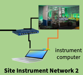
The advent of modern, high-speed electron detectors has made the collection of multidimensional hyperspectral transmission electron microscopy datasets, such as 4D-STEM, a routine. However, many microscopists find such experiments daunting since analysis, collection, long-term storage, and networking of such datasets remain challenging. Some common issues are their large and unwieldy size that often are several gigabytes, non-standardized data analysis routines, and a lack of clarity about the computing and network resources needed to utilize the electron microscope. The existing computing and networking bottlenecks introduce significant penalties in each step of these experiments, and thus, real-time analysis-driven automated experimentation for multidimensional TEM is challenging. One solution is to integrate microscopy with edge computing, where moderately powerful computational hardware performs the preliminary analysis before handing off the heavier computation to high-performance computing (HPC) systems. Here we trace the roots of computation in modern electron microscopy, demonstrate deep learning experiments running on an edge system, and discuss the networking requirements for tying together microscopes, edge computers, and HPC systems.
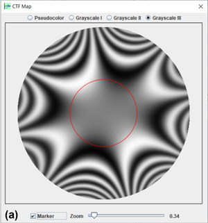
Contrast transfer function (CTF) is a vital function in transmission electron microscopy (TEM). It expresses to what extent amplitudes converted from the phase changes of the diffracted waves contribute to the TEM image, including the effects of lens aberrations. Simulation is very helpful to understand the application of the function thoroughly. In this work, we develop the CTFscope as a component in the Landyne software suite, to calculate the CTF with temporal and spatial dumping envelopes for conventional TEM and to extend it to various aberrations (up to fifth order) for aberration-corrected (AC)-TEM. It also includes effects on the CTF and imaging due to the objective aperture and image drift for tutorial purposes. The CTFscope has a user-friendly graphical interface for simulation, visualization, saving, and loading of the microscopy conditions and results. It can be used to explore instrumental performance information due to optical aberrations, effects on the resolution of a TEM and AC-TEM, and for teaching electron microscopy courses.
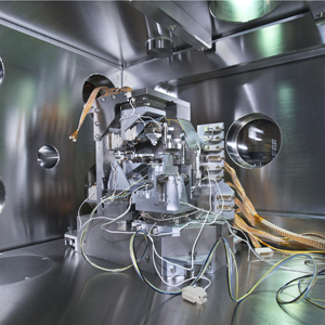
This article introduces the 2D multilayer Laue lens (MLL) nanofocusing optics recently developed for high-resolution hard X-ray microscopy. The new optics utilized a micro-electro-mechanical-system (MEMS)-based template to accommodate two linear MLL optics in a pre-aligned configuration. Angular misalignment between the two lenses was controlled in tens of millidegrees, and the lateral position error was on a micrometer scale. Using the developed 2D MLLs, an astigmatism-free point focus of approximately 14 nm by 13 nm in horizontal and vertical directions, respectively, at 13.6 keV photon energy was obtained. The success of 2D MLL optics with an approaching 10 nm resolution is a significant step forward for the development of high-resolution hard X-ray microscopy and applications of MLL optics in the hard X-ray community.
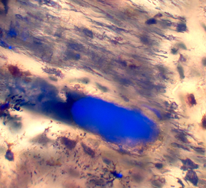
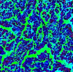
Theropod dinosaurs have captured the imagination of the public and paleontologists alike. Histology of the bones of theropods has revealed much about dinosaur physiology, behavior, and growth. Histology and ultraviolet fluorescence (UVFL) microscopy of one controversial dinosaur, Nanotyrannus lancensis, reveals the presence of blood clots in post-fixed vessel canals of claw, vertebra, and other isolated post-cranial elements collected at Hell Creek, MT. These clots are thicker, more closely adherent to canal walls, and more reactive to 347 nm UVFL incident light than unfixed specimens. Theropod histology images in the literature display similar clots, and those should be subjected to UVFL for confirmation. In addition, nematodes are evidently preserved in vessel canals of dinosaurs.
This is the third article within a three-part series on Fourier ptychography, which is a computational microscopy technique for high-resolution, large field-of-view imaging. While the previous articles introduced the working principles of the technique, in this article we focus on the practical benefits that it brings to the imaging community. We present a didactic overview of the most important and well-established practical use-cases such as gigapixel imaging, quantitative phase contrast, thick sample imaging, and aberration metrology. We also discuss how Fourier ptychography can leave the visible light domain and venture into the realm of smaller wavelengths such as X-rays and electrons, among other topics.