Skip to main content Accessibility help
×
Hostname: page-component-8448b6f56d-jr42d Total loading time: 0 Render date: 2024-04-24T06:04:25.040Z Has data issue: false hasContentIssue false

Chapter 9 - Meta-analysis of optical coherence tomography in multiple sclerosis

Published online by Cambridge University Press:  05 May 2015

Peter A. Calabresi
Affiliation:
Department of Neurology, Johns Hopkins University Hospital, Baltimore
Laura J. Balcer
Affiliation:
Department of Neurology, NYU Langone Medical Center, New York
Elliot M. Frohman
Affiliation:
Department of Neurology, UT Southwestern Medical Center, Dallas
Get access

Summary

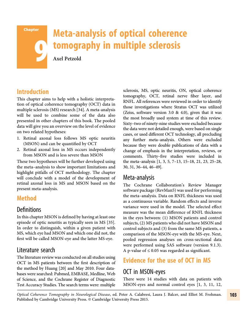
Image of the first page of this content. For PDF version, please use the ‘Save PDF’ preceeding this image.'
Type
Chapter
Information
Publisher: Cambridge University Press
Print publication year: 2015

Access options

Get access to the full version of this content by using one of the access options below. (Log in options will check for institutional or personal access. Content may require purchase if you do not have access.)

References

Albrecht, Philipp et al. “Optical coherence tomography measures axonal loss in multiple sclerosis independently of optic neuritis.” eng. In: J Neurol 254.11 (Nov. 2007), pp. 15951596.CrossRefGoogle ScholarPubMed
Balk, Lisanne J. et al. “The physiological variation of the retinal nerve fiber layer thickness and macular volume in humans as assessed by spectral domain-optical coherence tomography.” eng. In: Invest Ophthalmol Vis Sci 53.3 (Mar. 2012), pp. 12511257.CrossRefGoogle ScholarPubMed
Bock, Markus et al. “Patterns of retinal nerve fiber layer loss in multiple sclerosis patients with or without optic neuritis and glaucoma patients.” eng. In: Clin Neurol Neurosurg (May 2010).CrossRefGoogle Scholar
Budenz, Donald L et al. “Determinants of normal retinal nerve fiber layer thickness measured by Stratus OCT.” eng. In: Ophthalmology 114.6 (June 2007), pp. 10461052.CrossRefGoogle ScholarPubMed
Cheng, Han et al. “The relationship between visual field and retinal nerve fiber layer measurements in patients with multiple sclerosis.” eng. In: Invest Ophthalmol Vis Sci 48.12 (Dec. 2007), pp. 57985805.CrossRefGoogle ScholarPubMed
Choi, Stacey S et al. “Changes in cellular structures revealed by ultra-high resolution retinal imaging in optic neuropathies.” eng. In: Invest Ophthalmol Vis Sci 49.5 (May 2008), pp. 21032119.CrossRefGoogle ScholarPubMed
Costello, Fiona et al. “Differences in retinal nerve fiber layer atrophy between multiple sclerosis subtypes.” eng. In: J Neurol Sci 281.1–2 (June 2009), pp. 7479.CrossRefGoogle ScholarPubMed
Costello, Fiona et al. “Quantifying axonal loss after optic neuritis with optical coherence tomography.” eng. In: Ann Neurol 59.6 (June 2006), pp. 963969.CrossRefGoogle ScholarPubMed
Costello, Fiona et al. “Retinal nerve fiber layer and future risk of multiple sclerosis.” eng. In: Can J Neurol Sci 35.4 (Sept. 2008), pp. 482487.CrossRefGoogle ScholarPubMed
Costello, F. et al. “Tracking retinal nerve fiber layer loss after optic neuritis: a prospective study using optical coherence tomography.” eng. In: Mult Scler 14.7 (Aug. 2008), pp. 893905.CrossRefGoogle ScholarPubMed
Fisher, Jennifer B et al. “Relation of visual function to retinal nerve fiber layer thickness in multiple sclerosis.” eng. In: Ophthalmology 113.2 (Feb. 2006), pp. 324332.CrossRefGoogle ScholarPubMed
Frohman, Elliot M et al. “Relationship of optic nerve and brain conventional and non-conventional MRI measures and retinal nerve fiber layer thickness, as assessed by OCT and GDx: a pilot study.” eng. In: J Neurol Sci 282.1–2 (July 2009), pp. 96105.CrossRefGoogle ScholarPubMed
Garcia-Martin, Elena et al. “Progressive changes in the retinal nerve fiber layer in patients with multiple sclerosis.” eng. In: Eur J Ophthalmol 20.1 (2010), pp. 167173.CrossRefGoogle ScholarPubMed
Gelfand, Jeffrey M. et al. “Microcystic Inner Nuclear Layer Abnormalities and Neuromyelitis Optica.” eng. In: JAMA Neurol (Mar. 2013), pp. 15.Google Scholar
Gordon-Lipkin, E. et al. “Retinal nerve fiber layer is associated with brain atrophy in multiple sclerosis.” eng. In: Neurology 69.16 (Oct. 2007), pp. 16031609.CrossRefGoogle ScholarPubMed
Grazioli, Erica et al. “Retinal nerve fiber layer thickness is associated with brain MRI outcomes in multiple sclerosis.” eng. In: J Neurol Sci 268.1–2 (May 2008), pp. 1217.CrossRefGoogle ScholarPubMed
Gundogan, Fatih C, Demirkaya, Seref, and Sobaci, Gungor. “Is optical coherence tomography really a new biomarker candidate in multiple sclerosis?–A structural and functional evaluation.” eng. In: Invest Ophthalmol Vis Sci 48.12 (Dec. 2007), pp. 57735781.CrossRefGoogle ScholarPubMed
Henderson, Andrew P D et al. “An investigation of the retinal nerve fibre layer in progressive multiple sclerosis using optical coherence tomography.” eng. In: Brain 131.Pt 1 (Jan. 2008), pp. 277287.Google ScholarPubMed
Hoyt, W. F., Schlicke, B., and Eckelhoff, R. J.. “Fundoscopic appearance of a nerve-fibre-bundle defect.” eng. In: Br J Ophthalmol 56.8 (Aug. 1972), pp. 577583.CrossRefGoogle ScholarPubMed
Huang, D. et al. “Optical coherence tomography.” eng. In: Science 254.5035 (Nov. 1991), pp. 11781181.CrossRefGoogle ScholarPubMed
Jeanjean, L. et al. “[Retinal atrophy using optical coherence tomography (OCT) in 15 patients with multiple sclerosis and comparison with healthy subjects].” fre. In: Rev Neurol (Paris) 164.11 (Nov. 2008), pp. 927934.CrossRefGoogle ScholarPubMed
Jindahra, P., Petrie, A., and Plant, G. T.. “Retrograde trans-synaptic retinal ganglion cell loss identified by optical coherence tomography.” eng. In: Brain 132 (2009), pp. 628634.CrossRefGoogle ScholarPubMed
Klistorner, Alexander et al. “Axonal loss and myelin in early ON loss in postacute optic neuritis.” eng. In: Ann Neurol 64.3 (Sept. 2008), pp. 325331.CrossRefGoogle ScholarPubMed
Klistorner, Alexander et al. “Multifocal VEP and OCT in optic neuritis: a topographical study of the structure-function relationship.” eng. In: Doc Ophthalmol 118.2 (Apr. 2009), pp. 129137.CrossRefGoogle ScholarPubMed
Kochkorov, A. et al. “Rigidity of retinal vessels in patients with multiple sclerosis.” eng. In: Klin Monatsbl Augenheilkd 226.4 (Apr. 2009), pp. 276279.CrossRefGoogle ScholarPubMed
Laron, Michal et al. “Comparison of multifocal visual evoked potential, standard automated perimetry and optical coherence tomography in assessing visual pathway in multiple sclerosis patients.” eng. In: Mult Scler 16.4 (Apr. 2010), pp. 412426.CrossRefGoogle ScholarPubMed
Merle, Harold et al. “Retinal nerve fiber layer thickness and spatial and temporal contrast sensitivity in multiple sclerosis.” eng. In: Eur J Ophthalmol 20.1 (2010), pp. 158166.CrossRefGoogle ScholarPubMed
Merle, Harold et al. “Retinal peripapillary nerve fiber layer thickness in neuromyelitis optica.” eng. In: Invest Ophthalmol Vis Sci 49.10 (Oct. 2008), pp. 44124417.CrossRefGoogle ScholarPubMed
Naismith, R. T. et al. “Optical coherence tomography differs in neuromyelitis optica compared with multiple sclerosis.” eng. In: Neurology 72.12 (Mar. 2009), pp. 10771082.CrossRefGoogle ScholarPubMed
Nakamura, Masahiko et al. “Early high-dose intravenous methylprednisolone is effective in preserving retinal nerve fiber layer thickness in patients with neuromyelitis optica.” eng. In: Graefes Arch Clin Exp Ophthalmol (Mar. 2010).CrossRefGoogle Scholar
Oreja-Guevara, C. et al. “Optic neuritis, multiple sclerosis-related or not: structural and functional study.” engspa. In: Neurologia 25.2 (Mar. 2010), pp. 7882.CrossRefGoogle ScholarPubMed
Petzold, A and Plant, GT. “Chronic Relapsing Inflammatory Optic Neuropathy: a Systematic Review of 122 cases reported.” In:J Neurol ([Epub ahead of print]).Google Scholar
Petzold, Axel. “Neurofilament phosphoforms: surrogate markers for axonal injury, degeneration and loss.” eng. In: J Neurol Sci 233.1–2 (June 2005), pp. 183198.CrossRefGoogle ScholarPubMed
Petzold, Axel et al. “Optical coherence tomography in multiple sclerosis: a systematic review and meta-analysis.” eng. In: Lancet Neurol 9.9 (Sept. 2010), pp. 921932.CrossRefGoogle ScholarPubMed
Petzold, A. et al. “The investigation of acute optic neuritis: a review and proposed protocol.” Nature Rev Neurol 2014; 10: 447458.CrossRefGoogle ScholarPubMed
Pueyo, Victoria et al. “Sub-clinical atrophy of the retinal nerve fibre layer in multiple sclerosis.” eng. In: Acta Ophthalmol (June 2009), pp. 15.Google Scholar
Pueyo, V. et al. “Axonal loss in the retinal nerve fiber layer in patients with multiple sclerosis.” eng. In: Mult Scler 14.5 (June 2008), pp. 609614.CrossRefGoogle ScholarPubMed
Pulicken, M. et al. “Optical coherence tomography and disease subtype in multiple sclerosis.” eng. In: Neurology 69.22 (Nov. 2007), pp. 20852092.CrossRefGoogle ScholarPubMed
Quelly, Amanda et al. “Comparison of Optical Coherence Tomography and Scanning Laser Polarimetry Measurements in Patients with Multiple Sclerosis.” eng. In: Optom Vis Sci (May 2010).CrossRefGoogle Scholar
Ratchford, J. N. et al. “Optical coherence tomography helps differentiate neuromyelitis optica and MS optic neuropathies.” eng. In: Neurology 73.4 (July 2009), pp. 302308.CrossRefGoogle ScholarPubMed
Salter, A. R. et al. “Retinal architecture predicts pupillary reflex metrics in MS.” eng. In: Mult Scler 15.4 (Apr. 2009), pp. 479486.CrossRefGoogle ScholarPubMed
Sepulcre, Jorge et al. “Diagnostic accuracy of retinal abnormalities in predicting disease activity in MS.” eng. In: Neurology 68.18 (May 2007), pp. 14881494.CrossRefGoogle ScholarPubMed
Siepman, Theodora A M, Bettink-Remeijer, Marijke Wefers, and Hintzen, Rogier Q. “Retinal nerve fiber layer thickness in subgroups of multiple sclerosis, measured by optical coherence tomography and scanning laser polarimetry.” eng. In: J Neurol (May 2010).CrossRefGoogle Scholar
Siger, Malgorzata et al. “Optical coherence tomography in multiple sclerosis: thickness of the retinal nerve fiber layer as a potential measure of axonal loss and brain atrophy.” eng. In: J Neurol 255.10 (Oct. 2008), pp. 15551560.CrossRefGoogle ScholarPubMed
Sotirchos, Elias S. et al. “In vivo identification of morphologic retinal abnormalities in neuromyelitis optica.” eng. In: Neurology 80.15 (Apr. 2013), pp. 14061414.CrossRefGoogle ScholarPubMed
Spain, Rebecca I et al. “Thickness of retinal nerve fiber layer correlates with disease duration in parallel with corticospinal tract dysfunction in untreated multiple sclerosis.” eng. In: J Rehabil Res Dev 46.5 (2009), pp. 633642.CrossRefGoogle ScholarPubMed
Talman, Lauren S et al. “Longitudinal study of vision and retinal nerve fiber layer thickness in multiple sclerosis.” eng. In: Ann Neurol 67.6 (June 2010), pp. 749760.CrossRefGoogle ScholarPubMed
Trip, S. Anand et al. “Retinal nerve fiber layer axonal loss and visual dysfunction in optic neuritis.” eng. In: Ann Neurol 58.3 (Sept. 2005), pp. 383391.CrossRefGoogle ScholarPubMed
Zaveri, Maulik S et al. “Retinal imaging by laser polarimetry and optical coherence tomography evidence of axonal degeneration in multiple sclerosis.” eng. In: Arch Neurol 65.7 (July 2008), pp. 924928.CrossRefGoogle ScholarPubMed

Save book to Kindle

To save this book to your Kindle, first ensure coreplatform@cambridge.org is added to your Approved Personal Document E-mail List under your Personal Document Settings on the Manage Your Content and Devices page of your Amazon account. Then enter the ‘name’ part of your Kindle email address below. Find out more about saving to your Kindle.

Note you can select to save to either the @free.kindle.com or @kindle.com variations. ‘@free.kindle.com’ emails are free but can only be saved to your device when it is connected to wi-fi. ‘@kindle.com’ emails can be delivered even when you are not connected to wi-fi, but note that service fees apply.

Find out more about the Kindle Personal Document Service.

Available formats
×

Save book to Dropbox

To save content items to your account, please confirm that you agree to abide by our usage policies. If this is the first time you use this feature, you will be asked to authorise Cambridge Core to connect with your account. Find out more about saving content to Dropbox.

Available formats
×

Save book to Google Drive

To save content items to your account, please confirm that you agree to abide by our usage policies. If this is the first time you use this feature, you will be asked to authorise Cambridge Core to connect with your account. Find out more about saving content to Google Drive.

Available formats
×