Skip to main content Accessibility help
×
  • Cited by 9
Publisher:
Cambridge University Press
Online publication date:
May 2010
Print publication year:
2010
Online ISBN:
9780511730238

Book description

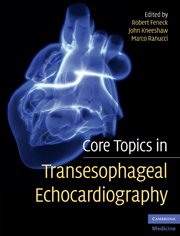
Core Topics in Transesophageal Echocardiography is a highly illustrated, full color, comprehensive clinical text reviewing all aspects of TEE. The text has been written particularly for those who are seeking accreditation in TEE. Section 1 includes chapters on first principles including cardiovascular anatomy, safety issues, indications and contraindications for use, US technology and physics and the details of image acquisition and interpretation in a variety of routine pathologies. Section 2 chapters discuss the use of TEE in a variety of more demanding clinical conditions including valve disease, complex ischaemic heart disease, the use of TEE in critical care and emergency settings, new echocardiography technologies, and TEE reporting. An outstanding free companion website (www.cambridge.org/feneck) contains numerous TEE video clips showing both normal and pathological states. Written by leading TEE experts from EACTA and EAE, this is an invaluable practical resource for all clinicians involved in the care of cardiac patients.

Reviews

'… a reasonably comprehensive, well-written and well-illustrated review of TOE practice. This textbook will be of interest to both novice and experienced practitioners, and to those preparing for accreditation examinations …'

Source: Ultrasound

'For the past month I have taken Core Topics to the clinic rather than my usual reference text. The book has answered all questions that have arisen during the course of the list … I have no hesitation in recommending this book. It should be of interest to both cardiac anaesthetists and cardiologists, particularly but by no means exclusively, those working towards an accreditation exam.'

Source: British Journal of Anaesthesia

Refine List

Actions for selected content:

Select all | Deselect all
  • View selected items
  • Export citations
  • Download PDF (zip)
  • Save to Kindle
  • Save to Dropbox
  • Save to Google Drive

Save Search

You can save your searches here and later view and run them again in "My saved searches".

Please provide a title, maximum of 40 characters.
×

Contents


Page 1 of 2


  • 9 - Left ventricular systolic and diastolic function
    pp 145-162
  • View abstract

    Summary

    Transesophageal echocardiography (TEE) is a relatively recent development in imaging. In 1997 the American College of Cardiology (ACC) and the American Heart Association (AHA) published guidelines for the clinical application of echocardiography. Routine intraoperative use of TEE has been shown to lead to an improvement in both surgical and anesthetic management, which presumably is translated into better and more consistent outcomes for patients. The use of TEE as a diagnostic tool in the echo lab, and as a diagnostic and monitoring tool during and after cardiac or other major operations, has increased significantly. In Europe, the problem of training and accreditation in perioperative TEE has been addressed, during the last decade, by the European Association of Echocardiography (EAE), which is a formal association of the European Society of Cardiology (ESC), and the European Association of Cardiothoracic Anaesthesiologists (EACTA).
  • 10 - The right ventricle
    pp 163-179
  • View abstract

    Summary

    This chapter provides a basic understanding of the most important physical principles and an introduction into the basics of ultrasound techniques. It is important to consider that the three-dimensional ultrasound beam is not an ideal, linear beam, but has a cylindrical near zone and a cone-shaped far field. Live moving 3D displays are now possible with this technique, which has accordingly been named 4D echocardiography or real-time 3D echocardiography. Lowering the pulse repetition frequency suddenly gives the impression that the cyclic movement is running backwards at a much lower frequency. This is called signal aliasing. In Doppler echocardiography, aliasing occurs if the frequency of the Doppler shift is higher than half of the pulse repetition frequency. Imaging artifacts occur with all imaging modalities. Electronic interference artifacts in the 2D and Doppler modes result from other electric instruments with inadequate shielding, for example electric cauterizing and continuous cardiac output devices.
  • 11 - Cardiac masses and pericardial disease
    pp 180-192
  • View abstract

    Summary

    This chapter deals with safety and complications, and probe maintenance in transesophageal echocardiography (TEE). TEE is frequently undertaken outside the operating room. The usual sedation offered includes topical anesthesia to the mouth and oropharynx, and one or more intravenous sedatives. Intravenous benzodiazepines have been the mainstay of intravenous sedative techniques over the years, particularly for non-anesthetists, and they are still frequently used. TEE has been described as a semi-invasive procedure, presumably to identify it as less invasive than intravascular monitoring and diagnostic procedures. TEE is a relatively safe procedure that can be undertaken on a wide range of patients and with minimal complications. Close attention to probe care, maintenance, and disinfection also plays an important role in maintaining the effectiveness of each study and of the TEE service, whether in the echo lab or in the operating room.
  • 12 - The thoracic aorta
    pp 193-204
  • View abstract

    Summary

    Intraoperative transesophageal echocardiography (TEE) is increasingly being used to assess ventricular function in patients about to undergo coronary revascularization, and it should always be used to assess the result of surgery and cardiac function both following bypass and/or at the time of chest closure. The development of standard chocardiographic views, coupled with agreed terminology for describing manipulation of the TEE probe, has been essential for the development of TEE into an effective imaging modality. Th e 20 standard views described in 1999 form the basis of the TEE study. Echocardiographic examination of left ventricular function can reveal useful information when the heart is at rest or under stress with a pharmacological agent such as dobutamine. In order to evaluate each part accurately, the left ventricle has been divided into a number of segments. TEE is particularly useful for examining the mitral valve.
  • 13 - Hemodynamic assessment
    pp 205-222
  • View abstract

    Summary

    This chapter presents a standardized approach to assess the four valves of the heart: the mitral valve (MV); aortic valve; tricuspid valve; pulmonary valve. The MV is examined through four mid-esophageal and two transgastric views. The aortic valve is a semilunar valve with three cusps of similar size. The aortic root is usually easy to visualize with transesophageal echocardiography (TEE) and needs three cross-sections for a complete evaluation. The tricuspid valve consists of three leaflets, attached via multiple chordae tendineae to three small papillary muscles, a supporting annular ring, and a portion of the myocardium of the right ventricular (RV) free wall. The pulmonary valve and the main pulmonary artery can be assessed with TEE in the basal view. The standardized approach outlined in the chapter provides a useful framework for a TEE assessment of the cardiac valves.
  • 14 - Ischemic heart disease
    pp 225-246
  • View abstract

    Summary

    Aortic valve (AV) disease is very common in Western populations. Aortic sclerosis is diagnosed when there is an ejection systolic murmur present in the AV region due to calcification in the ascending aorta, with associated turbulent flow. Aortic stenosis (AS) is differentiated from sclerosis when significant restriction of cusp movement and a raised transaortic peak velocity is seen on echocardiography. Transesophageal echocardiography (TEE) evaluation of AS starts with a 2D examination of the mid-esophageal short-axis (ME SAX) and long-axis (LAX) views of the valve. Continuous-wave Doppler (CWD) is applied to measure flow velocity across the valve and then calculate a pressure gradient using the Bernoulli equation. Aortic regurgitation (AR) results from a primary valve lesion, an abnormal aortic root and/or ascending aorta, or a combination of both. TEE is valuable in revealing important aspects of AV disease.
  • 15 - Cardiomyopathies
    pp 247-267
  • View abstract

    Summary

    This chapter deals with mitral valve (MV) disease. Mitral stenosis is obstruction of left ventricular inflow at the level of the MV, as a result of structural abnormalities of the MV apparatus that limit proper opening during diastole. Mitral annular calcification is a degenerative process, and is a common incidental finding in the elderly. Rheumatic carditis is the commonest cause of mitral stenosis in both developed and developing countries. Mitral regurgitation (MR) is the most commonly encountered valvular lesion in modern clinical practice. Primary MV prolapse syndrome (MVPS) refers to a disease spectrum with frank myxomatous degeneration at one extreme. The transesophageal echocardiography (TEE) variables used in the assessment of MR severity can be classified as semi-quantitative or quantitative. The vena contracta (VC) refers to the narrowest portion or neck of the regurgitant jet, which occurs at or just beyond the regurgitant orifice.
  • 16 - Mitral valve repair
    pp 268-274
  • View abstract

    Summary

    The tricuspid valve (TV) apparatus consists of three leaflets: a large anterior leaflet, a septal leaflet, and a smaller posterior leaflet. Tricuspid stenosis (TS) is a relatively uncommon valvular lesion in North America and Western Europe. Unlike TS, tricuspid regurgitation (TR) is common and can be primary or secondary to annular dilation. Right-sided valvular heart disease occurs frequently in patients with carcinoid syndrome, but left-sided involvement, pericardial effusion, and myocardial metastases may also occur. The pulmonary valve (PV) is a trileaflet semilunar valve with an anterior, a left, and a right leaflet, and is similar to the aortic valve in basic structure and function. The pulmonary artery (PA) trunk is approximatively 4-5 cm long and 2-3 cm wide in normal adults. Severe pulmonary regurgitation (PR) is usually caused by dilation of the PA and PV annulus, for example as a result of acute or chronic pulmonary hypertension.
  • 17 - Aortic valve surgery
    pp 275-293
  • View abstract

    Summary

    Ejection fraction (EF) is a well accepted and useful index of quantitative left ventricular (LV) function, but it is influenced by changes in preload, afterload, and contractility. Stroke volume (SV) can be obtained with 2D transesophageal echocardiography (TEE) by measuring LV end-systolic and end-diastolic volumes for measuring EF. TEE evaluation of LV systolic function, both global and regional, provides insight into hemodynamic impairment in a variety of situations. Doppler echocardiography is the principal diagnostic tool to assess LV diastolic function non-invasively. TEE facilitates a complete evaluation of diastolic LV function by assessing diastolic phases and elucidating structural causes of altered diastole. The use of this tool has relevant implications in the management of hemodynamic derangement due to impaired diastolic function, in vasoactive drugs titration, in the detection of myocardial ischemia, and in performing prognostic stratification.
  • 18 - Infective endocarditis
    pp 294-309
  • View abstract

    Summary

    The long- and short-axis views of the right ventricle (RV) are defined by the corresponding views of the left ventricle (LV), but these two standard echocardiographic imaging planes often transect the RV in an oblique way. Discrete probe manipulations and the proper use of the multiplane capacity of transesophageal echocardiography (TEE) are often necessary to fully visualize the RV. Continuous-wave Doppler plays an important role in the study of pathological conditions of the RV and pulmonary circulation. Abnormalities in the shape and motion of the interventricular septum (IVS) reflect the altered pressure differences between the LV and RV. Tricuspid annular plane systolic excursion (TAPSE) corresponds to wall shortening of the RV free wall along its long axis. The total ejection isovolume (TEI) index or myocardial performance index is a Doppler derived measurement combining systolic and diastolic time intervals as a parameter of global ventricular function.
  • 19 - Prosthetic valves
    pp 310-323
  • View abstract

    Summary

    Transesophageal echocardiography (TEE) is a useful modality for imaging cardiac masses. Atrial myxomas account for 90% of all cardiac tumors found at operation, and 50% of tumors found at autopsy. Pericardial cysts are frequently asymptomatic and may be an incidental finding. Mediastinal cysts, including bronchogenic and duplication cysts, may occur. These have a more echodense appearance on TEE, and may contain thicker mucilaginous material that may layer differently with postural change. Cardiac tamponade may be defined as the decompensated phase of cardiac compression resulting from increased intrapericardial pressure. Constrictive pericarditis is characterized by a fibrotic, inflamed, or calcified pericardial sac. In patients with a suspected cardiac mass, transesophageal echocardiography is recommended for determining its nature, and the assessment of sources of cardiac embolism. It may be particularly useful in patients with loculated or trapped pericardial fluid, for example in the post-operative setting.
  • 20 - Adult congenital heart disease
    pp 324-338
  • TEE guidance of interventional catheter-delivered devices
  • View abstract

    Summary

    Transesophageal echocardiography (TEE) is a valuable means of assessing diseases of the thoracic aorta. The portion of descending aorta between the left subclavian artery proximally and the ligamentum arteriosum distally is known as the aortic isthmus. A number of the branches of the aorta can be imaged using TEE. Coarctation of the aorta may present as a partial narrowing, or rarely a complete occlusion, of the aorta. The ductus arteriosus connects the pulmonary artery to the aorta in the fetal circulation. Supravalvular aortic stenosis is a narrowing of the aorta usually at the level of the border of the sinus of Valsalva. The etiology of a true aortic aneurysm has much in common with aortic dissection. True tumors of the aorta, including leiomyosarcoma, fibroelastoma, and epithelioma, are fortunately extremely rare. Traumatic injury to the aorta usually occurs as a result of rapid deceleration injury or blunt chest trauma.
  • 21 - Critical and emergency care
    pp 339-347
  • View abstract

    Summary

    The quantification of hemodynamics by Doppler echocardiography using transesophageal windows may be useful in providing rapid information useful in the acute management of some patients. Three main equations form the basis of hemodynamic calculations: the flow equation, the Bernoulli equation, and the continuity equation. According to the continuity equation, the blood flow entering a cardiac chamber in diastole is equal to the blood flow leaving that chamber in systole. If people apply this concept to regurgitant valves, they can measure the volume of the regurgitant blood during the cardiac cycle. The assessment of the severity of regurgitant valves is more complex. The vena contracta, the area of the regurgitant jet, and the proximal isovelocity surface area (PISA) method may be useful. The data obtained should always be interpreted with caution and in association with data obtained from other modalities.

Page 1 of 2


Metrics

Altmetric attention score

Full text views

Total number of HTML views: 0
Total number of PDF views: 0 *
Loading metrics...

Book summary page views

Total views: 0 *
Loading metrics...

* Views captured on Cambridge Core between #date#. This data will be updated every 24 hours.

Usage data cannot currently be displayed.