Skip to main content Accessibility help
×
Hostname: page-component-848d4c4894-xfwgj Total loading time: 0 Render date: 2024-06-14T10:18:15.387Z Has data issue: false hasContentIssue false

Chapter Eighteen - Aneurysms and Subarachnoid Hemorrhage

from Types of Stroke

Published online by Cambridge University Press:  13 December 2022

Louis R. Caplan
Affiliation:
Beth Israel Deaconess Medical Centre
Aishwarya Aggarwal
Affiliation:
John F. Kennedy Medical Center
Get access

Summary

This chapter reviews the history of the diagnosis and evolution of knowledge about subarachnoid hemorrhage (SAH) and brain aneurysms. Chapter 57 discusses management.

Type
Chapter
Information
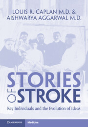
Stories of Stroke
Key Individuals and the Evolution of Ideas
, pp. 138 - 156
Publisher: Cambridge University Press
Print publication year: 2022

Access options

Get access to the full version of this content by using one of the access options below. (Log in options will check for institutional or personal access. Content may require purchase if you do not have access.)

References

Notes and References

Two detailed biographies describe the main life events and accomplishments of Harvey Cushing: Fulton, J. Harvey Cushing: A Biography – The Story of a Great Medical Pioneer. Springfield, IL: Charles C. Thomas, 1946.Google ScholarGoogle Scholar
Cushing, H. Surgery of the head. In Keen, WW (ed.), Surgery: Its Principles and Practice. Philadelphia: WB Saunders, 1911.Google Scholar
Cushing, H. Tumors of the Nervus Acusticus and the Syndrome of the Cerebellopontile Angle. Philadelphia: WB Saunders, 1917.Google ScholarGoogle Scholar
Biographical information about Sir Charles Symonds was derived from the entry for Charles Symonds in Wikipedia. Available at https://en.wikipedia.org/wiki/Charles_Symonds. Shorvon, S, Compston, A. Queen Square: A History of the Natil Hospital and Its Institute of Neurology. Cambridge: Cambridge University Press, 2019, pp. 239249.Google ScholarGoogle Scholar
Symonds, C. Contributions to the clinical study of intracranial aneurysms, with Harvey Cushing. Guy’s Hospital Reports 1923;73:139158.Google ScholarGoogle Scholar
Symonds, C. Spontaneous subarachnoid haemorrhage. Quarterly Journal of Medicine 1924;18:93122.Google ScholarGoogle ScholarGoogle Scholar
Quincke, HI. Verhandlungen des Congresses Innere Medizin. Wiesbaden, 1891, 10:321331.Google ScholarGoogle Scholar
Froin, G. Les hémorrhagies sous-arachnoidïennes et le méchanism de l’hématolyse en général. Thesis, Hôpitaux de Paris, 1904.Google Scholar
Hippocrates’s work is described in Chapter 1 of this book. Clark, E. Apoplexy in the Hippocratic writings. Bulletin of the History of Medicine 1963;37:301314.Google ScholarGoogle ScholarGoogle Scholar
Wepfer, JJ. Observationes anatomicae ex cadaveribus eorum, quos sustulit apoplexia, cum exercitatione de ejus loco affecto. Schaffhausen: J. Caspari Suteri, 1658. Wepfer’s contributions are described in Chapter 8.Google Scholar
Chapter 7 contains much more detail about Morgagni and his life, works, and influence. Morgagni, GB. The Seats and Causes of Diseases Investigated by Anatomy. Trans. Alexander, B.. London: Miller and Cadell, 1769.Google Scholar
Cheyne, J. Cases of Apoplexy and Lethargy with Observations upon the Comatose Diseases. London: J. Moyes printer, 1812. Reprinted in The Classics of Neurology and Neurosurgery Library, Division of Gryphon editions, New York, 1986. Cheyne’s work is discussed in Chapter 8.Google Scholar
Abercrombie, J. Pathological and Practical Researches on Diseases of the Brain and Spinal Cord. Edinburgh: Waugh and Innes, 1828. Reprinted in The Classics of Neurology and Neurosurgery Library, Division of Gryphon editions, New York, 1993.Google Scholar
Bright, R. Reports of Medical Cases, Selected with a View of Illustrating the Symptoms and Cure of Diseases by a Reference to Morbid Anatomy. vol. II: Diseases of the Brain and Nervous System. London: Longman, Rees, Orme, Brown, and Green, 1831. Bright and his work is described in more detail in Chapter 9.Google Scholar
Bright, R. Cases illustrative of the effects produced when the arteries and brain are diseased. Guy’s Hospital Reports 1836;1:916.Google Scholar
Biumi, F. Observations anatomicae, scholiis ilustratae. Observatio V. In Sandifort, E (ed.), Thesaurus dissertationum, vol. 3. Leyden: S et J Luchtmans, 1778, pp. 373379.Google Scholar
Blane, C. Case of aneurisms of the carotid arteries. Trans. Soc. Improv. Med. Chir. Knowledge (London) 1800;2.Google Scholar
The quote is from Gull, WW. Cases of aneurism of the cerebral vessels. Guy’s Hospital Reports 1859; 5:281304.Google ScholarGoogle Scholar
The topic of cerebral angiography is discussed in more detail in Chapter 25 and in Krayenbühl, H. History of cerebral angiography and its development since Egaz Moniz. In Egas Moniz Centenary: Scientific Reports. Lisbon: Comissao Executiva das Comemoracoes do Centenario do Nascimento do Prof. Egas Moniz, 1977, pp. 6374.Google ScholarGoogle ScholarGoogle Scholar
Seldinger, SI. Catheter replacement of the needle in percutaneous arteriography. Acta Radiol. 1953;39:368376.Google Scholar
Reviews of the natural history of aneurysmal SAH are found in Locksley, HB. Report of the Cooperative Study of Intracranial Aneurysms and Subarachnoid Hemorrhage: Sec. V, part II. Natural history of subarachnoid hemorrhage, intracranial aneurysms, and arteriovenous malformation. J. Neurosurg. 1966;25:321368.Google ScholarGoogle ScholarGoogle ScholarGoogle Scholar
Hunt, WE, Meagher, JN, Hess, RM. Intracranial aneurysm: A nine-year study. Ohio State Med. J. 1966 Nov;62(11):11681171.Google ScholarGoogle Scholar
Broderick, JP, Brott, TG, Duldner, JE, et al. Initial and recurrent bleeding are the major causes of death following subarachnoid hemorrhage. Stroke 1994;25:13421347.Google ScholarGoogle Scholar
Mullan, S, Dawley, J. Antifibrinolytic therapy for intracranial aneurysms. J. Neurosurg. 1968 Jan;28(1):2123.Google ScholarGoogle Scholar
Crompton, MR. The pathogenesis of cerebral infarction following rupture of cerebral berry aneurysms. Brain 1964;87:491510.Google Scholar
Crompton, MR. Hypothalamic lesions following the rupture of cerebral berry aneurysms. Brain 1963;86:301314.Google Scholar
Chapter 29 discusses Fisher’s life and contributions to stroke. Fisher’s life story is described in Caplan, LR. C. Miller Fisher: Stroke in the 20th Century. New York: Oxford University Press, 2020.Google Scholar
Fisher, CM, Roberson, GH, Ojemann, RG. Cerebral vasospasm with ruptured saccular aneurysm: The clinical manifestations. Neurosurgery 1977;1:245248.Google ScholarGoogle Scholar
Kistler, JP, Crowell, RM, Davis, KR, Heros, R, Ojemann, RG, Zervas, NT, Fisher, CM. The relation of cerebral vasospasm to the extent and location of subarachnoid blood visualized by CT scan: A prospective study. Neurology 1983;33:424437.Google Scholar
Conway, LW, McDonald, LW. Structural changes of the intradural arteries following subarachnoid hemorrhage. J. Neurosurg. 1972;37:715723.Google ScholarGoogle ScholarGoogle Scholar
Heros, RC, Zervas, NT, Varsos, V. Cerebral vasospasm after subarachnoid hemorrhage: An update. Ann. Neurol. 1983;14:599608.Google ScholarGoogle ScholarGoogle ScholarGoogle Scholar
Pickard, JD, Murray, GD, Illingworth, R, et al. Effect of oral nimodipine in cerebral infarction and outcome after subarachnoid hemorrhage: British aneurysm nimodipine trial. BMJ 1981;298:636642.Google ScholarGoogle Scholar
Feigin, VL, Rinkel, GJE, Algra, A, et al. Calcium antagonists in patients with aneurysmal subarachnoid hemorrhage: A systematic review. Neurology 1998;50:876883.Google Scholar
Higashida, RT, Halbach, VV, Cahan, LD, et al. Transluminal angioplasty for treatment of intracranial arterial vasospasm. J. Neurosurg. 1989;71:648653.Google ScholarGoogle Scholar
Chapter 34 discusses the history of ultrasound and of transcranial Doppler imaging. Aaslid, R (ed.). Transcranial Doppler Sonography. Wien: Springer-Verlag, 1986.CrossRefGoogle Scholar
Sloan, MA, Haley, EC, Kassell, NF, et al. Sensitivity and specificity of transcranial Doppler ultrasonography in the diagnosis of vasospasm following subarachnoid hemorrhage. Neurology 1989;391:15141518.Google ScholarGoogle ScholarGoogle Scholar
Graff-Radford, NR, Torner, J, Adams, HP, Kassell, NF. Factors associated with hydrocephalus after subarachnoid hemorrhage. Arch. Neurol. 1989;46:744752.Google Scholar
Caplan, LR, Hurst, JW. Cardiac and cardiovascular findings in patients with nervous system diseases. In Caplan, LR, Hurst, JW, Chimowitz, MI (eds.), Clin. Neurocardiol. New York: Marcel Dekker, 1999, pp. 298312.Google Scholar
Lee, VH, Connolly, HM, Fulgham, JR, Manno, EM, Brown, RD Jr, Wijdicks, EF. Takotsubo cardiomyopathy in aneurismal subarachnoid hemorrhage: An underappreciated ventricular dysfunction. J. Neurosurg. 2006;105:264270.Google Scholar
Ciongoli, AK, Poser, CM. Pulmonary edema secondary to subarachnoid hemorrhage. Neurology 1972;22:867870.Google ScholarGoogle Scholar
Takaku, A, Shindo, K, Tanaki, S, et al. Fluid and electrolyte disturbances in patients with intracranial aneurysms. Surg. Neurol. 1979;11:349356.Google ScholarGoogle ScholarGoogle Scholar
Van Gijn, J, Kerr, RS, Rinkel, GJE. Subarachnoid haemorrhage. Lancet 2007;369:306318.Google ScholarGoogle ScholarGoogle ScholarGoogle ScholarGoogle Scholar
Rinkel, GJE, Djibuti, M, Algra, A, van Gijn, J. Prevalence and risk of rupture of intracranial aneurysms: A systematic review. Stroke 1998;29:251256.Google ScholarGoogle ScholarGoogle Scholar
Van Gijn, J, van Dongen, KJ, Vermeulan, M, et al. Perimesencephalic hemorrhage: A nonaneurysmal and benign form of subarachnoid hemorrhage. Neurology 1985;35:483487.Google ScholarGoogle Scholar
Schievink, WI, Wijdicks, EFM. Pretruncal subarachnoid hemorrhage: An anatomically correct description of the perimesencephalic subarachnoid hemorrhage. Stroke 1997;28:2572.Google Scholar
Kumar, S, Goddeau, RP, Selim, MH, Thomas, A, Schlaug, G, Alhazzani, A, Searls, DE, Caplan, LR. Atraumatic convexal subarachnoid hemorrhage: Clinical presentation, imaging patterns, and etiologies. Neurology 2010;74:893899.Google Scholar

Save book to Kindle

To save this book to your Kindle, first ensure coreplatform@cambridge.org is added to your Approved Personal Document E-mail List under your Personal Document Settings on the Manage Your Content and Devices page of your Amazon account. Then enter the ‘name’ part of your Kindle email address below. Find out more about saving to your Kindle.

Note you can select to save to either the @free.kindle.com or @kindle.com variations. ‘@free.kindle.com’ emails are free but can only be saved to your device when it is connected to wi-fi. ‘@kindle.com’ emails can be delivered even when you are not connected to wi-fi, but note that service fees apply.

Find out more about the Kindle Personal Document Service.

Available formats
×

Save book to Dropbox

To save content items to your account, please confirm that you agree to abide by our usage policies. If this is the first time you use this feature, you will be asked to authorise Cambridge Core to connect with your account. Find out more about saving content to Dropbox.

Available formats
×

Save book to Google Drive

To save content items to your account, please confirm that you agree to abide by our usage policies. If this is the first time you use this feature, you will be asked to authorise Cambridge Core to connect with your account. Find out more about saving content to Google Drive.

Available formats
×