Hostname: page-component-848d4c4894-ndmmz Total loading time: 0 Render date: 2024-05-28T08:28:54.121Z Has data issue: false hasContentIssue false

Seizure in Neurodegeneration with Brain Iron Accumulation: A Systematic Review

Published online by Cambridge University Press:  24 January 2022

Maziar Emamikhah
Affiliation:
Department of Neurology, Rasool Akram Hospital, Iran University of Medical Sciences, Tehran, Iran
Parisa Saiyarsarai
Affiliation:
Department of Pharmacoeconomics and Pharmaceutical Administration, Faculty of Pharmacy, Tehran University of Medical Sciences, Tehran, Iran Evidence-Based Evaluation of Cost-Effectiveness and Clinical Outcomes Group, Pharmaceutical Sciences Research Center (PSRC), and the Pharmaceutical Management and Economics Research Center (PMERC), the Institute of Pharmaceutical Sciences (TIPS), Tehran University of Medical Sciences, Tehran, Iran
Susanne A. Schneider
Affiliation:
Department of Neurology, University Hospital of the Ludwig Maximilians University Munich, München, Germany
Alfonso Fasano
Affiliation:
Edmond J. Safra Program in Parkinson’s Disease, Morton and Gloria Shulman Movement Disorders Clinic, Toronto Western Hospital, UHN, Toronto, Ontario, Canada Division of Neurology, University of Toronto, Toronto, Ontario, Canada Krembil Brain Institute, Toronto, Ontario, Canada CenteR for Advancing Neurotechnological Innovation to Application (CRANIA), Toronto, Ontario, Canada
Nahid Mohammadzadeh
Affiliation:
Department of Neurology, Rasool Akram Hospital, Iran University of Medical Sciences, Tehran, Iran
Mohammad Rohani*
Affiliation:
Department of Neurology, Rasool Akram Hospital, Iran University of Medical Sciences, Tehran, Iran Skull Base Research Center, Five Senses Health Institute, Iran University of Medical Sciences, Tehran, Iran
*
Corresponding author: Mohammad Rohani, Division of Neurology, Rasool Akram Hospital, Iran University of Medical Sciences, Niyayesh St, Sattarkhan Ave, Tehran 1445613131, Iran. E-mail: mohammadroohani@gmail.com, rohani.m@iums.ac.ir
Rights & Permissions [Opens in a new window]

Abstract:

Background:

Neurodegeneration with brain iron accumulation (NBIA) is a rare genetic disorder. Its clinical manifestations comprise a wide spectrum mainly movement disorders. Seizure as a clinical manifestation is known to occur in some NBIAs, but the exact prevalence of epilepsy in each individual disorder is not well elucidated. The aim of this review was to investigate the frequency of seizures in NBIA disorders as well as to determine the associated features of patients with seizures.

Method:

The electronic bibliographic databases PubMed, Scopus, Embase, and Google Scholar were systematically searched for all cases in any type of article from inception to December 16, 2019. All the reported cases of NBIA (with or without genetic confirmation) were identified. Case reports with an explicit diagnosis of any types of NBIA, which have reported occurrence (or absence) of any type of seizure or epilepsy, in the English language, were included. Seizure incidence rate, type, and age of onset were reported as frequencies and percentages.

Result:

1698 articles were identified and 51 were included in this review. Of 305 reported cases, 150 (49.2%) had seizures (phospholipase A2-associated neurodegeneration (PLAN) = 64 (50.8%), beta-propeller protein-associated neurodegeneration (BPAN) = 57 (72.1%), pantothenate kinase-associated neurodegeneration (PKAN) = 11 (23.4%), and others = 18 (very variable proportions)). The most frequent seizure type in NBIA patients was generalized tonic-clonic seizure with the mean age of seizure onset between 2 and 36 years. However, most of these papers had been published before the new classification of epilepsy became accessible. Affected patients were more likely to be females.

Conclusion:

Seizures are common in NBIA, particularly in PLAN and BPAN. In PKAN, the most common type of NBIA, around 10% of patients are affected by seizures. BPAN is the most possible NBIA accompanying seizure. Most of the findings regarding the seizure characteristics in the NBIAs are biased due to the huge missing data. Therefore, any conclusions should be made with caution and need further investigations.

Résumé :

RÉSUMÉ :

Crises convulsives associées à la neuro-dégénérescence avec accumulation de fer dans le cerveau : une revue systématique.

Contexte :

La neuro-dégénérescence avec accumulation intracérébrale de fer (« NBIA » en anglais) constitue un trouble génétique peu fréquent. Ses manifestations cliniques composent un large spectre, pour l’essentiel des troubles du mouvement. De plus, on sait désormais que des crises convulsives sont une manifestation clinique de certains cas de NBIA ; toutefois, la prévalence exacte de l’épilepsie pour chaque cas individuel n’a pas encore été bien élucidée. L’objectif de cet article est donc d’étudier la fréquence des crises convulsives associées à la NBIA et de déterminer les caractéristiques des patients présentant de telles crises.

Méthodes :

Nous avons ainsi interrogé de façon systématique les bases de données bibliographiques électroniques PubMed, Scopus, Embase et Google Scholar pour tous les cas de NBIA et dans tous les types d’articles, et ce, de la création de ces bases jusqu’au 16 décembre 2019. Tous les cas signalés de NBIA, avec ou sans confirmation génétique, ont été identifiés. Nous avons en outre inclus des rapports de cas rédigés en anglais qui incluaient à la fois un diagnostic explicite de NBIA (tout type) et qui avaient rapporté des crises convulsives de tout type ou l’absence de telles crises. Enfin, soulignons que le taux d’incidence, le type et l’âge d’apparition des crises convulsives ont été compilés sous forme de fréquences et de pourcentages.

Résultats :

Nous avons identifié un total de 1698 articles. De ce nombre, 51 ont été inclus dans notre étude. Sur un total de 305 cas de NBIA signalés, 150 patients (49,2 %) ont donné à voir des crises convulsives. Chez 64 patients, ces crises ont été associées à une neuro-dégénérescence liée à la phospholipase A2 (50,8 %) ; chez 57 patients, à une neuro-dégénérescence liée à la protéine bêta-propulseur (β-propeller) (72,1 %) ; chez 11 patients, à une neuro-dégénérescence associée à la pantothénate kinase (23,4 %) ; chez 18 autres, à une proportion très variable. Le type de crise convulsive le plus fréquent chez les patients atteints de NBIA était la crise tonico-clonique généralisée, l’âge moyen d’apparition des premières crises se situant entre 2 et 36 ans. Cependant, rappelons que la plupart de ces articles avaient été publiés avant que la nouvelle classification de l’épilepsie ne devienne accessible. Enfin, les patients affectés étaient le plus souvent de sexe féminin.

Conclusion :

Les crises convulsives sont fréquentes chez les patients atteints de NBIA, en particulier quand il est question de neuro-dégénérescence liée à la phospholipase A2 et de neuro-dégénérescence liée à la protéine bêta-propulseur. Dans le cas des neuro-dégénérescences associées à la pantothénate kinase, le type le plus courant de NBIA, environ 10 % des patients sont affectés par des crises convulsives. Les neuro-dégénérescences liées à la protéine bêta-propulseur sont par ailleurs celles qui sont les plus susceptibles de s’accompagner de crises convulsives. Cela dit, la plupart des résultats concernant les caractéristiques des crises convulsives liées aux NBIA sont biaisés en raison de l’importance de données manquantes. Par conséquent, toute conclusion devrait être tirée de façon prudente et s’appuyer sur des recherches supplémentaires.

Type
Original Article
Copyright
© The Author(s), 2022. Published by Cambridge University Press on behalf of Canadian Neurological Sciences Federation

Introduction

Neurodegeneration with brain iron accumulation (NBIA) is a heterogeneous group of genetic disorders affecting both children and adults with the unifying feature of iron accumulation in specific brain structures, especially globus pallidus. Main defective proteins in different NBIAs have no known direct role in iron homeostasis, but similar pathologic changes in consistent brain areas, mostly basal ganglia, have led to this assumption that iron accumulation is a common pathophysiologic feature among NBIAs.

Different types of NBIA are recognized, labeled according to the molecular finding, replacing the historical term Hallervorden–Spatz disease. The most common subtype is pantothenate kinase-associated neurodegeneration (PKAN) designating the defective enzyme discovered to cause it. Soon after PKAN was described, other malfunctioning enzymes and their responsible genes were discovered and the NBIA group began to expand, today encompassing at least 10 distinct disorders (Table 1). Reference Louis, Mayer and Rowland1Reference Marras, Lang and van de Warrenburg4 This category is expected to divide further into more subcategories, as still a substantial proportion of “sporadic disorders of uncertain origin” continue to exist as an unclassified heterogeneous subclass of NBIAs. Reference Marras, Lang and van de Warrenburg4

Table 1: The distinct disorders and their responsible proteins and genes comprising the neurodegeneration with brain iron accumulation group

AR = autosomal recessive; Chr = chromosome; HSP = hereditary spastic paraparesis; INAD = infantile neuroaxonal dystrophy; Inher = inheritance; MDS = Movement Disorder Society; NBIA = neurodegeneration with brain iron accumulation; OMIM = Online Mendelian Inheritance in Man; PARK = Parkinson’s disease; SENDA = static encephalopathy of childhood with neurodegeneration in adulthood; SPG = spastic gait gene.

NBIA disorders are rare with an overall estimated prevalence of 1 in 500,000. Their clinical manifestations comprise a wide range of the spectrum (Table 2) mainly movement disorders (especially dystonia and Parkinsonism) but also neuropathy, retinopathy, optic neuropathy, oculomotor abnormalities, mental disability, behavioral, and systemic disorders. Reference Louis, Mayer and Rowland1Reference Marras, Lang and van de Warrenburg4 Seizure as a clinical manifestation is known to occur in some NBIAs such as phospholipase A2-associated neurodegeneration (PLAN) and beta-propeller protein-associated neurodegeneration (BPAN), but the exact prevalence of epilepsy in each individual disorder is not well elucidated. Reference Hayflick, Kurian and Hogarth2

Table 2: Clinical and brain MRI characteristics of NBIAs

MRI = magnetic resonance imaging.

We reviewed the literature with a systematic method to establish the frequency of seizures reported in NBIA disorders as well as to determine the associated features of patients with seizure.

Method

Search Strategy

All details about search methodology regarding databases, keywords, time and language limits, and inclusion criteria were considered. Reference Ferrari5 The electronic bibliographic databases Pubmed, Scopus, Embase, and Google Scholar were searched for all cases in any type of article from inception to December 16, 2019. The medical subject headings words “seizures” and “epilepsy” and keywords “neurodegeneration with brain iron accumulation,” “Idiopathic NBIA,” “Seitelberger disease,” “infantile neuroaxonal dystrophy” (INAD), “Woodhouse–Sakati syndrome” (WSS), “Kufor–Rakeb syndrome” (KRS), “beta-propeller protein-associated neurodegeneration” (BPAN), “PLA2G6-associated neurodegeneration” (PLAN), “hereditary aceruloplasminemia” (HA), “fatty acid hydroxylase-associated neurodegeneration” (FAHN), “fatty acid 2-hydroxylase” (FA2H), “pantothenate kinase-associated neurodegeneration” (PKAN), “mitochondrial membrane-associated neurodegeneration” (MPAN), and “static encephalopathy of childhood with neurodegeneration in adulthood” (SENDA) were used for the search.

Endnote software X8 was used for the review and inclusion of studies.

Eligibility Criteria

Title and abstract screening and full-text screening were conducted independently by two reviewers. Case reports with an explicit diagnosis of any types of NBIA which were assessed for the incidence of any type of seizure or epilepsy were included. Further cases were identified via cross-referencing between papers. The articles, which were not available in full text or have not been written in English, were excluded. Detailed reasons for the exclusion of papers in each step are provided in the PRISMA diagram (Figure 1).

Figure 1: PRISMA diagram.

Quality Assessment

Two reviewers assessed the quality of the reports using the methodological quality of case reports/series tool. Reference Murad, Sultan, Haffar and Bazerbachi6 This tool proposes an approach to evaluate the quality based on the domains of selection, ascertainment, causality, and reporting. Any disagreements were settled through discussion with a third reviewer.

Data Extraction

Two reviewers independently screened the titles and abstracts of all records. Then, the selection of full texts was accomplished based on the eligibility criteria. The reviewers independently extracted data on study characteristics, type of NBIA and demographic information, clinical characteristics, in particular seizure data including incidence rate, type of seizure, and age of seizure onset. Afterward, a third reviewer cross-checked the extracted data. In order not to miss any cases, we also reviewed cases in the references of all papers, including reviews, but during cross-checking, we ensured to omit the duplicate cases.

Statistical Analysis

All results presented as frequencies and percentages of reported data using Excel® 2016.

Results

A total of 1698 articles was retrieved from the electronic database search. Through the process of duplicate record elimination, title and abstract screening, and eligibility assessments, 71 articles fulfilled the inclusion criteria. After the full-text screening, 51 studies comprising of 305 cases were included (Figure 1, Table 3).

Table 3: Studies’ characteristics, sorted alphabetically by author

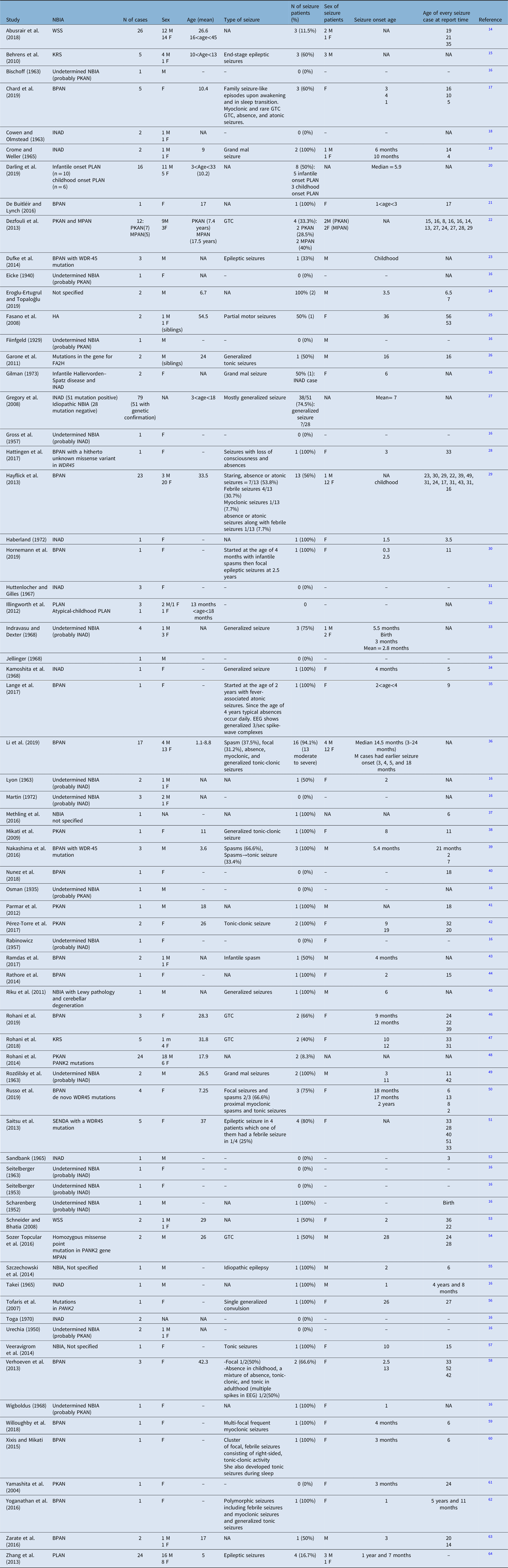
BPAN = beta-propeller protein-associated neurodegeneration; EEG = electroencephalography; FA2H = fatty acid 2-hydroxylase; GTC = generalized tonic-clonic; HA = hereditary aceruloplasminemia; INAD = infantile neuroaxonal dystrophy; KRS = Kufor–Rakeb syndrome; MPAN = mitochondrial membrane-associated neurodegeneration; NBIA = neurodegeneration with brain iron accumulation; PKAN = pantothenate kinase-associated neurodegeneration; PLAN = PLA2G6-associated neurodegeneration; SENDA = static encephalopathy of childhood with neurodegeneration in adulthood; WSS = Woodhouse–Sakati syndrome.

Among these 305 cases in whom the seizure was mentioned as an investigated symptom, 150 (49.2%) had seizures. The incidence of seizure depending on the subtype of NBIA and the demographic characteristics of patients with seizures are presented in Table 4. To summarize, among all NBIA syndromes, seizures were most frequently reported in PLAN (64/126, 50.8%) followed by BPAN (57/79, 72.1%), PKAN (11/47, 23.4%), WSS (4/28, 14.3%), KRS (5/10, 50%), MPAN (2/5, 40%), HA (1/2, 50%), and FAHN (1/2, 50%).

Table 4: The prevalence of epilepsy in each NBIA disorder and the available features related to these patients

BPAN = beta-propeller protein-associated neurodegeneration; FA2H = fatty acid 2-hydroxylase; GTC = generalized tonic-clonic; HA = hereditary aceruloplasminemia; INAD = infantile neuroaxonal dystrophy; KRS = Kufor–Rakeb syndrome; MPAN = mitochondrial membrane-associated neurodegeneration; NBIA = neurodegeneration with brain iron accumulation; PKAN = pantothenate kinase-associated neurodegeneration; PLAN = PLA2G6-associated neurodegeneration; SENDA = static encephalopathy of childhood with neurodegeneration in adulthood; WSS = Woodhouse–Sakati syndrome.

Patients with seizures were more likely to be females and the mean age of seizure onset ranged from 2 to 36 years. The most frequent seizure type in NBIA patients was generalized tonic-clonic seizure, although in BPAN a variety of other seizure types (including infantile spasms, absence, atonic, febrile, myoclonic, focal, proximal myoclonic spasms, and tonic seizures) and in HA focal motor seizure had been reported. However, we should emphasize that most of these papers had been published before the new classification of epilepsy became accessible. So, the type of seizure is based on the previous version of the classification of epilepsy when these papers were released. In most instances, except in some cases of BPAN, seizure was not an early symptom of NBIA. The data regarding the natural history of seizures per se is scarce in the literature. Most of these patients were controlled with one or two antiepileptic drugs.

Discussion

Epilepsy is a frequent neurological disorder, affecting 0.7% of the general population (lifetime prevalence per person is 7/1000 Reference Fiest, Sauro and Wiebe7 ). Causes of seizure in childhood and young patients in order of frequency are genetic (including hereditary neurodegenerative disorders), structural lesions, metabolic disorders, and infections. Reference Sokka, Olsen and Kirjavainen8 In the late-onset sporadic neurodegenerative disorders, such as Alzheimer’s disease and Parkinson’s disease, seizures are reported to occur in about 4.1% and 2.6% of cases, respectively. Reference Vossel, Beagle and Rabinovici9,Reference Feddersen, Rémi, Einhellig, Stoyke, Krauss and Noachtar10 In early-onset neurodegenerative disease, seizures occur more frequently and further complicate the nature of these disorders. One example is NBIA, a group of syndromes clinically characterized by the combination of dystonia, parkinsonism, retinal or optic nerve pathology, and other neurological signs including seizure. Given the lack of systematic data about seizure frequency and characteristics, Reference Hayflick, Kurian and Hogarth2,Reference Vercueil11 we systematically reviewed the literature. Our systematic review revealed that NBIAs as a subcategory of hereditary neurodegenerative disorders are an important cause of epilepsy in children and young adults. Seizure in NBIAs is generally more frequent compared to Wilson’s disease, another metal storage disease, where seizures have been reported in 6.2% of cases. Reference Vercueil11,Reference Dening, Berrios and Walshe12

Among the NBIA disorders, the ones most frequently reported to have epilepsy as a consistent manifestation are PLAN (especially the INAD subtype), BPAN, FAHN, and WSS. Reference Hayflick, Kurian and Hogarth2,Reference Marras, Lang and van de Warrenburg4 By contrast, PKAN – the most common NBIA – is thought to be an infrequent cause of epilepsy. Reference Hayflick, Kurian and Hogarth2

In the present study, in each NBIA disorder, the proportion of patients with seizures was reported to be between 14% (for WSS) and 72% (for BPAN). However, these numbers are biased due to exclusion of papers in which the occurrence of seizure has not been reported and also due to the limited number of case reports for some NBIAs, such as FAHN and HA. These considerations make the estimation of the proportion of patients presenting with seizure very fragile and highlight the importance of complete clinical descriptions, for example, the total number of reported cases of FAHN in papers which alluded to the presence or absence of seizure was two, one of them had seizures, resulting in 50% seizure prevalence in this disorder. Obviously, these percentages cannot be compared to the percentages of more frequent NBIAs such as PLAN with 126 reported cases with seizure annotation in the papers, 64 of them having seizures, resulting in a prevalence of 50% in this disorder. The obvious fact, however, is that PLAN is the most reported NBIA (considering crude numbers) with seizures and this is less biased by the low prevalence, as this disease accounts for nearly 20% of all NBIAs. The other interesting finding of this review is that seizure is not an infrequent finding in PKAN, as 23% of 47 reported PKAN patients happened to be suffering from epilepsy. The other salient result from the current review is that seizure occurrence is more frequent in BPAN (72%) than in any other NBIA and this seems to be a solid result. In spite of the lower prevalence of BPAN compared to PLAN (2% vs. 20% of all NBIAs), the crude number of BPAN patients with seizures stand just next to PLAN (57 and 64 cases, respectively). We estimate based on the present study that BPAN is the most probable NBIA with seizure included in the clinical picture (nearly 72%) followed by PLAN (50%) and PKAN (23%). The other NBIAs, which may be commonly accompanied by seizures, would be WSS, KRS, MPAN, FAHN, and HA, but the exact proportion of patients with seizures and the order of frequency among these disorders cannot be judged with available data.

As already outlined, the findings of our systematic need to be interpreted with caution as no study were specifically addressed to screen the NBIA population for the occurrence of seizures. Our assumptions are indeed based on individual case reports and case series. In addition, the data regarding the natural history of seizures and treatment response were scarce in the literature, thus making it hard to draw conclusions also in terms of the relationship with the natural history of these progressive disorders. Likewise, the data regarding seizure onset age, sex, seizure type, and phenomenology was insufficient, and the results might be distorted by excessive missing data. On the other hand, one cannot be sure that all the reported seizures in the previous reports have been epileptic in nature, for example, the possibility of including provoked seizures should be considered as a bias which cannot be sorted out retrospectively. In addition, the papers were from a diverse time period and almost all of them were published before the new classification of epilepsy. Therefore, the new classification could not be applied to the cases. Reference Fisher, Cross and French13 For instance, based on these data, we were not able to differentiate between generalized tonic-clonic seizures and bilateral tonic-clonic ones which is a new concept. Another problem regarding the classification was that the seizure type, which was not the goal of most reports, was not mentioned or clearly understandable from the reports (see Table 3). So, the readers are advised to consider this issue as a major limitation. Another limitation to our review that readers should be aware of is that this study solely reviewed the English literature on NBIAs which can make the conclusion more biased.

Nevertheless, our study is the first study in this regard, and future studies with more precise information will be needed to clarify this issue.

Disclosures

ME, PS, NM, and MR have no disclosures. SAS was supported by the LMU Clinician Scientist Programme and the Stiftung Verum, receives royalties from Thieme Publisher, receives consulting fees from Medavante Prophase, and receives honoraria from Movement Disorders Society. AF receives consultancy fees from Apple, Abbvie, Abbott, Medtronic, Boston Scientific, Sunovion, Chiesi farmaceutici, UCB, Ipsen, and Rune Labs, receives honoraria from the American Academy of Neurology, Abbott, Abbvie, Medtronic, Boston Scientific, Sunovion, Chiesi farmaceutici, UCB, Ipsen, Paladin Lab, and Movement Disorders Society, receives grant support from the University of Toronto, Abbvie, Medtronic, Boston Scientific, MSA Coalition, and McLaughlin Centre, and receives royalties from springer and sits on advisory boards for Abbvie, Boston Scientific, and Ipsen.

Authors Contributions

ME: writing of the first draft; PS: design of the study and writing of the first draft; SAS: review and critique; AF: conception, review, and critique; NM: review and critique; MR: conception, design of the study, review, and critique.

References

Louis, ED, Mayer, SA, Rowland, LP. Merritt’s neurology. Lippincott Williams & Wilkins; 2015.Google Scholar
Hayflick, SJ, Kurian, MA, Hogarth, P. Neurodegeneration with brain iron accumulation. Handb Clin Neurol. 2018;147:293305.Google ScholarPubMed
Levi, S, Cozzi, A, Santambrogio, P. Iron pathophysiology in neurodegeneration with brain iron accumulation. Adv Exp Med Biol. 2019;1173: 15377.Google ScholarPubMed
Marras, C, Lang, A, van de Warrenburg, BP, et al. Nomenclature of genetic movement disorders: recommendations of the international Parkinson and movement disorder society task force. Mov Disord. 2016;31:43657.CrossRefGoogle ScholarPubMed
Ferrari, R. Writing narrative style literature reviews. Med Writ. 2015;24:23035.CrossRefGoogle Scholar
Murad, MH, Sultan, S, Haffar, S, Bazerbachi, F. Methodological quality and synthesis of case series and case reports. BMJ Evid Based Med. 2018;23:6063.CrossRefGoogle ScholarPubMed
Fiest, KM, Sauro, KM, Wiebe, S, et al. Prevalence and incidence of epilepsy: a systematic review and meta-analysis of international studies. Neurology. 2017;88:296303.CrossRefGoogle ScholarPubMed
Sokka, A, Olsen, P, Kirjavainen, J, et al. Etiology, syndrome diagnosis, and cognition in childhood-onset epilepsy: a population-based study. Epilepsia Open. 2017;2:7683.CrossRefGoogle ScholarPubMed
Vossel, KA, Beagle, AJ, Rabinovici, GD, et al. Seizures and epileptiform activity in the early stages of Alzheimer disease. JAMA Neurol. 2013;70:115866.CrossRefGoogle ScholarPubMed
Feddersen, B, Rémi, J, Einhellig, M, Stoyke, C, Krauss, P, Noachtar, S. Parkinson’s disease: less epileptic seizures more status epilepticus. Epilepsy Res. 2014;108:34954.CrossRefGoogle ScholarPubMed
Vercueil, L. Epilepsy and neurodegenerative diseases in adults: a clinical review. Epileptic Disord. 2006;8:4454.Google Scholar
Dening, T, Berrios, G, Walshe, J. Wilson’s disease and epilepsy. Brain. 1988;111:113955.CrossRefGoogle ScholarPubMed
Fisher, RS, Cross, JH, French, JA, et al. Operational classification of seizure types by the International League Against Epilepsy: Position Paper of the ILAE Commission for Classification and Terminology. Epilepsia. 2017;58:52230.CrossRefGoogle ScholarPubMed
Abusrair, A, Bohlega, S, Al-Ajlan, F, Al-Semari, A, Mohamed, B, AlDakheel, A. Brain MRI findings in Woodhouse-Sakati syndrome. Neurology. 2018;90(15 Supplement):P6.071.Google Scholar
Behrens, MI, Brüggemann, N, Chana, P, et al. Clinical spectrum of Kufor-Rakeb syndrome in the Chilean kindred with ATP13A2 mutations. Mov Disord. 2010;25:192937.CrossRefGoogle ScholarPubMed
Gilman, S, Barrett, RE. Hallervorden-Spatz disease and infantile neuroaxonal dystrophy. Clinical characteristics and nosological considerations. J Neurol Sci. 1973;19:189205.CrossRefGoogle ScholarPubMed
Chard, M, Appendino, JP, Bello-Espinosa, LE, et al. Single-center experience with Beta-propeller protein-associated neurodegeneration (BPAN); expanding the phenotypic spectrum. Mol Genet Metab Rep. 2019;20:100483.CrossRefGoogle ScholarPubMed
Cowen, D, Olmstead, EV. Infantile neuroaxonal dystrophy. J Neuropathol Exp Neurol. 1963;22:175236.Google ScholarPubMed
Crome, L, Weller, S. Infantile neuroaxonal dystrophy. Arch Dis Child. 1965;40:50207.CrossRefGoogle ScholarPubMed
Darling, A, Aguilera-Albesa, S, Tello, CA, et al. PLA2G6-associated neurodegeneration: new insights into brain abnormalities and disease progression. Parkinsonism Relat Disord. 2019;61:17986.CrossRefGoogle ScholarPubMed
De Buitléir, C, Lynch, J. Neurodegeneration with brain iron accumulation. Ir J Med Sci. 2016;185:S295.Google Scholar
Dezfouli, MA, Alavi, A, Rohani, M, et al. PANK2 and C19orf12 mutations are common causes of neurodegeneration with brain iron accumulation. Mov Disord. 2013;28:22831.CrossRefGoogle ScholarPubMed
Dufke, A, Grasshoff, U, Dufke, C, et al. NGS based whole X-exome analysis reveals a novel WDR45 missense mutation in a family with 3 males with non-syndromic intellectual disability. Med Genet. 2014;26:13334.Google Scholar
Eroglu-Ertugrul, N, Topaloğlu, H. P.86Depends on how you look: hereditary spastic paraplegia, leukodystrophy or brain iron accumulation. Neuromuscul Disord. 2019;29:S66.CrossRefGoogle Scholar
Fasano, A, Colosimo, C, Miyajima, H, Tonali, AP, Re, TJ, Bentivoglio, AR. Aceruloplasminemia: a novel mutation in a family with marked phenotypic variability. Mov Disord. 2008;23:75155.CrossRefGoogle Scholar
Garone, C, Pippucci, T, Cordelli, DM, et al. FA2H-related disorders: a novel c.270+3A>T splice-site mutation leads to a complex neurodegenerative phenotype. Dev Med Child Neurol. 2011;53:95861.CrossRefGoogle ScholarPubMed
Gregory, A, Westaway, SK, Holm, IE, et al. Neurodegeneration associated with genetic defects in phospholipase A2. Neurology. 2008;71:140209.CrossRefGoogle Scholar
Hattingen, E, Handke, N, Cremer, K, Hoffjan, S, Kukuk, GM. Clinical and imaging presentation of a patient with beta-propeller protein-associated neurodegeneration, a rare and sporadic form of neurodegeneration with brain iron accumulation (NBIA). Clin Neuroradiol. 2017;27:48183.CrossRefGoogle ScholarPubMed
Hayflick, SJ, Kruer, MC, Gregory, A, et al. Beta-propeller protein-associated neurodegeneration: a new X-linked dominant disorder with brain iron accumulation. Brain. 2013;136:170817.CrossRefGoogle ScholarPubMed
Hornemann, F, Le Duc, D, Roth, C, Pfäffle, R, Huhle, D, Merkenschlager, A. Childhood dystonia-parkinsonism following infantile spasms-clinical clue to diagnosis in early beta-propeller protein-associated neurodegeneration. Neuropediatrics. 2019; 51:229 . Google ScholarPubMed
Huttenlocher, PR, Gilles, FH. Infantile neuroaxonal dystrophy: clinical, pathologic, and histochemical findings in a family with 3 affected siblings. Neurology. 1967;17:1174.CrossRefGoogle Scholar
Illingworth, MA, Younis, R, Hardy, C, et al. Novel mutations in PLA2G6-associated neurodegeneration (PLAN). Dev Med Child Neurol. 2012;54:38.Google Scholar
Indravasu, S, Dexter, RA. Infantile neuroaxonal dystrophy and its relationship to Hallervorden-Spatz disease. Neurology. 1968;18:69399.CrossRefGoogle ScholarPubMed
Kamoshita, S, Neustein, HB, Landing, BH. Infantile neuroaxonal dystrophy with neonatal onset. Neuropathologic and electron microscopic observations. J Neuropathol Exp Neurol. 1968;27:300–23.CrossRefGoogle ScholarPubMed
Lange, J, Herberhold, T, Winkler, P, et al. Early onset absence epilepsy (EOAE) in a young girl with a heterozygous mutation in the WDR45 gene and pathognomonic changes on brain MRI for ß-propeller protein-associated neurodegeneration (BPAN). Zeitschrift fur Epileptol. 2017;30:S3940.Google Scholar
Li, WH, Chen, Q, Wang, H, et al. A phenotypic and genetic study on beta-propeller protein-associated neurodegeneration. Chin J Pediatr. 2019;57:83036.Google Scholar
Methling, B, Thiels, C, Heyer, C, Mengel, E, Lücke, T. α-fucosidase deficiency: a rare differential diagnosis of the eye-of-the-tiger sign and extrapyramidal movement disorders. Neuropediatrics. 2016;47(S 01):PS017.CrossRefGoogle Scholar
Mikati, MA, Yehya, A, Darwish, H, Karam, P, Comair, Y. Deep brain stimulation as a mode of treatment of early onset pantothenate kinase-associated neurodegeneration. Eur J Paediatr Neurol. 2009;13:6164.CrossRefGoogle ScholarPubMed
Nakashima, M, Takano, K, Tsuyusaki, Y, et al. WDR45 mutations in three male patients with West syndrome. J Hum Genet. 2016;61:65361.CrossRefGoogle ScholarPubMed
Nunez, E, Neumann, T, Brauner, M, Fedorcak, M, Kohlhase, J. A novel mutation in WDR45 in a girl with developmental delay, loss off speech and motor skills. Med Genet. 2018;30:16970.Google Scholar
Parmar, A, Khare, S, Srivastav, V. Pantothenate - Kinase associated neurodegeneration. J Assoc Physicians India. 2012;60:7476.Google ScholarPubMed
Pérez-Torre, P, Escobar Villalba, A, Martínez Ulloa, P, et al. PLA2G6-associated neurodegeneration: report of a novel mutation in two siblings with strikingly different clinical presentation. Mov Disord Clin Pract. 2017;4:12931.Google ScholarPubMed
Ramdas, S, Blair, E, Jayawant, S. WDR45 gene-a cause of neurodegeneration with brain iron accumulation. Dev Med Child Neurol. 2017;59:109.Google Scholar
Rathore, GS, Schaaf, CP, Stocco, AJ. Novel mutation of the WDR45 gene causing beta-propeller protein-associated neurodegeneration. Mov Disord. 2014;29:57475.CrossRefGoogle ScholarPubMed
Riku, Y, Mano, K, Mimuro, M, Hashizume, Y, Yoshida, M. An autopsied case of neurodegeneration with brain iron accumulation (NBIA). Neuropathology. 2011;31:328.Google Scholar
Rohani, M, Fasano, A, Akhoundi, FH, et al. Beta-propeller protein associated neurodegeneration (BPAN); the first report of three patients from Iran with de novo novel mutations. Parkinsonism Relat Disord. 2019;61:23133.CrossRefGoogle ScholarPubMed
Rohani, M, Lang, AE, Sina, F, et al. Action myoclonus and seizure in Kufor-Rakeb syndrome. Mov Disord Clin Pract. 2018;5:19599.CrossRefGoogle ScholarPubMed
Rohani, M, Shahidi, GA, Tonekaboni, SH, Elahi, E, Rezvani, M, Miri, S. Genotype and clinical presentation of pantothenate kinaseassociated neurodegeneration: case series of 24 patients from Iran. Mov Disord. 2014;29:S61.Google Scholar
Rozdilsky, B, Cumings, JN, Huston, AF. Hallervorden-Spatz disease. Late infantile and adult types, report of two cases. Acta Neuropathol. 1968;10:116.Google ScholarPubMed
Russo, C, Ardissone, A, Freri, E, et al. Substantia nigra swelling and dentate nucleus t2 hyperintensity may be early magnetic resonance imaging signs of β-propeller protein-associated neurodegeneration. Mov Disord Clin Pract. 2019;6:5156.CrossRefGoogle ScholarPubMed
Saitsu, H, Nishimura, T, Muramatsu, K, et al. De novo mutations in the autophagy gene WDR45 cause static encephalopathy of childhood with neurodegeneration in adulthood. Nat Genet. 2013;45:44549.Google ScholarPubMed
Sandbank, U. Infantile neuroaxonal dystrophy. Arch Neurol. 1965;12:15559.Google ScholarPubMed
Schneider, SA, Bhatia, KP. Dystonia in the woodhouse Sakati syndrome: a new family and literature review. Mov Disord. 2008;23:59296.CrossRefGoogle ScholarPubMed
Sozer Topcular, N, Cagirici, S, Bajrami, A, Demir, E, Akçakaya, H, Yayla, V. Neurodegeneration with brain iron accumulation (NBIA): two cases with different subtypes and a rare mutation. Mov Disord. 2016;31:S228.Google Scholar
Szczechowski, L, Smilowska, K, Smilowski, M, Grechuta, M, Jalowiecki, P. Diagnostic difficulties in neurodegeneration with brain iron accumulation (NBIA) in the 6-year old child. Mov Disord. 2014;29:S40203.Google Scholar
Tofaris, GK, Revesz, T, Jacques, TS, Papacostas, S, Chataway, J. Adult-onset neurodegeneration with brain iron accumulation and cortical α-synuclein and tau pathology: a distinct clinicopathological entity. Arch Neurol. 2007;64:28082.CrossRefGoogle Scholar
Veeravigrom, M, Desudchit, T, Chomtho, K, Pongpunlert, W. Neurodegeneration with brain iron accumulation disorder mimics autism. Pediatr Neurol. 2014;51:86263.CrossRefGoogle ScholarPubMed
Verhoeven, WMA, Egger, JIM, Koolen, DA, et al. Beta-propeller protein-associated neurodegeneration (BPAN), a rare form of NBIA: novel mutations and neuropsychiatric phenotype in three adult patients. Parkinsonism Relat Disord. 2014;20:33236.CrossRefGoogle ScholarPubMed
Willoughby, J, Duff-Farrier, C, Desurkar, A, Kurian, M, Raghavan, A, Balasubramanian, M. Functional mRNA analysis reveals aberrant splicing caused by novel intronic mutation in WDR45 in NBIA patient. Am J Med Genet A. 2018;176:104954.CrossRefGoogle ScholarPubMed
Xixis, KI, Mikati, MA. Epileptic spasms: a previously unreported manifestation of WDR45 gene mutation. Epileptic Disord. 2015;17:46772.Google ScholarPubMed
Yamashita, S, Maeda, Y, Ohmori, H, et al. Pantothenate kinase-associated neurodegeneration initially presenting as postural tremor alone in a Japanese family with homozygous N245S substitutions in the pantothenate kinase gene. J Neurol Sci. 2004;225:12933.CrossRefGoogle Scholar
Yoganathan, S, Arunachal, G, Sudhakar, SV, Rajaraman, V, Thomas, M, Danda, S. Beta propellar protein-associated neurodegeneration: a rare cause of infantile autistic regression and intracranial calcification. Neuropediatrics. 2016;47:12327.Google ScholarPubMed
Zarate, YA, Jones, JR, Jones, MA, et al. Lessons from a pair of siblings with BPAN. Eur J Hum Genet. 2016;24:108083.Google ScholarPubMed
Zhang, P, Gao, Z, Jiang, Y, et al. Follow-up study of 25 Chinese children with PLA2G6-associated neurodegeneration. Eur J Neurol. 2013;20:32230.CrossRefGoogle ScholarPubMed
Figure 0

Table 1: The distinct disorders and their responsible proteins and genes comprising the neurodegeneration with brain iron accumulation group

Figure 1

Table 2: Clinical and brain MRI characteristics of NBIAs

Figure 2

Figure 1: PRISMA diagram.

Figure 3

Table 3: Studies’ characteristics, sorted alphabetically by author

Figure 4

Table 4: The prevalence of epilepsy in each NBIA disorder and the available features related to these patients