Hostname: page-component-848d4c4894-wg55d Total loading time: 0 Render date: 2024-05-31T02:53:42.283Z Has data issue: false hasContentIssue false

Fast and Low-dose Electron Ptychography

Published online by Cambridge University Press:  01 August 2018

Jiamei Song
Affiliation:
National Laboratory of Solid State Microstructures, College of Engineering and Applied Sciences and Collaborative Innovation Center of Advanced Microstructures, Nanjing University, Nanjing, People's Republic of China.
Biying Song
Affiliation:
National Laboratory of Solid State Microstructures, College of Engineering and Applied Sciences and Collaborative Innovation Center of Advanced Microstructures, Nanjing University, Nanjing, People's Republic of China.
Liqi Zou
Affiliation:
National Laboratory of Solid State Microstructures, College of Engineering and Applied Sciences and Collaborative Innovation Center of Advanced Microstructures, Nanjing University, Nanjing, People's Republic of China.
Christopher Allen
Affiliation:
Department of Materials, University of Oxford, Parks Road, Oxford, UK. Electron Physical Sciences Imaging Centre, Diamond Lightsource Ltd., Diamond House, U.K.
Hidetaka Sawada
Affiliation:
JEOL Ltd, Akishima, Tokyo, Japan.
Fucai Zhang
Affiliation:
Department of Electrical and Electronic Engineering, Southern University of Science and Technology, Shenzhen, China.
Xiaoqing Pan
Affiliation:
Department of Chemical Engineering and Materials Science and Department of Physics and Astronomy, University of California, Irvine, CA, USA.
Angus. I. Kirkland
Affiliation:
Department of Materials, University of Oxford, Parks Road, Oxford, UK. Electron Physical Sciences Imaging Centre, Diamond Lightsource Ltd., Diamond House, U.K.
Peng Wang
Affiliation:
National Laboratory of Solid State Microstructures, College of Engineering and Applied Sciences and Collaborative Innovation Center of Advanced Microstructures, Nanjing University, Nanjing, People's Republic of China.

Abstract

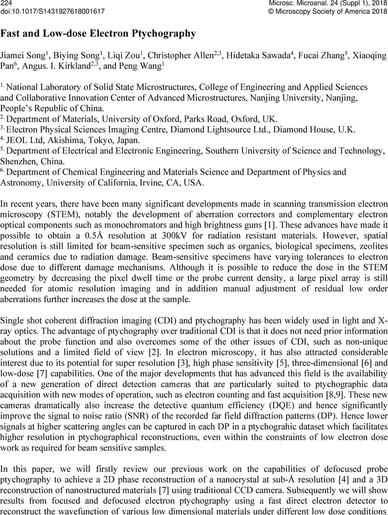
Image of the first page of this content. For PDF version, please use the ‘Save PDF’ preceeding this image.'
Type
Abstract
Copyright
© Microscopy Society of America 2018 

References

[1] Sawada, H, et al, J Electron Microsc (Tokyo) 58 2009) p. 357.Google Scholar
[2] Maiden, AM Rodenburg, JM Ultramicroscopy 109 2009) p. 1256.Google Scholar
[3] Nellist, PD, McCallum, BC Rodenburg, JM Nature 374 1995) p. 630.Google Scholar
[4] Wang, P, et al., Scientific Reports 7 2017) p. 2857.Google Scholar
[5] Yang, H, et al, Nat. Comm 7 2016) p. 12532.Google Scholar
[6] D'Alfonso, AJ, et al., Journal of Applied Physics 119 2016) p. 054302.Google Scholar
[7] Gao, S, et al, Nature Communications 8 2017) p. 163.Google Scholar
[8] Mir, JA, et al, Ultramicroscopy 182 2017) p. 44.Google Scholar
[9] Tate, MW, et al, Microsc. Microanal. 22 2016) p. 237.Google Scholar
[10] The authors acknowledge funding from the National Natural Science Foundation of China (11474147), and the National Basic Research Program of China, (Grant No. 2015CB654901).Google Scholar