


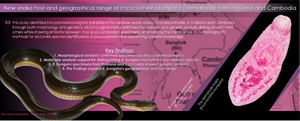
Introduction
Encyclometra Baylis & Cannon, 1924, the genus in the Encyclometridae Mehra, 1931, is classified under the superfamily Gorgoderoidea Loose, 1901. Encyclometra is one of the two genera in the family Encyclometridae, where they parasitise the oesophagus, stomach and intestine of snakes (Tkach, Reference Tkach, Bray, Gibson and Jones2008; Saito et al., Reference Saito, Hayashi, Suse, Kuroki, Shibahara and Takashima2022). Encyclometra is characterised by well-developed oral and ventral suckers, with the oral sucker being smaller or similar in size to the ventral sucker. It is also differentiated from Encyclobrephus Sinha, 1949, the other genus in Encyclometridae, by having a tegument without spines (Tkach, Reference Tkach, Bray, Gibson and Jones2008).
The species of Encyclometra include Encyclometra colubrimurorum Rudolphi, Reference Rudolphi1819, Encyclometra japonica Yoshida and Ozaki, Reference Yoshida and Ozaki1929, Encyclometra asymmetrica Wallace, Reference Wallace1936 and Encyclometra bungara Srivastava and Ghosh, Reference Srivastava and Ghosh1968 (Rudolphi, Reference Rudolphi1819; Yoshida and Ozaki, Reference Yoshida and Ozaki1929; Wallace, Reference Wallace1936; Yeh, Reference Yeh1958; Srivastava and Ghosh, Reference Srivastava and Ghosh1968). The type species E. colubrimurorum was first discovered in a grass snake (Natrix natrix) in Europe, and it was initially morphologically described by having tandem testes and caeca that are equal in length (Rudolphi, Reference Rudolphi1819). Encyclometra japonica was first discovered in a Japanese striped snake (Elaphe quadrivirgata) in Japan and was described as a new species due to the testes positioned diagonally relative to each other (Yoshida and Ozaki, Reference Yoshida and Ozaki1929). The third species, E. asymmetrica, has been described in various snake species and is characterised by intestinal caeca that are significantly unequal in length (Wallace, Reference Wallace1936). Finally, E. bungara was first described in Bungarus fasciatus in India (Srivastava and Ghosh, Reference Srivastava and Ghosh1968) and has also been documented in southeast Asian countries (Thailand and Lao People's Democratic Republic) in Xenochrophis piscator and Enhydris plumbea (Scholz and Ditrich, Reference Scholz and Ditrich1991; Wongsawad et al., Reference Wongsawad, Sey, Rojanapaibul, Chariyahpongpun, Suwattanacoupt, Marayong, Wongsawad and Rojtinnakorn1991). Species discrimination within Encyclometra has primarily relied on morphological differences in the length of the caeca and position of the testes. However, the criteria for species discrimination among the 4 valid species remain unclear. For example, Yeh (Reference Yeh1958) provided the key to distinguish species of Encyclometra based on differences in caeca length (equal, subequal or very unequal), while Gupta and Mehrotra (Reference Gupta and Mehrotra1977) amended the key and suggested the testes position can be used for species discrimination (Gupta and Mehrotra, Reference Gupta and Mehrotra1977).
Molecular methods for species identification are not widely utilised for Encyclometra. To date, there are limited sequences in reference databases, with sequences only available for E. colubrimurom and E. japonica. Here, in order to identify the Encyclometra species found in Enhydris enhydris from Thailand and Cambodia, a comparison of morphological characters among the other valid species of Encyclometra was performed. Additionally, molecular identification using both nuclear and mitochondrial genetic markers was used to obtain novel sequences for E. bungara.
Materials and methods
Parasite isolation
A total of 18 rainbow water snakes (E. enhydris) were obtained from southern Thailand in Nakhon Si Thammarat and adjacent provinces, north-eastern Thailand in Mahasarakham province and Kampong Chhnang province in Cambodia. The snakes were dissected to examine parasites present in the oesophagus. The adult trematodes were isolated, counted and then preserved in 70% ethanol.
Morphological analysis
Thirty adult trematodes were selected, stained in acetic carmine and mounted in permount for morphological analysis. Morphological characters were observed and measured using an inverted compound microscope equipped with a camera and software (ZEISS primovert, Germany). These characters included body length, maximum body width, oral and ventral sucker dimensions, testes dimensions and position, intestinal caeca symmetry and egg length and width. The morphological characters and measurements were then compared with those from previous studies on Encyclometra (Yeh, Reference Yeh1958; Gupta and Mehrotra, Reference Gupta and Mehrotra1977; Scholz and Ditrich, Reference Scholz and Ditrich1991; Wongsawad et al., Reference Wongsawad, Sey, Rojanapaibul, Chariyahpongpun, Suwattanacoupt, Marayong, Wongsawad and Rojtinnakorn1991; Saito et al., Reference Saito, Hayashi, Suse, Kuroki, Shibahara and Takashima2022). Morphological drawings of the specimens were performed under a compound light microscope to illustrate the morphological characters.
Molecular analysis
Four trematodes (2 from Thailand and 2 from Cambodia) were selected and individually placed into 1.7 mL microcentrifuge tubes and washed thoroughly with sterile distilled water. Total genomic DNA was isolated using the DNeasy Blood & Tissue kit (Qiagen, Hilden, Germany) following the manufacturer's recommendations. Polymerase chain reaction (PCR) amplifications of the partial regions of the nuclear 18S rRNA gene (C for: 5′-ATGGCTCATTAAATCAGCTAT – 3′, A rev: 5′ – TGCTTTGAGCACTCAAATTTG – 3′), 28S rRNA gene (Digl2: 5′ – AAGCATATCACTAAGCGG – 3′, 1500R: 5′ – GCTATCCTGAGGGAAACTTCG – 3′) and the mitochondrial cytochrome c oxidase subunit 1 (COI) gene (JB3: 5′ – TTTTTTGGGCATCCTGAGGTTTAT – 3′, JB4.5: 5′ – TAAAGAAAGAACATAATGAAAATG – 3′) were performed in a final PCR volume of 30 μL (Bowles et al., Reference Bowles, Blair and McManus1992; Curran et al., Reference Curran, Tkach and Overstreet2011; Routtu et al., Reference Routtu, Grunberg, Izhar, Dagan, Guttel, Ucko and Ben-Ami2014). The lengths of the partial regions of the 18S rRNA, 28S rRNA and COI genes are 800, 1200 bp and 440 bp, respectively. Each reaction contained 15 μL of 2× i-Taq™ mastermix (iNtRON Biotechnology, Gyeonggi, South Korea), 0.1–0.5 μM of each primer and 1 ng μL−1 of DNA. PCR was conducted in a T100™ thermocycler (Bio-Rad, California, The United States of America), and the thermocycling conditions followed the respective publications of the primer sequences. PCR amplicons were visualized on 1% agarose gel stained with SYBR™ Safe (Invitrogen, Massachusetts, The United States of America). DNA products were subsequently sent for Barcode Taq sequencing (Celemics, Seoul, South Korea).
Electropherograms of the partial 18S rRNA, 28S rRNA and COI genes were manually checked using Bioedit 7.0 (Hall, Reference Hall1999). A dataset containing the concatenated 18S + 28S rRNA genes was also analysed by manually merging the partial 18S and 28S rRNA gene sequences. Multiple sequence alignment with reference sequences obtained from the NCBI database (Supplementary Table S1) was performed using ClustalX 2.1 for each genetic marker (Thompson et al., Reference Thompson, Gibson and Higgins2002). The aligned sequences were checked, and a suitable nucleotide substitution model was selected for maximum-likelihood (ML) phylogenetic analysis. The substitution model selection and ML phylogenetic analysis was carried out in MEGA X (Kumar et al., Reference Kumar, Stecher, Li, Knyaz and Tamura2018). Schistosoma mansoni and Fasciola hepatica were used as outgroups to root the phylogenetic trees. The phylogenetic trees were visualized and labelled using FigTree 1.3.1 (Rambaut, Reference Rambaut2009). Pairwise inter- and intra-species genetic distances were calculated using p-distance as the model for each genetic marker in MEGA X (Kumar et al., Reference Kumar, Stecher, Li, Knyaz and Tamura2018).
Results
Morphological identification
Out of the 18 E. enhydris examined, 12 were found to be infected with Encyclometra, indicating a prevalence of 66.7%. The infection was in the oesophagus, yielding a total of 103 adult Encyclometra specimens. Morphological identification and measurements confirmed that the specimens obtained from both Thailand and Cambodia are E. bungara. These diagnostic characters include the diagonally placed testes and equal-length intestinal caeca. The morphological measurements (n = 30) obtained from these specimens are presented in Table 1 and Supplementary Table S2. Slight differences in the shape of the posterior end were observed between E. bungara specimens from Thailand and Cambodia. The former consistently exhibited a broad posterior end, while the latter consistently displayed a more pointed posterior end. Figure 1 depicts the differences between E. bungara obtained from the 2 countries.
Table 1. Comparison of Encyclometra morphological measurements

Note: measurements are represented as the minimum–maximum (mean) values in micrometres (μm).
VS, ventral sucker; OS, Oral sucker.

Figure 1. Morphological drawing of adult Encyclometra bungara obtained from Enhydris enhydris in (a) Thailand and (b) Cambodia (right).
The description of E. bungara obtained in this study is as follows: whole-body shape flat and lanceolate, posterior end either broad or pointed, cuticle lacking spines. Body length 771–2996 μm, maximum width 267–926 μm. Oral and ventral suckers well-developed, approximately equal in size. Subterminal oral sucker 120–263 μm ×109–257 μm, ventral sucker 124–316 μm×122–297 μm, positioned in anterior quarter to a third of the body. Ratio of ventral sucker to oral sucker 1.1:1. Pharynx distinct, oesophagus very short or absent. Intestinal caeca arise near the pharynx and extend to the posterior end, being equal in length. Curved cirrus sac at anterior border of ventral sucker. Ovary oval, near the posterior border of the ventral sucker, next to seminal vesicle. Two oval or slightly lobed testes, always positioned obliquely in the posterior half of body, 56–192 μm × 41–170 μm. Vitelline glands extend along the side of intestinal caeca, starting from the position between the ovary and testes, and extending to the posterior end of the body. Uterus fully filled, with oval-shaped eggs measuring 60–78 μm×32–44 μm. Excretory bladder Y-shaped.
Taxonomic summary
Host: Rainbow water snake, Enhydris enhydris (Schneider, 1799) (Squamata: Homalopsidae)
Locality: Thailand (Southern region: Nakhon Si Thammarat and adjacent provinces; and north-eastern region: Mahasarakham province) and Cambodia (Kampong Chhnang province)
Site of infection: Oesophagus
Prevalence: 66.7% (12 out of 18 host infected)
Specimens deposited: Department of Helminthology, Faculty of Tropical Medicine, Mahidol University
Molecular and phylogenetic analysis
Three molecular genetic markers – nuclear 18S rRNA gene, 28S rRNA gene, mitochondrial COI gene and the concatenated 18S + 28S rRNA genes were used for analysis. Firstly, based on the phylogenies obtained, all 3 genetic markers supported that the E. bungara specimens obtained from E. enhydris in this study were genetically different from E. colubrimurorum and E. japonica (Fig. 2A–D). Inter-species genetic distances also support that E. bungara are distinct from E. colubrimurorm and E. japonica. The genetic distances between E. colubrimurorum or E. japonica with E. bungara ranged from 0.2–0.4, 1.1–1.2 and 12.0–14.7% using the 18S, 28S and COI genes, respectively. Moreover, these genetic distance values obtained were higher than the inter-species genetic distances between E. colubrimurorum and E. japonica. Table 2 presents the comparison of genetic distances for the 3 genetic markers.

Figure 2. Maximum-likelihood phylogeny using the (a) nuclear 18S rRNA gene (K2 + G + I), (b) nuclear 28S rRNA gene (GTR + G), (c) concatenated nuclear 18S with 28S rRNA genes (GTR + G + I), and (d) mitochondrial COI gene (HKY + G). Numbers at nodes indicate bootstrap values. Representative sequences generated from this study are indicated with an ‘*’. The families in the superfamily Gorgoderoidea are colour-coded.
Table 2. Inter- and intra-species genetic distances of Encyclometra

The values are expressed as the percentage of nucleotide differences.
Secondly, the E. bungara specimens from Thailand and Cambodia exhibited genetic similarity, providing evidence that the specimens obtained are the same species. Genetic distances between the specimens using the 18S (0.0–0.13%), 28S (0%) and COI (0.3–6.3%) genes were lower than the inter-species genetic distance observed in Encyclometra. Additionally, using the 18S rRNA gene, no sequence variation was observed between E. colubrimurorum and E. japonica. Contrarily, the use of the 28S rRNA gene and the concatenated 18S + 28S rRNA gene showed slight sequence variations of 0.8 and 0.5%, respectively.
Despite low sequence variation using the nuclear genetic markers (Figs 2A–C), the phylogeny obtained was well-resolved, with the monophyly of Encyclometridae and its sister group relationship with the family Dicrocoeliidae. Moreover, phylogenetic relationships within the superfamily Gorgoderoidea (comprising 8 families) were similar with both the nuclear 18S and 28S rRNA genes, except for Callodistomidae and Orchipedidae.
Discussion
Through morphological and molecular identification of the Encyclometra specimens found in E. enhydris from Thailand and Cambodia, we provide a record of E. bungara in a new host and new locality, along with novel molecular sequences. Secondly, the similarities in morphological characters among the 4 valid species in Encyclometra demonstrate the importance of using molecular information for species identification.
Encyclometra bungara was previously found in India, as well as in Southeast Asia, including Lao People's Democratic Republic (PDR) and Thailand. Although, to date, this species is not among the Encyclometra species that are listed as valid, the morphological descriptions match with previous records of E. bungara found in Southeast Asia. Currently, with the results obtained from this study, E. bungara can be found infecting 4 snake species. These 4 species are B. fasciatus, X. piscator, E. plumbea and E. enhydris. The first record of E. bungara in Southeast Asia was reported by Scholz and Ditrich (Reference Scholz and Ditrich1991) in the rice paddy snake (E. plumbea) from Vientiane Province in Lao PDR (Scholz and Ditrich, Reference Scholz and Ditrich1991). Subsequently, in Thailand, E. bungara was found in the checkered keelback snake X. piscator during a survey of trematodes in reptiles and amphibians from Doi Suthep-Pui National Park and suburban areas in Chiang Mai Province (Wongsawad et al., Reference Wongsawad, Sey, Rojanapaibul, Chariyahpongpun, Suwattanacoupt, Marayong, Wongsawad and Rojtinnakorn1991). Aside from host species, another difference between our specimens and previous studies lies in the site of infection. Our specimens were exclusively found in the oesophagus, whereas previous studies found E. bungara in the intestine of hosts. However, Encyclometra has also been found to infect various organs of hosts, including the oesophagus, stomach and intestine (Yeh, Reference Yeh1958; Tkach, Reference Tkach, Bray, Gibson and Jones2008; Saito et al., Reference Saito, Hayashi, Suse, Kuroki, Shibahara and Takashima2022). Although no molecular information on E. bungara was available for comparison, our specimens can be considered conspecific with E. bungara due to similar morphological characters such as the diagonally placed testes and equal-length intestinal caeca. Phylogenetic evidence also supported the differentiation of the Encyclometra specimens obtained in this study from E. colubrimurorum and E. japonica, suggesting that the Encyclometra specimens obtained are distinct from other Encyclometra species.
The rainbow water snake, E. enhydris, is endemic in Southeast Asia and has expanded its range to include parts of China, India and Australia (Murphy et al., Reference Murphy, Voris, Karns, Chan-ard and Suvunart1999; Karns et al., Reference Karns, Voris, Chan-ard, Goodwin and Murphy2000; Lim and D'Rozarlo, Reference Lim and D'Rozarlo2009; Karns et al., Reference Karns, Lukoschek, Osterhage, Murphy and Voris2010a). In the southern and central regions of Thailand, previous surveys demonstrated the dominance of E. enhydris, comprising more than 80% of the snake species surveyed (Karns et al., Reference Karns, Voris, Chan-ard, Goodwin and Murphy2000; Karns et al., Reference Karns, Murphy and Voris2010b). Enhydris enhydris can thrive in various habitats, including rice paddies, canals and artificial fishponds. Since their diet primarily consists of fish and amphibians (both adults and juveniles), the life cycle of Encyclometra can be completed with the rainbow water snake as the final host (Voris and Murphy, Reference Voris and Murphy2010). Based on evidence from morphological similarities, geographic localities, host characteristics and molecular data, the specimens obtained in our study expand the known geography and host species of E. bungara, thereby affirming the presence of E. bungara in semi-aquatic snakes from Southeast Asia.
Comparing the species within the genus Encyclometra, morphological similarities are present, and key diagnostic characters such as the intestinal caeca and testes’ position are uninformative for species discrimination. Only E. asymmetrica can be successfully differentiated by having unequal intestinal caeca and a slightly larger oral sucker compared to the ventral sucker (Wallace, Reference Wallace1936). Furthermore, there are morphological variations within species that can complicate species identification. For instance, E. colubrimurorum was initially described as having tandem testes, but a redescription by Gupta and Mehrotra (Reference Gupta and Mehrotra1977) showed that E. colubrimurorum can present both tandem and diagonally testes (Rudolphi, Reference Rudolphi1819; Gupta and Mehrotra, Reference Gupta and Mehrotra1977; Tkach, Reference Tkach, Bray, Gibson and Jones2008). A recent discovery by Saito et al. (Reference Saito, Hayashi, Suse, Kuroki, Shibahara and Takashima2022) also revealed morphological variations within E. japonica, including both caeca types with equal and subequal lengths (Saito et al., Reference Saito, Hayashi, Suse, Kuroki, Shibahara and Takashima2022). With the overlap of morphological characters among E. bungara (presenting diagonal testes and equal caeca lengths), E. colubrimurorum (diagonal or tandem testes and equal caeca lengths) and E. japonica (diagonal or tandem testes and equal or subequal caeca lengths), the use of molecular data is valuable.
Previous studies have demonstrated the importance of molecular data for species discrimination among trematodes, especially among closely related and morphologically similar species (Nolan and Cribb, Reference Nolan and Cribb2005; Thaenkham and Waikagul, Reference Thaenkham and Waikagul2008; Nadler and Pérez-Ponce de León., Reference Nadler and Pérez-Ponce de León2011; Thaenkham et al., Reference Thaenkham, Nuamtanong, Vonghachack, Yoonuan, Sanguankiat, Dekumyoy, Prommasack, Kobayashi and Waikagul2011). Given the presence of morphological variations and overlap of morphological characters within Encyclometra, the use of molecular information coupled with morphology is recommended (Saito et al., Reference Saito, Hayashi, Suse, Kuroki, Shibahara and Takashima2022). Furthermore, our results also revealed that selecting appropriate genetic markers is crucial for species discrimination. As the nuclear 18S rRNA gene is highly conserved, this genetic marker may not provide sufficient sequence variation for species discrimination (Blasco-Costa et al., Reference Blasco-Costa, Cutmore, Miller and Nolan2016; Chan et al., Reference Chan, Chaisiri, Saralamba, Morand and Thaenkham2021). Our results showed no sequence variation between E. colubrimurorum and E. japonica using the 18S rRNA gene. Similarly, this genetic marker also provided no sequence variation between the sister species of Paragonimus heterotremus and Paragonimus pseudoheterotremus (Chan et al., Reference Chan, Saralamba, Saralamba, Ruangsittichai and Thaenkham2022). However, albeit the low sequence variation of the nuclear rRNA genes, they provide robust phylogenetic inferences at higher taxonomic levels (e.g. family level and above) for Digenea, rendering them suitable markers for molecular systematics (Olson et al., Reference Olson, Cribb, Tkach, Bray and Littlewood2003; Chan et al., Reference Chan, Chaisiri, Saralamba, Morand and Thaenkham2021).
Molecular analysis for this study was constrained by the limited number of sequences and genetic markers employed for Encyclometra. Nevertheless, the sequences generated from the 3 genetic markers in this study can hopefully provide a more comprehensive insight for future studies on Encyclometra.
Conclusion
This study focused on identifying Encyclometra species parasitising E. enhydris in Thailand and Cambodia using both morphological and molecular methods. Morphological analysis confirmed the specimens as E. bungara based on diagnostic traits like diagonally positioned testes and equal-length intestinal caeca. Molecular analysis, employing nuclear and mitochondrial genetic markers, supported differentiation from other Encyclometra species, with significant genetic distances. The research also revealed genetic similarity between E. bungara specimens from Thailand and Cambodia, affirming their conspecific status. This work underscores the importance of combining molecular data with morphology for accurate species identification within Encyclometra and highlights the need for selecting appropriate genetic markers, and extending the knowledge in wildlife parasites.
Supplementary material
The supplementary material for this article can be found at https://doi.org/10.1017/S0031182023001166.
Data availability statement
The data that support the findings of this study are available from the first and corresponding authors upon reasonable request.
Acknowledgements
We wish to acknowledge all supporting staffs from the Department of Helminthology, Faculty of Tropical Medicine, the Department of Biology, Faculty of Science, Mahidol University, and the Snake Farm, Queen Saovabha Memorial Institute, for facilitating working spaces and conveyances
Author's contribution
A. H. E. C. served as the principal investigator and played a pivotal role in data investigation and curation, formal analyses, and contributed significantly to methodology, conceptualization, visualization, and drafting the original article. U. T., N. R. and V. C. were actively involved in research conceptualization, methodology, specimen preparation and collection, visualization, validation, and both writing and editing of the article. A. P., S. B. and P. N. made significant contributions to specimen preparation, collection and data investigation. P. L., T. T. and N. P. B. were instrumental in host specimen collection and processing, provided valuable resources and offered recommendations throughout the project.
Financial support
This project is funded by National Research Council of Thailand (NRCT) and Mahidol University (Contact ID: N42A660912).
Competing interests
None.
Ethical standards
All procedures performed by researchers, snake handlers and veterinarians in handling snakes were approved by the Safety Committee of Queen Saovabha Memorial Institute (Document No. SN001). The authors confirm that the field studies did not involve endangered or protected species. The study was also approved by the Ethics Committee of Queen Saovabha Memorial Institute (Approval Protocol Number: QSMI-ACUC-11-2021).


 Open access