


Methylmercury (MeHg) is a neurotoxic substance known to affect visual functions(Reference Lebel, Mergler and Branches1–Reference Murata, Grandjean and Dakeishi3). Visual impairments were studied in Japan, where people ingested contaminated fish following industrial releases of Hg and MeHg into Minamata Bay. Persons suffering from Minamata disease with cortical lesions showed restricted visual fields(Reference Korogi, Takahashi and Hirai4) and impaired spatial contrast sensitivity(Reference Mukuno, Ishikawa and Okamura5). However, Mukuno et al.(Reference Mukuno, Ishikawa and Okamura5) noted that visual acuity (VA) was not sensitive for detecting these cortical lesions.
More recent studies have looked at visual functions in adults exposed to lower levels of MeHg over their lifetime. In the Amazon, hair Hg levels were associated with acquired colour vision loss and reduced near visual contrast sensitivity, particularly in the middle and higher spatial frequencies(Reference Lebel, Mergler and Branches1, Reference Lebel, Mergler and Lucotte6). In fish consumers of the St. Lawrence River region, acquired colour vision loss was linked to monoamine oxidase-B activity, which, in turn was negatively associated with blood Hg(Reference Stamler, Mergler and Abdelouahab7, Reference Stamler, Abdelouahab and Vanier8). In these studies, VA was used as an independent variable to control for the possible effects of visual loss on the different visual functions.
In many areas of the Brazilian Amazon, fish is a dietary mainstay and fish-eating populations have among the highest Hg exposures observed today(Reference Berzas Nevado, Rodriguez Martin-Doimeadios and Guzman Bernardo9, Reference Passos and Mergler10). In this region, fish consumption is positively associated with n-3 fatty acids (FA) in plasma phospholipids (A Philibert, unpublished results). n-3 FA can have beneficial effects of on the adult and ageing visual system(Reference Jeffrey and Neuringer11–Reference Weisinger, Armitage and Jeffrey13), notably for near VA(Reference Cheatham, Colombo and Carlson14). DHA plays an important structural role in the eye and brain, where it can influence the physical nature of cell membranes, eicosanoid generation, cell signalling and gene expression(Reference Calder and Yaqoob15).
Other elements, identified in communities in the Brazilian Amazon, may likewise affect visual functions. Recently, elevated blood Pb levels were reported in communities of the Tapajós region(Reference Barbosa, Fillion and Lemire16). A possible source is farinha, a flour derived from roasted manioc; Pb may be present in the roasting metal plates and incorporated into the farinha during the artisanal transformation(Reference Barbosa, Fillion and Lemire16). In primates, Pb has been associated with diminished temporal visual function, with no evidence for accelerated decline in contrast sensitivity(Reference Rice and Hayward17).
Biomarkers of Se vary from normal to very high in this Amazonian region(Reference Lemire, Mergler and Fillion18–Reference Pinheiro, Muller and Sarkis20); local foods, such as Brazil nuts, are naturally rich in Se(Reference Lemire, Fillion and Barbosa21). Se may play a protective role in age-related eye diseases such as cataracts and maculopathies(Reference Flohe22–Reference Head24). In the Tapajós region, Se offsets Hg-related cataractogenesis(Reference Lemire, Fillion and Frenette23). However, no studies have examined Se and VA.
The objective of the present study was to examine biomarkers of Hg, Pb, n-3 FA and Se in relation to near and distant VA in a fish-eating population from the Tapajós region of the Brazilian Amazon.
Materials and methods
Study population
The present study is part of an interdisciplinary project on Hg exposure and its effects on human health in the Lower Tapajós River Basin (State of Pará, Brazil; see Fig. 1)(25). A cross-sectional study whose objective was to examine factors that may influence Hg toxicity was carried out from May to July 2006. A total of 448 participants were recruited in twelve communities using a convenience sampling procedure since it is difficult to apply a random sampling strategy in this setting(Reference Passos, Mergler and Fillion26). Recruitment is described elsewhere(Reference Lemire, Fillion and Frenette23).
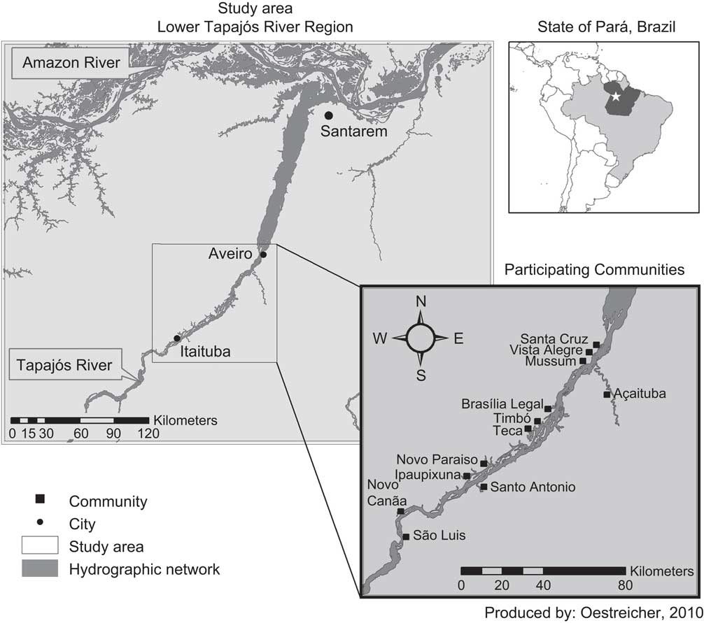
Fig. 1 Map of the study area showing the location of participating communities
The study was approved by the Ethics Review Boards of the University of Quebec at Montreal, the Federal University of Rio de Janeiro and the Faculty of Pharmaceutical Sciences of the University of São Paulo–Ribeirão Preto. All participants signed an informed consent form, which was read to them. There was no remuneration for study participation.
Sociodemographics and medical history
An interview-administered questionnaire was used to collect information on sociodemographics, occupational and residential history. Dietary information was collected with a 7 d FFQ(Reference Passos, Mergler and Fillion26). A trained nurse administered the questionnaire on medical history. Current medication was noted. None of the interviewers were aware of the participants’ exposure levels.
Assessment of biomarkers of Hg, Pb and Se
Hair
Hair Hg has often been used as a biomarker for current and retrospective exposure(Reference Bastos, Malm and Pfeiffer27); it reflects Hg intake from fish consumption (for a review, see Mergler et al.(Reference Mergler, Anderson and Chan28)). Hair strands from the occipital region were cut at the root and stored in plastic bags. The first two centimetres from the root were used to determine total hair Hg concentration (H-Hg) by cold vapour atomic absorption spectrometry, according to the method described in Farant et al.(Reference Farant, Brissette and Moncion29). Analyses were carried out at the laboratories of the First Nations and Inuit Health Laboratory of Health Canada (Ottawa, Canada). Analytical quality control was ensured with certified reference hair samples, provided by the Hair Mercury Inter-laboratory Comparison Program of Health Canada, Ottawa, Canada.
Blood
A Brazilian phlebotomist collected 6-ml blood samples in ‘trace metal free’ evacuated tubes (BD Vacutainer®) containing heparin as anticoagulant. Total Hg in blood (B-Hg) and plasma (P-Hg), total Pb in blood (B-Pb) and total Se in blood (B-Se) and plasma (P-Se) were determined by inductively coupled plasma–mass spectrometry (Perkin Elmer DRC II). For Hg determination in whole blood and plasma, the method proposed by Palmer et al. (Reference Palmer, Lewis and Geraghty30) was adopted. For the other elements, the method proposed by Batista et al.(Reference Batista, Rodrigues and Nunes31) was used. Samples were analysed at the Laboratório de Toxicologia e Essencialidade de Metais, Departamento de Análises Clínicas, Toxicológicas e Bromatológicas, Faculdade de Ciências Farmacêuticas de Ribeirão Preto, Universidade de São Paulo, Ribeirão Preto, Brazil. Quality control was guaranteed by analysing secondary reference materials, either provided by the New York State Department of Health's PT programme for trace elements in whole blood, or obtained from the external quality assessment scheme (EQAS) for trace elements operated by the Toxicology Center of the Quebec Public Health Institute (CTQ-INSP), Canada. Reference materials were analysed before and after ten ordinary samples.
Plasma phospholipid fatty acid analysis
Plasma total phospholipid FA were assessed for a subgroup of 349 people. For plasma separation, blood samples were centrifuged and pipetted into Eppendorf tubes previously cleaned in a class 100 clean room and immediately frozen at −20°C before analysis. Total phospholipid FA were measured in plasma using a modified method of Bondia-Pons et al.(Reference Bondia-Pons, Morera-Pons and Castellote32). Analyses were carried out at McGill University, Ste-Anne-de-Bellevue, Canada. EPA and DHA were considered in the present study. The concentration of each phospholipid FA was expressed as an absolute concentration (mg/ml) and as a percentage of total plasma phospholipid FA.
Visual functions assessment
Visual and ocular health examinations were carried out by four optometrists from the School of Optometry of the University of Montreal, trained prior to the study to minimize inter-observer bias. None were involved in assessment of toxins or nutrients.
The anterior segment of the eye was examined using slit-lamp biomicroscopy. The posterior segment of the retina and its periphery were examined during pupillary dilation using two mydriatic agents (tropicamide 1 % w/v and phenylephrine 2·5 % w/v). This procedure provided information on ocular pathologies for post hoc exclusion. The presence of pterygia, a corneal eye disease attributed to chronic UV-B exposure(Reference West and Munoz33, Reference Bradley, Yang and Bradley34), common in this region, was noted by the examiners.
Near and distant VA were assessed with the Allen and Tumbling E Charts, respectively for the right eye (OD) and the left eye (OS). For the purpose of statistical analysis, near and distant VA were log-transformed to obtain the minimum angle of resolution (Log MAR) to convert the geometric sequence of a traditional chart into a linear scale. The different functional categories for near and distant VA defined by the International Council of Ophthalmology(35) are presented in Table 2. All examinations were made under the same conditions.
Statistical analyses
Descriptive statistics were used to illustrate the study population's general characteristics, the distribution of the biomarkers, as well as near and distant VA. Data normality was assessed with the Shapiro–Wilk W test. Since exposure data and VA measures were skewed, both non-parametric (Wilcoxon/Kruskall–Wallis rank-sums test and Spearman's ρ) and parametric analyses (Student's t and Pearson's r correlation) were performed. When non-parametric and parametric results were in agreement, only the former are presented. For the multiple regression models, variables were log-transformed when necessary.
Multiple linear regressions were used to build five models for near and distant VA (Log MAR). The first model (M1) examined the relevant covariables: age, sex, smoking (yes v. no), drinking (yes v. no) and examiner (2 v. 1). In model 2 (M2), in addition to the previous covariables, we separately tested the biomarkers of Hg (B-Hg and H-Hg) and retained the one showing the strongest association (H-Hg). In model 3 (M3), we added B-Pb. In model 4 (M4), we retained the previous explanatory variables and separately tested EPA, DHA and EPA+DHA, as well as their percentage in the total plasma phospholipid FA (%EPA, %DHA and %EPA+DHA). The n-3 FA variable with the strongest association (%DHA) was retained in model 5 (M5), and we separately tested biomarkers of Se (B-Se and P-Se). Finally, model 6 (M6) included age, sex, smoking, drinking, examiner, H-Hg, B-Pb, %DHA and B-Se. The contribution of pterygia was verified in each of the models. All models were tested for OD and OS; the results are presented for OD. Homoscedasticity was verified using Studentized residuals and outliers were excluded at >|3|.
Since delayed neurotoxicity has been demonstrated in animal studies(Reference Rice and Hayward17), the multiplicative interaction term between age and Hg was likewise tested. The multiplicative interaction terms between Hg and Se, Hg and Pb and Hg and n-3 FA were also tested. The only significant interaction term was age × Log H-Hg. Data were stratified with respect to age category and the above multiple linear regression models were re-run.
For those over 40 years of age, multivariate logistic regression models (OR and 95 % CI) were used to examine the associations between near and distant visual impairment and biomarkers of Hg, Pb, Se and n-3 FA. H-Hg was categorized at 15 μg/g and B-Pb at 10 μg/dl, while Se was categorized at the median and n-3 FA at the first tertile. Age, sex, current smoking habit (yes v. no), alcohol consumption (yes v. no) and examiner were included in all of the models.
Results were defined as statistically significant at P < 0·05. Analyses were performed using the statistical software packages JMP version 8·0·1 (SAS Institute Inc., Cary, NC, USA) and SPSS version 16·0 (SPSS Inc., Chicago, IL, USA).
Results
The analyses considered 349 people for whom there were complete data on exposure and FA. Near and distant VA were not available in at least one eye for sixteen and four persons, respectively. Persons reporting health conditions, diseases or medications considered as possible aetiological factors for poor near and distant VA or influencing Hg and/or Se metabolism were excluded from the analyses: pregnancy (n 5), breast-feeding (n 5), reported diagnosed diabetes (n 5), psychotropic drugs (n 5), reported cerebrovascular accident (n 11). Further exclusions were made with respect to diagnosed eye pathology in either eye (n 21 for OD and n 23 for OS). Cataracts were diagnosed in forty-five people, who were likewise excluded from the analyses.
Analyses considered 243 persons whose sociodemographic characteristics are presented in Table 1. Alcohol, mostly beer, was consumed mainly during the weekend and on special occasions. Significantly more men reported drinking than women (73·5 % v. 48·4 %, P < 0·0001). Among smokers, cigarette consumption was low (median: 8·7 cigarettes/d). There was no association between age and alcohol or tobacco consumption. Most (90 %) had eaten at least one fish meal in the week preceding the interview.
Table 1 Sociodemographic characteristics, biomarkers of exposure, nutrients and visual acuity among the study population: adults (n 243) from fish-eating communities of Tapajós River in the Brazilian Amazon

H, hair; B, blood; P, plasma; FA, fatty acids; OD, right eye; OS, left eye; MAR, minimum angle of resolution.
Mean and median values of the biomarkers are presented in Table 1. H-Hg concentrations were higher in men than in women (median 14·3 v. 9·4 μg/g, Wilcoxon χ 2P < 0·0001) and H-Hg levels tended to increase with age (Spearman's ρ = 0·117, P = 0·07). B-Se was similar in men and women men and age did not influence B-Se status. B-Pb was higher in men than in women (14·4 v. 7·3 μg/dl, Wilcoxon χ 2P < 0·0001) and tended to increase with age (Spearman's ρ = 0·111, P = 0·08). All n-3 FA biomarkers were positively associated with age (Wilcoxon χ 2P < 0·0001) and were similar for men and women, except for the sum of EPA and DHA, which was significantly lower in men than in women (median: 0·024 v. 0·031 mg/ml, Wilcoxon χ 2P = 0·03).
Participants excluded for age-related cataracts were older than the participants included in the analyses (median: 58·5 v. 35·9 years, P < 0·0001). Indeed, the oldest study subjects here were 66 years old. As expected, both near and distant VA were better in participants compared with those excluded. Biomarkers of Hg, Pb and n-3 FA were similar in both groups, but B-Se was significantly lower in the excluded group (median: 202·6 v. 250·6 μg/l, P < 0·0001).
Near VA was similar for men and women in both eyes and decreased with age (OD: Spearman's ρ = 0·805, P < 0·0001; OS: Spearman's ρ = 0·753, P < 0·0001). Distant VA was the same for men and women and likewise decreased with age (OD: ρ = 0·5630, P < 0·0001; OS: ρ = 0·5925, P < 0·0001). Approximately 25 % of the study group presented moderate near VA impairment, and 7 % moderate distant VA impairment (Table 1). Pterygia was observed in at least one eye in eighty-seven persons (36 %).
The classification of VA according to functional category differed between age groups (Table 2). Most of those under 40 years (85 %) presented near VA in the normal range, while for the older group (40 to 66 years) it was the inverse: 85·9 % had mild to profound near visual loss. For distant VA, 13 % of the younger group presented mild to severe vision loss, while 55 % of the older group were in these categories. Pterygia was likewise more prevalent in the older persons compared with the younger (53 % v. 26 %, P < 0·0001).
Table 2 Near and distant visual acuity and their functional category in the right eye among adults (n 243) from fish-eating communities of Tapajós River in the Brazilian Amazon

MAR, minimum angle of resolution; ICO, International Council of Ophthalmology; N/A, not applicable.
The associations between near VA and the relevant covariables and biomarkers are presented in Table 3a. In the first model (M1), which contains only the covariables, age was strongly and positively associated with near VA loss. When Log H-Hg was added to the model (M2), it was likewise positively associated with near VA loss. In the following models (M3 to M6), %DHA showed a negative association with near VA loss but Log B-Pb and Log B-Se did not show any association with the outcome. When %DHA was included in the model, the β estimate for Log H-Hg increased by almost 30 %. The interaction term age × Log H-Hg was highly significant (P = 0·002).
Table 3 Results of multiple linear regression models for visual acuity loss in the right eye, showing the β estimates and their level of significance, among adults (n 243) from fish-eating communities of Tapajós River in the Brazilian Amazon

M, model; H, hair; B, blood.
Significance of the β estimate: (*)P < 0·10; *P < 0·05; **P < 0·01; ***P < 0·0001.
Since there was a highly significant interaction between age and Log H-Hg, results were stratified by age at 40 years old. No association was observed between Log H-Hg and near VA loss in younger people, whereas for older persons, there was a highly significant association with Log H-Hg (Table 3b). Within this age group, age still entered significantly in the model and near VA loss was negatively associated with %DHA. Entering %DHA in the model increased the β estimate for Log H-Hg by 50 % and when Log B-Se was entered into the model, the β estimate for Log H-Hg further increased by 12 % although Log B-Se did not reach the significance level (β = −0·19, P = 0·15). Twenty-four per cent (24 %) of the variance of near VA was explained by the final model; Log H-Hg explained 6·5 % of the variance, while age explained 3·5 %. Similar results were found for the left eye.
In the group over 40 years of age, multivariate logistic regression analyses showed that, for the right eye, higher H-Hg (≥15 μg/g) was associated with increased odds of presenting clinical near visual impairment (OR = 3·93, 95 % CI 1·25, 14·18), whereas higher %DHA (≥1st tertile concentration of 1·88 %) was associated with better vision (OR = 0·37, 95 % CI 0·11, 1·11). Similar results were obtained for the left eye.
The same process was followed for distant VA. In the entire study group, age, sex and examiner showed significant positive associations (P < 0·05) with distant VA in both eyes. For the right eye, there was a tendency for Log B-Pb to be positively associated with distant VA loss (β = 0·074, P = 0·08) while none of the n-3 FA measures nor Log B-Se were significantly associated to distant VA. For Log H-Hg, all of the estimates were in the positive direction but did not reach significance level (P < 0·05). In the left eye, the association with Log B-Pb was not present. In both models, the interaction term between Log B-Pb and age was not significant.
In the population between 40 and 66 years old, age and sex presented significant positive associations with distant VA loss (Table 3c). In the right eye, Log B-Pb showed a positive association with a decrease in distant VA (β = 0·19, P = 0·05). Twenty-eight per cent (28 %) of the variance of distant VA was explained by the final model; Log B-Pb explained 4·9 % of the variance, while age explained 14·6 % and sex explained 3·5 %.
For distant visual impairment among the older group, higher B-Pb (≥10 μg/dl) was associated with an adjusted OR for clinical moderate visual impairment of 5·39 (95 % CI 1·10, 34·55). Similar results were found for the left eye.
Discussion
In this fish-eating population in the Brazilian Amazon, those under 40 years old presented good near and distant VA. Quite the opposite was observed for those between 40 and 66 years old, who presented at least mild visual loss for near and distant VA (85 % and 55 %, respectively). Our findings suggest that after 40 years of age, near VA loss progresses more quickly with higher Hg exposure, while DHA may slow down this process. In this same age range, distant VA loss was associated with Pb exposure, but results were less consistent.
Presbyopia, the normal age-related loss of near VA, results from the eye's reduced ability accommodate and depends upon lens elasticity and ciliary muscle functioning(Reference Noback, Strominger and Demarest36). It occurs around 40 years of age, develops relatively rapidly in the first decade and tends to stabilize around 60 years(Reference Morgan37). In the population investigated in the present study, presbyopia likewise began around 40 years old, but there was a fourfold increased risk for clinical near visual impairment for those with H-Hg levels of 15 μg/g and higher.
Hg may contribute to the progressive decrease of power of accommodation by interfering with lens and/or ciliary muscle function. With ageing, the lens increases in size and becomes less flexible and transparent(Reference Pokorny, Smith and Verriest38). Some reports suggest that Hg accumulates in the lens and may be involved in cataract formation(Reference Winder, Astbury and Sheraidah39, Reference Gabal and Raslan40). In a study of this Amazon population, those above 65 years old showed and a very high prevalence of age-related cataracts, with an Hg-related increase in risk(Reference Lemire, Fillion and Frenette23). Although persons with diagnosed cataracts were excluded from the present analysis, it is possible that the observed loss of near VA may be due to initial subclinical changes in the lens crystalline structure that conventional techniques could not identify. Hg may also interfere with some of the processes involved in the contraction of ciliary muscles. Both animal and human studies have demonstrated effects of Hg on motor functions(Reference Clarkson and Magos41), but no study has specifically investigated ciliary muscles.
To our knowledge, the present study is the first to demonstrate that near VA is associated with DHA in adults. It has been suggested that dietary DHA is needed for the optimum functional maturation of the retina and visual cortex, with VA and mental development seemingly improved by extra DHA(Reference Uauy and Dangour42). Several studies have established a link between prenatal DHA and visual functions(Reference Hoffman, Boettcher and Diersen-Schade43–Reference Jacobson, Jacobson and Muckle47). The fact that DHA seems to play a role in near VA and not in distant VA suggests that DHA may influence ocular structures rather than the retina or visual cortex.
In the present study, Se did not appear to be protective for near VA loss. An experimental study in adult zebrafish showed that developmental co-exposure to selenomethionine could reduce MeHg-induced visual deficits associated with visual responses to a rotating black bar under low light conditions(Reference Weber, Connaughton and Dellinger48). It should be noted that there were few people with Se deficiency in the present population and it is possible that normal Se levels are sufficient to maintain visual functions. For age-related cataracts, in this same population, Lemire et al.(Reference Lemire, Fillion and Barbosa21) reported a synergistic effect of plasma Se and blood Hg, with a prevalence OR of 16·4 (95 % CI 3·04, 87·9) for those with low plasma Se and high blood Hg.
Our results suggest that distant VA loss may be associated to B-Pb levels in these middle-aged people, in whom we observed a fivefold increase in the probability of presenting clinical moderate visual impairment at B-Pb levels of 10 μg/dl or higher. Experiments on monkeys showed that selective lesions to the parvocellular pathway lead to deficits in distant VA(Reference Merigan, Katz and Maunsell49, Reference Merigan and Eskin50). Long-term developmental effects of Pb exposure on the visual system have been documented in animal models. Adult monkeys exposed to Pb pre- and postnatally showed a decrease in tyrosine hydroxylase in retinal neurons after a 35-month period of a Pb-free diet, suggesting a persisting effect of Pb exposure on dopaminergic retinal amacrine cells(Reference Kohler, Lilienthal and Guenther51). In rats, high-level gestational Pb exposure produced rod-selective toxicity, revealing the sensitivity and vulnerability of the developing retina to gestational Pb exposure(Reference Fox, Kala and Hamilton52). In the present study, no association between distant VA and blood Pb was observed for participants under 40 years of age. For the older group, since Pb exposure in the study region was unknown until recently(Reference Barbosa, Fillion and Lemire16), we do not know at what age exposure to Pb began, and whether the effect is due to cumulative, past or present exposure.
Since riverside people spend the major part of the day working outside, we also examined whether pterygia, a condition also known in this region as carne crescida and exacerbated by long-term exposure to sunlight (especially UV rays) and dust, influenced the results. As expected, the prevalence of pterygia was high in this population and increased with age, but it did not contribute to loss in near and distant VA in these mid-age people, nor did it affect the relationship between any of the biomarkers and VA loss.
The degree of age-related loss in distant VA in the present study is noteworthy. Although distant VA declines with age in people without any eye pathology, population studies indicate that the majority of people maintain at least a fair distant acuity (20/40 or better) into their 80s(Reference Gittings and Fozard53, Reference Vitale, Ellwein and Cotch54). It has been suggested that this decline cannot be attributed to optical changes and must therefore be due to changes in the retina or central visual pathway; however, anatomical studies in people and monkeys suggest that ageing has only relatively minor effects on the retino-geniculo-striate pathway(Reference Spear55). In the present study, since people presenting with any type of ocular pathology were excluded from the analyses, it is not likely that the age-related visual impairments observed were of ocular pathological origin.
Although this was not a random sample and one cannot extrapolate the prevalence data from the present study to the entire population, it is interesting to compare the distribution of VA loss with data reported from other countries. The distribution of distant visual loss with age is different from that reported in the US National Health and Nutrition Examination Survey (NHANES)(Reference Vitale, Ellwein and Cotch54), where 33 % of persons under 40 years of age presented myopia (≤−0·5D or 6/7·5). In the present study, only 12 % of the people below 40 years old presented myopia. For those over 40 years old, 41 % in NHANES had myopia, while in this Amazonian population, 55 % presented with distant VA poorer than 6/7·5. This higher prevalence of VA deficits in the Amazon population could be due to their elevated Hg levels, but could also be due to a selection bias: participation rate for the older group may have been higher among those with poor vision compared with those who did not require visual corrections.
In the Brazilian Amazon, where people have a limited access to health services, visual impairment often remains uncorrected. Other authors from Brazil have already pointed out the uncorrected refractive errors as the main cause of visual impairment in Brazil(Reference Salomao, Mitsuhiro and Belfort56). In the present study, we were able to adequately refract 85 % for near VA and 74 % for distant VA. These findings support the assertion that uncorrected refractive errors are often overlooked and need to be assessed and reported as a cause of visual impairment(Reference Resnikoff, Pascolini and Mariottia57).
Acknowledgements
This work was financially supported by the Canadian Institutes of Health Research and the Fundação de Amparo à Pesquisa do Estado de São Paulo from Brazil; Health Canada provided the facilities for hair Hg analyses. There are no conflicts of interest. M.F. participated in the design of the study, conducted the fieldwork and data collection, analysed the data and wrote the manuscript. M.L. participated in the design of the study, participated to the fieldwork and data collection and reviewed the manuscript. A.P. participated in the statistical analyses and reviewed the manuscript. B.F. designed the visual system evaluation and reviewed the manuscript. H.A.W. designed the methods for analysing fatty acids in plasma phospholipids and reviewed the manuscript. J.R.D. analysed the fatty acids in plasma phospholipids and reviewed the manuscript. J.R.D.G. participated to the fieldwork and reviewed the manuscript. F.L. participated in the statistical analyses and reviewed the manuscript. F.B. analysed Hg, Se and Pb in blood and urine samples and reviewed the manuscript. D.M. designed the study, analysed the data and participated in writing the manuscript. The authors are grateful to the villagers of the Tapajós region and to Marie-Ève Thibault for administrative work.


