


Introduction
Schistosomiasis impacts both the health and economics of regions worldwide, although poorly developed regions are mainly affected (WHO, 2018). This tropical disease is often associated with poverty. Schistosoma mansoni is the main causative agent of intestinal schistosomiasis and it has 2 main larval stages: miracidia and cercaria. Infective cercaria larvae are released from snails into fresh water, and this can lead to human infection. Adult worms can live inside portal-mesenteric venous vessels of humans and they release their eggs into feces. The ability of miracidia to infect snails as an intermediate host completes the life cycle of this parasite. The body of miracidia is covered with cilia to facilitate swimming. Non-feeding larvae use glycogen acquired during egg development as an energy source when searching for their snail host (WHO, 2018).
Cilia activity promotes whole body linear dislocation, while rotation of miracidia along their axis and a contraction–stretching movement are also used. Temperature influences the velocity and longevity of S. mansoni miracidia. High temperatures increase their velocity and reduce their lifespan, while low temperatures decrease their velocity and increase their lifespan (Benex and Deschiens, Reference Benex and Deschiens1963; Samuelson et al., Reference Samuelson, Quinn and Caulfield1984) and mortality is compensated by an increased probability of successfully finding their intermediate host, especially at temperatures greater than 20°C (Wang et al., Reference Wang, Russel, Wyeth, Bose, Ni, McManus and Cummins2019). High water salinity and acidity also reduce egg hatching and motility (Donnelly et al., Reference Donnelly, Appleton and Schutte1984).
When miracidia are present at high concentrations and are exposed to snail-conditioned water, an increase in both angular and linear velocities of miracidia is observed (Samuelson et al., Reference Samuelson, Quinn and Caulfield1984). At this higher velocity, miracidia exhibit a random circulatory movement which increases their probability of finding a snail (Wang et al., Reference Wang, Russel, Wyeth, Bose, Ni, McManus and Cummins2019).
In the present study, we explore miracidia mobility and their lifespan, with a focus on morphological and motility analyses. Motility is used as a proxy for infectivity to analyse miracidia survival and discuss their ability to potentially infect snail hosts. We used 4 observational criteria (degree of activity, locomotion, body shape and body transparency) to classify larvae into 6 categories of motility. The results obtained are compared with a review of literature regarding miracidia motility and lifespan. By optimizing the variables involved in modelling the interactions between miracidia and snails, a threshold which triggers spontaneous extinction of transmission may be identified to achieve local control.
Materials and methods
Obtaining S. mansoni miracidia
Eggs of S. mansoni, LE strain, were obtained from experimentally infected livers of Swiss mice at 40 days post-infection. Briefly, hatching was induced by incubating liver fragments in distilled water in a climatic chamber at 26°C under artificial light. After 50 min, the suspension was transferred to a 250 mL bottle which was covered with cardboard, except for the bottleneck which was exposed to light. Miracidia-positive phototropism facilitated the collection of parasites from the light-exposed water column. Handling of parasites and protocols for experimental infections in the laboratory was approved by the Ethics Committee on Animal Experimentation at PUCRS.
Criteria and classification of miracidia according to motility
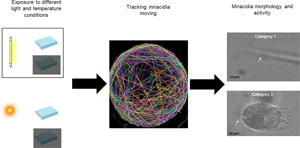
Based on subjective observations of miracidia moving in water that were collected in a pilot study, criteria were proposed based on body appearance (shape, transparency) and intensity of body and cilia activity. Six categories were established: (1) maximum activity and body dislocation; (2) fast movements but not a fully stretched body; (3) slow movements and rounding body tendency; (4) very slow movements and a well-defined oval body shape (lack of body stretching); (5) immobile body, yet cilia remain active and there is a small loss of body transparency and (6) no detectable body or cilia activity and an opaque miracidia body (Table 1).
Table 1. Definitions of criteria for classification of miracidia according to motility and body characteristics

Miracidia classified in categories 1–5 were considered alive as the endpoint for determination of their life span. Motility, either high (categories 1 and 2) or slow (category 3), was taken as a proxy for infectivity. Absence of movement defined lack of infectivity.
Monitoring miracidia motility over time
Groups of 5 miracidia that were obtained 1 h post-hatching were placed in a concave well plate with 300 μL dechlorinated water. Replicates (n = 3) were exposed to the following conditions: (A) a climate chamber with constant light and air temperature (25–26°C); (B) an open environment with uncontrolled air temperature and sunlight and (C) an open environment with an excavated glass plate covered with an opaque lid (dark) with uncontrolled air temperature. Motility and shape were observed every 60 min for 9 h and again after 24 h.
In the first experiment, survival time was longer than the total observation time (9 h). Therefore, in subsequent experiments, 60-min observation intervals (longer intervals, referred to as Li experiments), a shorter interval (30 min, referred to as Si experiments) and a longer total observation time (10 h) were implemented. An additional condition, D, was also introduced: an excavated glass plate covered with an opaque lid in a climate chamber (dark) with controlled temperature (25–26°C) (Table 2). The number of replicates was reduced to 2, with 5 miracidia per replicate for each experimental condition. Total numbers of miracidia observed in Li and Si experiments were, respectively, 450 and 500 (Li experiments: 5 miracidia at each of 3 replica for 1 experiment, 10 experiments for each of 3 conditions: A, B and C; Si experiments: 5 miracidia at each of 2 replica, 10 experiments for each of 4 conditions: A, B, C and D).
Table 2. Condition characteristics of environments which miracidia was conditioned during movements observations

Miracidia were visualized with a stereomicroscope at 6.3× magnification. Detailed observations were recorded at 10×, 16×, 25× or 40× magnifications. During the Li experiments, room temperature was monitored. During the Si experiments, water temperature was also recorded. A digital lux meter was used to record the intensity of light at each time point and water pH was measured in each experiment.
Statistical analysis
Data normality was tested by applying the Shapiro–Wilk test, with P < 0.05. Kaplan–Meier survival analysis was used to determine lifespan. Survival and infectivity between groups were compared by using the Wilcoxon unpaired test (significant differences had P < 0.05). Mann–Whitney and Kolmogorov–Smirnov tests were employed to investigate how light intensity and environment conditions affect survival. Data normality, comparison tests and survival tests were performed with RStudio 4.0.3 (RStudio, Boston, MA, USA). Graphs were generated with GraphPad Prism, version 8.4.3 (GraphPad, San Diego, CA, USA).
Results
Survival
The average time of miracidia longevity was 5.83 h (±0.13) and 3.59 h (±0.075), at time intervals of 60 min (Li) and 30 min (Si), respectively (Supplementary Tables S1 and S2). In the Li experiments, lifespan was longer (7.83 ± 0.235 h) when miracidia were maintained in a climatic chamber (condition A) vs in the dark (condition C, 5.48 ± 0.214 h) or under environmental light (condition B, 5.38 ± 0.224 h) (Fig. 1a). In the Si experiments, the longest average survival was observed under condition C (3.75 ± 0.15 h), followed by condition A (3.65 ± 0.15 h), condition B (3.61 ± 0.142 h) and condition D (3.38 ± 0.151 h). The latter involved a climate chamber under semi-dark conditions with the excavated plate containing miracidia covered with a lid. The highest median values were observed in the groups that remained in the environment (3.5 h) (Fig. 1b).

Fig. 1. Lifespan and infectivity of Schistosoma mansoni miracidia. (a) Estimated lifespan (S) and infectivity (I) data are presented from observations made in 60-min interval experiments (Li) in the closed environment (climatic chamber, condition A), open environment with sunlight (light environment, condition B) and open environment with sunlight blocked by using a lid (dark environment, condition C). (b) Lifespan (S) and infectivity (I) data are presented from observations made in 30-min interval experiments (Si) under the same A, B and C conditions described in panel a. In addition, data from condition D (climatic chamber with artificial light blocked by using a lid) (dark environment) are included. Standard deviation values are indicated with a dotted line, while median values are indicated with solid lines. The width of each shape is determined by the frequency of surviving (S) and infective (I) individuals at each time point.
Infectivity
Light and temperature did not significantly affect the infectivity of miracidia when longer observation intervals were used (Li) (P > 0.05). The infectivity period was estimated to last for 4.44 ± 0.123 h (Supplementary Table S3). It was also observed that the median values were higher for individual miracidia in the climatic chamber (5 h, condition A) compared to those under bright (condition B) or dark (condition C) open environment (4 h for both conditions) (Fig. 1a). When the miracidia were observed at shorter time intervals (Si), the overall infectivity was estimated to be 2.57 ± 0.06 h (Supplementary Table S4). Moreover, despite no statistically significant differences in the duration of infectivity among the various environmental conditions (P = 0.14) when compared with Si, miracidia exposed to light and observed at longer intervals (Li) exhibited higher median durations (2.5 h) than miracidia placed in the dark (2 h).
Frequency of movements over time
Under both Li and Si conditions, most miracidia showed an initial rapid decrease in movement. For example, more than 50% of miracidia became immobile within 5 or 3.3 h, respectively (Fig. 2). At 24 h, 100% of miracidia were immobile in both the Li and Si experiments.

Fig. 2. Frequency of motility categories observed over various time intervals. The categories are represented by a colour gradient from light grey to black which represents immobile dead miracidia (category 6) through activity (category 1), with increasing activity observed with darker colouring, respectively. Movement observations were recorded during long interval experiments (60 min, Li) (a) and short interval experiments (30 min, Si) (b).
In the Li experiments, the consolidated frequency for categories 1–3 was 55.3% up to 4 h, and less than 50% at the final time point (5 h). There was a small fraction (1.7%) of miracidia that was classified as category 1 at 8 h. At 9 h, 7.1% of the most active miracidia were classified as categories 2 and 3, while the least active miracidia were classified as categories 4 (3.3%), 5 (11.5%) and 6 (78.0%).
In the Si experiments, motility denoting infectivity (categories 1–3) was detected in 52.3% of miracidia up to 2.5 h, then it rapidly decreased to 10% after 4.5 h. Category 1 was recorded for 0.25% of miracidia up to 5.5 h, while both categories 2 and 3 represented 0.25% of motility classifications at 9 h. Only 0.75% of the total miracidia exhibited any activity after 9 h. Furthermore, only 3 miracidia survived up to 9.5 h, 2 under condition A and 1 under condition C.
Comparative analysis
To investigate the influence of light on motility, infectivity and survival, 2 nonparametric tests were performed (Mann–Whitney and Kolmogorov–Smirnov) after it was determined that the data did not exhibit a normal distribution. Data from the Li and Si experiments were compared by applying the Mann–Whitney test. There were no statistically significant differences between the results of the experiments conducted in the presence or absence of light (P > 0.05). However, there were statistically significant differences between the average and median estimates obtained in the Li and Si experiments (P < 0.001). The frequency of movements between the 2 observation intervals significantly differed between the following categories: 1 (P = 0.007), 2 (P = 0.007), 3 (P = 0.015) and 6 (P = 0.027). In contrast, the frequency of movements for categories 4 and 5 did not significantly differ (P = 0.812 and P = 0.382, respectively).
Discussion
To maintain their life cycle and survival, parasites must successfully and sequentially establish infections in one or more hosts. For transmission of schistosomiasis, the infection of snails by miracidia precedes the production of cercaria. With reproduction and transmission being key to survival, parasites predominantly invest their energy in the search, identification and infection of a host. Miracidia can recognize traces of snail mucus with apical papilla, and they change their behaviour immediately after recognizing these molecules. These behavioural changes include an increase in velocity, particularly angular velocity, which can help find a suitable host by increasing the search area (Fingerut et al., Reference Fingerut, Ann Zimmer and Zimmer2003). Infectivity and motility rapidly decrease with the age of miracidia, as demonstrated by Anderson et al. (Reference Anderson, Mercer, Wilson and Carter1982).
Nguyen et al. (Reference Nguyen, Gemmell and Rohr2020) previously highlighted that the superior infection performances exhibited by miracidia and cercaria are related to a high-energy expenditure and a shorter lifespan in environments with temperatures greater than 20°C and a low viscosity medium (Nguyen et al., Reference Nguyen, Gemmell and Rohr2020). Mobility is mainly affected by velocity, and both increases and decreases in velocity affect the longevity and infectivity of S. mansoni miracidia. Level of activity is also highly influenced by temperature. Miracidia move more slowly at low temperatures, and this reduces their chances of finding a host (Stirewalt, Reference Stirewalt1954; Nguyen et al., Reference Nguyen, Gemmell and Rohr2020). However, miracidia also exhibit an increased lifespan at temperatures below 9°C (Samuelson et al., Reference Samuelson, Quinn and Caulfield1984). Temperatures between 20 and 25°C have been identified as optimal for miracidia performance. While their longevity decreases when velocity and energy expenditure increase, a higher infection rate compensates for this reduction (Nguyen et al., Reference Nguyen, Gemmell and Rohr2020).
The lifespan and infectivity of miracidia have been described in several reports from 1948 to 1984 (Table 3). However, the methodology descriptions in these reports usually lack important information. For example, details about the water used in experiments are not provided in any of the studies that have analysed the life cycle of miracidia (Chernin and Antolics, Reference Chernin and Antolics1975; Mason and Fripp, Reference Mason and Fripp1976). Water quality is another important factor in evaluations of miracidia lifespan since chlorinated and acidic waters reduce their lifespan. Moreover, in some of these studies, statistical analysis was neither detailed nor even mentioned, thereby preventing a sound interpretation of the results (Benex and Deschiens, Reference Benex and Deschiens1963; Prah and James, Reference Prah and James1977; James and Prah, Reference James and Prah1978). In older reports, this limitation can be justified by the lack of more precise technology to measure and evaluate larvae motility. Typically, infectivity is measured by comparing the number of miracidia exposed to snails to the number of subsequently infected snails (Maldonado and Acosta-Matienzo, Reference Maldonado and Acosta-Matienzo1948; Chernin, Reference Chernin1968; Prah and James, Reference Prah and James1977).
Table 3. Descriptions of life span, infectivity, velocity, ratio of velocity increase over each 10°C increment and angular velocity reported for miracidia

SCW, snail-conditioned water; ±, approximate.
When cited, temperature and type of water used are also included.
In the present study, infectivity was measured only based on miracidia movements, since miracidia must actively move in water to successfully find a host. Infectivity and motility rapidly decrease with the age of miracidia, as demonstrated by Anderson et al. (Reference Anderson, Mercer, Wilson and Carter1982). Previously, Prah and James (Reference Prah and James1977) established 3 types of miracidia locomotion based on individual observed velocities of miracidia: fast, lethargic and slow. Reduction or even absence of movement at transversal experimental observations of individual miracidia does not directly translate into precise assessment of infectivity. This is a fundamental limitation of studies not exposing miracidia to susceptible snails and directly assessing infectivity. Although motility is not ideally taken as proxy for infectivity, it is a useful approach for the kind of analysis now proposed and it has been previously applied in other studies (Prah and James, Reference Prah and James1977; Anderson et al., Reference Anderson, Mercer, Wilson and Carter1982).
In the present study, we propose 6 categories of motility based on 4 observational criteria: degree of activity, locomotion, body shape and body transparency. Categories 1–3 are considered infective, while immobile and slow-moving miracidia (categories 4–6) are considered non-infective. If a miracidium did not present any sign of body or cilia activity, it was considered dead (category 6). It is anticipated that these proposed categories based on observational criteria will improve methodology studies of parasitic larvae dynamics, especially since locomotion is the main factor in the search for a host and subsequent infectivity.
The longevity estimates for miracidia reported in the current study (3.5–5.8 h) are very similar to those described by Maldonado and Acosta-Matienzo (Reference Maldonado and Acosta-Matienzo1948) and Chernin (Reference Chernin1968) (5–6 and 4 h, respectively). However, our findings diverge from those of Benex and Deschiens (Reference Benex and Deschiens1963) and Prah and James (Reference Prah and James1977) (7–9 and >15 h, respectively). We detected a rapid decrease in miracidia velocity, especially during the observations recorded over 30-min intervals. For example, after 2.5 h, 50.25% of miracidia were already very slow and probably unable to infect a host. In comparison, a wide variance of survival times have been reported by Targett and Robinson (Reference Targett and Robinson1964) when different conditioned media are used: 19 h with Helix pomatia extract; 23 h with Lymnaea stagnalis extract; 25–49 h with Biomphalaria glabrata extract; 53 h with Limnaea and mussel-conditioned saline with addition of H. pomatia blood (Targett and Robinson, Reference Targett and Robinson1964).
When lifespan and average infectivity time were compared, the latter was 1 h shorter than the average lifetime for miracidia, possibly due to a rapid depletion of energy sources. After 6 h, only 3% of miracidia exhibited sufficient motility to be infective. In other studies, 66 and 30% of miracidia remained infective after 2 h, and only 33% of miracidia were infective after 7 h (Maldonado and Acosta-Matienzo, Reference Maldonado and Acosta-Matienzo1948; Chernin, Reference Chernin1968; Chernin and Antolics, Reference Chernin and Antolics1975). Prah and James (Reference Prah and James1977) reported much higher proportions after 8 h, with 60–100% of miracidia remaining infective.
In the present study, only 3% of miracidia showed motility indicating infectivity after 6 h.
A possible explanation for the statistically significant difference in the results obtained from 60-min and 30-min observation intervals is that there is a larger negative effect associated with the shorter observation intervals. If correct, this represents a limitation for miracidia observations conducted under laboratory conditions.
Motility is essential for S. mansoni miracidia to find intermediate snail hosts and establish infections. This process is modulated by extrinsic factors, particularly temperature. The present results demonstrate that light does not appear to directly affect the survival of miracidia, yet it does interfere with lifespan and infectivity. Miracidia behaviour and survival are very responsive to extrinsic factors; consequently snail–miracidia interactions are a fragile link in the transmission cycle. It is possible that this fragility represents an opportunity for targeted interventions to interrupt schistosomiasis transmission, as proposed in the World Health Organization 2030 Road Map (WHO, 2021).
Overall, the present observations and the data from previous studies on miracidia survival and infectivity provide a better understanding of the biological characteristics and parameters of S. mansoni larvae and their infection of snails. Moreover, the present results may facilitate a better calibration of mathematical modelling of schistosomiasis transmission to improve our understanding of the dynamics of this process and the necessary optimization of control strategies.
Supplementary material
The supplementary material for this article can be found at https://doi.org/10.1017/S0031182022000579
Data
Data are annexed in Supplementary material and can also be accessed by contacting the corresponding author.
Author contributions
R. P. S., C. G. T. and L. R. P. U. conceived and designed the study. R. P. S. conducted data gathering. R. P. S., T. N. V. and H. R. B. performed statistical analyses. R. P. S., C. G. T., L. R. P. U. and V. F. P. wrote the article.
Financial support
This research received no specific grant from any funding agency, commercial or not-for-profit sectors.
Conflict of interest
None.
Ethical standards
The use of animals and experimental protocols were approved by the Ethics Committee on Animal Experimentation at PUCRS (CEUA 15/00443).


