
Book contents
- Caplan’s Stroke
- Caplan’s Stroke
- Copyright page
- Contents
- Preface
- Contributors
- Part I General principles
- Chapter 1 Introduction and perspective
- Chapter 2 Pathology, anatomy, and pathophysiology of stroke
- Chapter 3 Diagnosis and the clinical encounter
- Chapter 4 Imaging and laboratory diagnosis
- Chapter 5 Genetics of stroke
- Chapter 6 Treatment
- Part II Stroke syndromes
- Part III Prevention, complications, and recovery–rehabilitation
- Index
- Plate section
- References
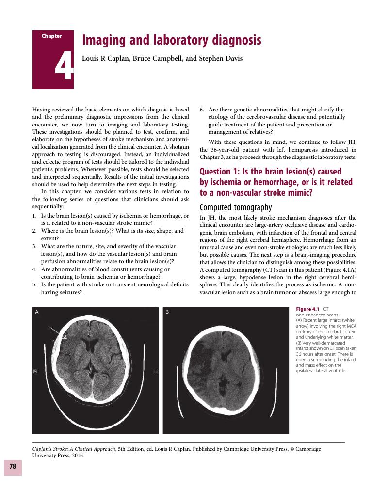
Chapter 4 - Imaging and laboratory diagnosis
from Part I - General principles
Published online by Cambridge University Press: 05 August 2016
Edited by

Book contents
- Caplan’s Stroke
- Caplan’s Stroke
- Copyright page
- Contents
- Preface
- Contributors
- Part I General principles
- Chapter 1 Introduction and perspective
- Chapter 2 Pathology, anatomy, and pathophysiology of stroke
- Chapter 3 Diagnosis and the clinical encounter
- Chapter 4 Imaging and laboratory diagnosis
- Chapter 5 Genetics of stroke
- Chapter 6 Treatment
- Part II Stroke syndromes
- Part III Prevention, complications, and recovery–rehabilitation
- Index
- Plate section
- References
Summary
A summary is not available for this content so a preview has been provided. Please use the Get access link above for information on how to access this content.
- Type
- Chapter
- Information
- Caplan's StrokeA Clinical Approach, pp. 78 - 128Publisher: Cambridge University PressPrint publication year: 2016
References
Eckert, B, Zeumer, H: Brain computed tomography. In Ginsberg, MD, Bogousslavsky, J (eds): Cerebrovascular Disease: Pathophysiology, Diagnosis, and Management, vol 2. Boston: Blackwell Science, 1998, pp 1241–1264.Google Scholar
von Kummer, R, Nolte, PN, Schnittger, H, et al: Detectability of cerebral hemisphere ischaemic infarcts by CT within 6 hours of stroke. Neuroradiology 1996;38:31–33.Google Scholar
Moulin, T, Cattin, F, Crepin-Leblond, T, et al: Early CT signs in acute middle cerebral artery infarction: Predictive value for subsequent infarct location and outcome. Neurology 1996;47:366–375.CrossRefGoogle ScholarPubMed
Norman, D, Price, D, Boyd, D, et al: Quantitative aspects of computed tomography of the blood and cerebrospinal fluid. Radiology 1977;7:223–228.Google Scholar
Caplan, LR, Flamm, ES, Mohr, JP, et al: Lumbar puncture and stroke: A statement for physicians by a committee of the Stroke Council of the American Heart Association. Stroke 1987;18:540A–544A.Google Scholar
Edlow, JA, Caplan, LR: Primary care: Avoiding pitfalls in the diagnosis of subarachnoid hemorrhage. N Engl J Med 2000;341:29–36.CrossRefGoogle Scholar
Beauchamp, NJ, Bryan, RN: Neuroimaging of stroke. In Welch, KMA, Caplan, LR, Reis, DJ, Siesjo, BK, Weir, B (eds): Primer on Cerebrovascular Diseases. San Diego: Academic Press, 1997, pp 599–611.Google Scholar
Baird, AE, Warach, S: Magnetic resonance imaging of acute stroke. J Cereb Blood Flow Metab 1998;18:583–609.Google Scholar
Brant-Zawadski, M, Atkinson, D, Detrick, M, et al: Fluid-attenuated inversion recovery (FLAIR) for assessment of cerebral infarction: Initial clinical experience in 50 patients. Stroke 1996;27:1187–1191.CrossRefGoogle Scholar
Warach, S, Chien, D, Li, W, et al: Fast magnetic resonance diffusion-weighted imaging of acute human stroke. Neurology 1992;42:1717–1723.Google Scholar
Warach, S, Gaa, J, Siewert, B, et al: Acute human stroke studied by whole brain echo planar diffusion-weighted magnetic resonance imaging. Ann Neurol 1995;37:231–241.Google Scholar
Lansberg, MG, Norbash, AM, Marks, MP, et al: Advantages of adding diffusion-weighted magnetic resonance imaging to conventional imaging for evaluating acute stroke. Arch Neurol 2000;57:1311–1316.Google Scholar
Engelter, ST, Wetzel, SG, Radue, EW, et al: The clinical significance of diffusion-weighted imaging in infratentorial strokes. Neurology 2004;62:574–580.Google Scholar
Kang, DW, Chalela, JA, Ezzeddline, MA, Warach, S: Association of ischemic lesion patterns on early diffusion-weighted imaging with TOAST stroke subtypes. Arch Neurol 2003;60:1730–1734.CrossRefGoogle ScholarPubMed
Bonati, LH, Lyrer, PA, Wetzel, SG, et al: Diffusion-weighted imaging, apparent diffusion coefficient maps and stroke etiology. J Neurol 2005;252:1387–1393.Google Scholar
Bonati, LH, Kessel-Schaefer, A, Linka, AZ, et al: Diffusion-weighted imaging in stroke attributable to patent foramen ovale. Stroke 2006;37:2030–2034.Google Scholar
Kidwell, CS, Saver, JL, Mattiello, J, et al: Thrombolytic reversal of acute human cerebral ischemic injury shown by diffusion/perfusion magnetic resonance imaging. Ann Neurol 2000;47:462–469.3.0.CO;2-Y>CrossRefGoogle ScholarPubMed
Chemmanam, T, Campbell, BCV, Christensen, S, et al: Ischemic diffusion lesion reversal is uncommon and rarely alters perfusion–diffusion mismatch. Neurology 2010;75:1040–1047.CrossRefGoogle ScholarPubMed
Campbell, BCV, Purushotham, A, Christensen, S, et al: The infarct core is well represented by the acute diffusion lesion: sustained reversal is infrequent. J Cereb Blood Flow Metab 2012; 32:50–56.Google Scholar
Patel, MR, Edelman, RR, Warach, S: Detection of hyperacute primary intraparenchymal hemorrhage by magnetic resonance imaging. Stroke 1996;27:2321–2324.Google Scholar
Linfante, I, Llinas, RH, Caplan, LR, Warach, S: MRI features of intracerebral hemorrhage within 2 hours from symptom onset. Stroke 2002;30:2263–2267.CrossRefGoogle Scholar
Chalela, JA, Latour, LL, Jeffries, N, et al: Hemorrhage and early MRI evaluation from the emergency room (HEME-ER): A prospective single center comparison of MRI to CT for the emergency diagnosis of intracranial hemorrhage in patients with suspected acute cerebrovascular disease. Stroke 2003;34:239–240.Google Scholar
Schellinger, PD, Fiebach, JB, Mohr, A, et al: The role of stroke MRI in intracranial and subarachnoid hemorrhage. Nervenarzt 2001;72:907–917.Google Scholar
Schellinger, PD, Jansen, O, Fiebach, JB, et al: A standardized MRI protocol comparison with CT in hyperacute intracerebral hemorrhage. Stroke 1999;30:765–768.Google Scholar
Assouline, E, Benziane, K, Reizine, D, et al: Intra-arterial thrombus visualized on T2 gradient echo imaging in acute ischemic stroke. Cerebrovasc Dis 2005;20:6–11.Google Scholar
Dul, K, Drayer, BP: CT and MR imaging of intracerebral hemorrhage. In Kase, CS, Caplan, LR (eds): Intracerebral Hemorrhage. Boston: Butterworth–Heinemann, 1994, pp 73–93.CrossRefGoogle Scholar
Rumboldt, Z, Kalousek, M, Castillo, M: Hyperacute subarachnoid hemorrhage on T2-weighted MR images. AJNR Am J Neuroradiol 2003;24:472–475.Google ScholarPubMed
Pexman, JHW, Barber, PA, Hill, MD, et al.: Use of the Alberta Stroke Program Early CT Score (ASPECTS) for Assessing CT Scans in Patients with Acute Stroke. AJNR Am J Neuroradiol 2001;22:1534–1542.Google Scholar
Becker, H, Desch, H, Hacker, H, et al: CT fogging effect with ischemic cerebral infarcts. Neuroradiol 1978;18:185–192.CrossRefGoogle Scholar
Nicolaides, AN, Kalodiki, E, Ramaswami, G, et al: The significance of cerebral infarcts on CT scans in patients with transient ischemic attacks. In Bernstein, EF, Callow, AD, Nicolaides, AN, Shifrin, EG (eds): Cerebral Revascularisation. London, Med-Orion, 1993, pp 159–178.Google Scholar
Inatomi, Y, Kimura, K, Yonehara, T, et al: DWI abnormalities and clinical characteristics in TIA patients. Neurology 2004;62:376–380.Google Scholar
Winbeck, K, Bruckmaier, K, Etgen, T, et al: Transient ischemic attack and stroke can be differentiated by analyzing early diffusion-weighted imaging signal intensity changes. Stroke 2004;35:1095–1099.Google Scholar
Lamy, C, Oppenheim, C, Calvet, D, et al: Diffusion-weighted MR imaging in transient ischaemic attacks. Eur Radiol 2006;16:1090–1095.Google Scholar
Bykowski, J, Latour, LL, Warach, S: More accurate identification of reversible ischemic injury in human stroke by cerebrospinal fluid suppressed diffusion-weighted imaging. Stroke 2004;35:1100–1106.Google Scholar
Prabhakaran, S, Chong, JY, Sacco, RL: Impact of abnormal diffusion-weighted imaging results on short-term outcome following transient ischemic attack. Arch Neurol 2007;64:1105–1109.Google Scholar
Redgrave, JNE, Coutts, SB, Schulz, UG, et al: Systematic review of associations between the presence of acute ischemic lesions on diffusion-weighted imaging and clinical predictors of early stroke risk after transient ischemic attack. Stroke 2007;38:1482–1488.CrossRefGoogle ScholarPubMed
Sylaja, PN, Coutts, SB, Subramaniam, S, et al: Acute ischemic lesions of varying ages predict risk of ischemic events in stroke/TIA patients. Neurology 2007;68:415–419.Google Scholar
Caplan, LR: Transient ischemic attack with abnormal diffusion-weighted imaging results. What’s in a name? Arch Neurol 2007; 64:1080–1082.Google Scholar
Adams, HP Jr, Kassell, NF, Turner, JC, et al: CT and clinical correlations in recent aneurysmal subarachnoid hemorrhage: A preliminary report of the Cooperative Aneurysm Study. Neurology 1983;33:981–988.CrossRefGoogle ScholarPubMed
Fishman, RA: Cerebrospinal fluid in cerebrovascular disorders. In Barnett, HJM, Mohr, JP, Stein, BM, Yatsu, FJ (eds): Stroke: Pathophysiology, Diagnosis, and Management. New York: Churchill Livingstone, 1986, pp 109–117.Google Scholar
Schluep, M, Bogousslavsky, J: Cerebrospinal fluid in cerebrovascular disease. In Ginsberg, MD, Bogousslavsky, J (eds): Cerebrovascular Disease: Pathophysiology, Diagnosis, and Management, vol 2. Boston: Blackwell Science, 1998, pp 1221–1226.Google Scholar
Van der Meulen, JP: Cerebrospinal fluid xanthrochromia: An objective index. Neurology 1966;16:170–178.CrossRefGoogle Scholar
Soderstrom, CE: Diagnostic significance of CSF spectrophotometry and computer tomography in cerebrovascular disease: A comparative study in 231 cases. Stroke 1977;8:606–612.Google Scholar
Davalos, A, Blanco, M, Pedraza, S, et al: The clinical-DWI mismatch: A new diagnostic approach to the brain tissue at risk of infarction. Neurology. 2004;62:2187–2192.Google Scholar
Caplan, LR: Significance of unexpected (silent) brain infarcts. In Caplan, LR, Shifrin, EG, Nicolaides, AN, Moore, WS (eds): Cerebrovascular Ischaemia: Investigation and Management. London: Med-Orion, 1996, pp 423–433.Google Scholar
Yamamoto, H, Bogousslavsky, J: Mechanisms of second and further strokes. J Neurol Neurosurg Psychiatry 1998;64:771–776.Google Scholar
Caplan, LR: Reperfusion of ischemic brain: Why and why not? In Hacke, W, del Zoppo, G, Hirschberg, M (eds): Thrombolytic Therapy in Acute Stroke. Berlin: Springer, 1991, pp 36–45.CrossRefGoogle Scholar
Ropper, AH: Lateral displacement of the brain and level of consciousness in patients with an acute hemispheral mass. N Engl J Med 1986;314:953–958.Google Scholar
Ropper, AH: A preliminary MRI study of the geometry of brain displacement and level of consciousness with acute intracranial masses. Neurology 1989;39:622–627.Google Scholar
Lansberg, MG, Thijs, VN, O’Brien, MW, et al: Evolution of apparent diffusion coefficient, diffusion-weighted, and T2-weighted signal intensity of acute stroke. AJNR Am J Neuroradiol 2001;22:637–644.Google Scholar
Weisberg, LA, Stazio, A, Shamsnia, M, et al: Nontraumatic parenchymal brain hemorrhages. Medicine (Baltimore) 1990;69:277–295.CrossRefGoogle ScholarPubMed
Delgado Almandoz, JE, Schaefer, PW, Forero, NP, et al: Diagnostic accuracy and yield of multidetector CT angiography in the evaluation of spontaneous intraparenchymal cerebral hemorrhage. AJNR Am J Neuroradiol 2009;30:1213–1221.Google Scholar
Kase, CS: Cerebral amyloid angiopathy. In Kase, CS, Caplan, LR (eds): Intracerebral Hemorrhage. Boston: Butterworth–Heinemann, 1994, pp 179–200.Google Scholar
Hauw, J-J, Seilhean, D, Duyckaerts, CH: Cerebral amyloid angiopathy. In Ginsberg, MD, Bogousslavsky, J (eds): Cerebrovascular Disease: Pathophysiology, Diagnosis, and Management. Boston: Blackwell, 1998, pp 1772–1794.Google Scholar
Kase, C, Robinson, R, Stein, R, et al: Anticoagulant-related intracerebral hemorrhages. Neurology 1985;35:943–948.Google Scholar
Kase, CS: Bleeding disorders. In Kase, CS, Caplan, LR (eds): Intracerebral Hemorrhage. Boston: Butterworth–Heinemann, 1994, pp 117–151.Google Scholar
Broderick, JP, Brott, TG, Duldner, JE, Tomsick, T, Huster, G. Volume of intracerebral hemorrhage: a powerful and easy-to-use predictor of 30-day mortality. Stroke 1993;24:987–993.CrossRefGoogle Scholar
Wada, R, Aviv, RI, Fox, A, et al: CT angiography “spot sign” predicts hematoma expansion in acute intracerebral hemorrhage. Stroke 2007;38:1257–1262.Google Scholar
Demchuk, AM, Dowlatshahi, D, Rodriguez-Luna, D, et al; and PREDICT Group: Prediction of haematoma growth and outcome in patients with intracerebral haemorrhage using the CT-angiography spot sign (PREDICT): A prospective observational study. Lancet Neurol 2012;11:307–314.CrossRefGoogle ScholarPubMed
Weisberg, L: Computed tomography in aneurysmal subarachnoid hemorrhage. Neurology 1979;29:802–808.Google Scholar
Adams, H, Kassell, N, Torner, J, et al: CT and clinical correlations in recent aneurysmal subarachnoid hemorrhage: A preliminary report of the cooperative aneurysm study. Neurology 1983;33:981–988.Google Scholar
van Gijn, J, van Dongen, K: Computerized tomography in subarachnoid hemorrhage: Difference between patients with and without an aneurysm on angiography. Neurology 1980;30:538–539.CrossRefGoogle ScholarPubMed
van Gijn, J, van Dongen, KJ, Vermeulen, M, et al: Perimesencephalic hemorrhage: A non-aneurysmal and benign form of subarachnoid hemorrhage. Neurology 1985;35:493–497.Google Scholar
Rinkel, GJ, Wijdicks, EF, Vermeulen, M, et al: Outcome in perimesencephalic (non-aneurysmal) subarachnoid hemorrhage: A follow-up study in 37 patients. Neurology 1990;40:1130–1132.Google Scholar
Kumar, S, Goddeau, RP Jr, Selim, MH, et al: Atraumatic convexal subarachnoid hemorrhage: clinical presentation, imaging patterns, and etiologies. Neurology 2010;74:893–899.Google Scholar
Kistler, JP, Crowell, R, Davis, K, et al: The relation of cerebral vasospasm to the extent and location of subarachnoid blood visualized by CT scan: A prospective study. Neurology 1983;33:424–437.CrossRefGoogle Scholar
Mohsen, F, Pominis, S, Illingworth, R: Prediction of delayed cerebral ischemia after subarachnoid hemorrhage by computed tomography. J Neurol Neurosurg Psychiatry 1984;47:1197–1202.CrossRefGoogle ScholarPubMed
Kern, R, Szabo, K, Hennerici, M, Meairs, S: Characterization of carotid artery plaques using real-time compound B-mode ultrasound. Stroke 2004;35:870–875.Google Scholar
Landry, A, Spence, JD, Fenster, A: Measurement of carotid plaque volume by 3-dimensional ultrasound. Stroke 2004;35:864–869.Google Scholar
O’Donnell, TF, Erdoes, L, Mackey, W, et al: Correlation of B-mode ultrasound imaging and arteriography with pathologic findings at carotid endarterectomy. Arch Surg 1985;120:443–449.Google Scholar
Hennerici, M, Baezner, H, Daffertshofer, M: Ultrasound of cervical arteries. In Caplan, LR, Manning, WJ (eds): Brain Embolism. New York: Informa Healthcare, 2006, pp 223–242.Google Scholar
Schenk, EA, Bond, G, Aretz, T, et al: Multicenter validation study of real-time ultrasonography, arteriography and pathology: Pathologic evaluation of carotid endarterectomy specimens. Stroke 1988;19:289–296.Google Scholar
Hennerici, M, Meairs, S: Imaging arterial wall disease. Cerebrovasc Dis 2000;10(Suppl 5):9–20.Google Scholar
Gronholdt, M-LM, Nordestgaard, BG, Nielsen, TG, Sillesen, H: Echolucent carotid artery plaques are associated with elevated levels of fasting and postprandial triglyceride-rich lipoproteins. Stroke 1996;27:2166–2172.Google Scholar
Geroulakos, G, Hobson, RW, Nicolaides, AW: Ultrasonic carotid plaque morphology. In Caplan, LR, Shifrin, EG, Nicolaides, AN, Moore, WS (eds): Cerebrovascular Ischaemia: Investigation and Management. London: Med-Orion, 1996, pp 25–32.Google Scholar
O’Leary, DH, Polka, JF, Kronmal, RA, et al: Thickening of the carotid wall: A marker for atherosclerosis in the elderly? Stroke 1996;27:224–231.Google Scholar
Bots, ML, Hoes, AW, Koudstaal, PJ, et al: Common carotid intima-media thickness and risk of stroke and myocardial infarction: The Rotterdam Study. Circulation 1997;96:1432–1437.Google Scholar
O’Leary, DH, Polak, JF, Kronmal, RA, et al: Carotid artery intima and media thickness as a risk factor for myocardial infarction and stroke risk in older adults. N Engl J Med 1999;340:14–22.CrossRefGoogle ScholarPubMed
Yakushiji, Y, Yasaka, M, Takada, T, Minematsu, K: Serial transoral carotid ultrasonographic findings in extracranial internal carotid artery dissection. J Ultrasound Med 2005;24:877–880.Google Scholar
Yakushijji, Y, Takase, Y, Kosugi, M, et al: Transoral carotid ultrasonography is useful for detection and follow-up of extracranial internal carotid artery dissecting aneurysm. Cerebrovasc Dis 2007;24:144–146.Google Scholar
Forteza, A, Krejza, J, Koch, S, Babikian, V: Ultrasound imaging of cerebrovascular disease. In Babikian, VL, Wechsler, LR, Higashida, RT (eds): Imaging Cerebrovascular Disease. Philadelphia: Butterworth–Heinemann, 2003, pp 3–35.Google Scholar
von Reutern, GM, von Budingen, HJ: Ultrasound Diagnosis of Cerebrovascular Disease. New York: Georg Thieme, 1993.Google Scholar
Bartels, E: Color-Coded Duplex Ultrasonography of the Cerebral Vessels. Stuttgart: Schattauer, 1998.Google Scholar
Steinke, W, Kloetzsch, C, Hennerici, M: Carotid artery disease assessed by color Doppler flow imaging: correlation with standard Doppler sonography and angiography. AJNR Am J Neuroradiol 1990;11:259–266.Google ScholarPubMed
Steinke, W, Hennerici, M, Rautenberg, W, Mohr, JP: Symptomatic and asymptomatic high-grade carotid stenosis in Doppler color-flow imaging. Neurology 1992;42;131–138.CrossRefGoogle ScholarPubMed
Steinke, W, Ries, S, Artemis, N, et al: Power Doppler imaging of carotid artery stenosis. Comparison with color Doppler flow imaging and angiography. Stroke 1997;28:1981–1987.CrossRefGoogle ScholarPubMed
Griewing, B, Doherty, C, Kessler, CH: Power Doppler ultrasound examination of the intracerebral and extracerebral vasculature. J Neuroimaging 1996;6:32–35.Google Scholar
Lenzi, GL, Vicenzini, E: The ruler is dead: An analysis of carotid plaque motion. Cerebrovasc Dis 2007;23:121–125.Google Scholar
Alexandrov, AV (ed): Cerebrovascular Ultrasound in Stroke Prevention and Treatment. New York: Futura Blackwell Publishing, 2003.Google Scholar
Molina, CA, Alexandrov, AV: Transcranial Doppler ultrasound. In Caplan, LR, Manning, WJ (eds): Brain Embolism. New York: Informa Healthcare, 2006, pp 113–128.Google Scholar
Babikian, VL, Wechsler, LR (eds): Transcranial Doppler Ultrasonography, 2nd ed. Boston: Butterworth–Heinemann, 1999.Google Scholar
Otis, SM, Ringelstein, EB: The transcranial Doppler examination: Principles and applications of transcranial Doppler sonography. In Tegeler, CH, Babikian, VL, Gomez, CR (eds): Neurosonology. St Louis: Mosby, 1996, pp 113–128.Google Scholar
Gomez, CR, Brass, LM, Tegeler, CH, et al: The trans-cranial Doppler standardization project. Phase 1 results. The TCD Study Group, American Society of Neuroimaging. J Neuroimaging 1993;3:190–192.Google Scholar
Caplan, LR, Brass, LM, DeWitt, LD, et al: Transcranial Doppler ultrasound: Present status. Neurology 1990;40:696–700.Google Scholar
Hennerici, M, Rautenberg, W, Sitzer, G, et al: Transcranial Doppler ultrasound for the assessment of intracranial arterial flow velocity. Surg Neurol 1987;27:439–448.Google Scholar
Hennerici, M, Rautenberg, W, Schwartz, A: Transcranial Doppler ultrasound for the assessment of intracranial arterial flow velocity. II. Evaluation of intracranial arterial disease. Surg Neurol 1987;27:523–532.Google Scholar
Demchuk, A, Christou, I, Wein, T, et al: Accuracy and criteria for localizing arterial occlusion with transcranial Doppler. J Neuroimaging 2000;10:1–12.CrossRefGoogle ScholarPubMed
Demchuk, AM, Christou, I, Wein, T, et al: Specific transcranial Doppler flow findings related to the presence and site of arterial occlusion. Stroke 2000;31:140–146.Google Scholar
Baumgartner, RW: Transcranial color duplex sonography in cerebrovascular disease: A systematic review. Cerebrovasc Dis 2003;16:4–13.Google Scholar
Krejza, J, Baumagartner, RW: Clinical applications of transcranial color-coded duplex sonography. J Neuroimaging 2004;14:215–225.Google Scholar
Burns, PN: Overview of echo-enhanced vascular ultrasound imaging for clinical diagnosis in neurosonology. J Neuroimaging 1997;7(Suppl 1):S2–S14.Google Scholar
Bogdahn, U, Becker, G, Schlief, R, et al: Contrast-enhanced transcranial color-coded real-time sonography. Stroke 1993;24:676–684.Google Scholar
Delcker, A, Turowski, B: Diagnostic value of three-dimensional transcranial contrast duplex sonography. J Neuroimaging 1997;7:139–144.Google Scholar
Stolz, E, Kaps, M: New techniques in ultrasound. In Babikian, VL, Wechsler, LR, Higashida, RT (eds): Imaging Cerebrovascular Disease. Philadelphia: Butterworth–Heinemann, 2003, pp 383–401.Google Scholar
Sharma, VK, Tsivgoulis, G, Lao, AY, Alexandrov, AV: Role of transcranial Doppler ultrasonography in evaluation of patients with cerebrovascular disease. Curr Neurol Neurosci Rep 2007;7:8–20.Google Scholar
Tsivgoulis, G, Sharma, VK, Lao, AY, et al: Validation of transcranial Doppler with computed tomography angiography in acute cerebral ischemia. Stroke 2007;38:1245–1249.Google Scholar
Sharma, VK, Tsivgoulis, G, Lao, AY, et al: Noninvasive detection of diffuse intracranaial disease. Stroke 2007;38:3175–3181.CrossRefGoogle Scholar
Caplan, LR: Posterior Circulation Disease: Clinical Findings, Diagnosis, and Management. Boston: Blackwell, 1996.Google Scholar
Sliwka, U, Rautenberg, W: Multimodal ultrasound versus angiography for imaging the vertebrobasilar circulation. J Neuroimaging 1998;8:182.Google Scholar
Seiler, RW, Grolimund, P, Asaslid, R, et al: Cerebral vasospasm evaluated by transcranial ultrasound correlated with clinical grade and CT-visualized subarachnoid hemorrhage. J Neurosurg 1986;64:594–600.Google Scholar
Becker, G, Greiner, K, Kaune, B, et al: Diagnosis and monitoring of subarachnoid hemorrhage by transcranial color-coded real time sonography. Neurosurgery 1991;28:814–820.Google Scholar
Chaudhuri, R, Padayachee, TS, Lewis, RR, et al: Non-invasive assessment of the circle of Willis using transcranial pulsed Doppler ultrasound with angiographic correlation. Clin Radiol 1992;46:193–197.Google Scholar
Anzola, GP, Gasparotti, R, Magoni, M, Prandini, F: Transcranial Doppler sonography and magnetic resonance angiography in the assessment of collateral hemispheric flow in patients with carotid artery disease. Stroke 1995;26:214–217.Google Scholar
Klotzsch, C, Popescu, O, Berlit, P: Assessment of the posterior communicating artery by transcranial color-coded duplex sonography. Stroke 1996;27:486–489.Google Scholar
Piepgras, A, Schmiedek, P, Leinsinger, G, et al: A simple test to assess cerebrovascular reserve capacity using transcranial Doppler sonography and acetazolamide. Stroke 1990;21:1306–1311.Google Scholar
Dahl, A, Russell, D, Rootwelt, K, et al: Cerebral vasoreactivity assessed with transcranial Doppler and regional cerebral blood flow measurements. Dose, concentration, and time of the response to acetazolamide. Stroke 1995;26:2302–2306.Google Scholar
Valdueza, JM, Draganski, B, Hoffman, O, et al: Analysis of CO2 vasomotor reactivity and vessel diameter changes by simultaneous venous and arterial Doppler recordings. Stroke 1999;30:81–86.Google Scholar
Yonas, H, Smith, HA, Durham, SR, et al: Increased stroke risk predicted by compromised cerebral blood flow reactivity. J Neurosurg 1993;79:483–489.Google Scholar
Markus, HS: Transcranial Doppler detection of circulating cerebral emboli: A review. Stroke 1993;24:1246–1250.Google Scholar
Markus, HS, Harrison, MJ: Microembolic signal detection using ultrasound. Stroke 1995;26:1517–1519.Google Scholar
Tong, DC, Albers, GW: Transcranial Doppler-detected microemboli in patients with acute stroke. Stroke 1995;26:1588–1592.Google Scholar
Sliwka, U, Job, F-P, Wissuwa, D, et al: Occurrence of transcranial Doppler high-intensity transient signals in patients with potential cardiac sources of embolism: A prospective study. Stroke 1995;26:2067–2070.Google Scholar
Daffertshofer, M, Ries, S, Schminke, U, Hennerici, M: High-intensity transient signals in patients with cerebral ischemia. Stroke 1996;27:1844–1849.Google Scholar
Sliwka, U, Lingnau, A, Stohlmann, W-D, et al: Prevalence and time course of microembolic signals in patients with acute strokes: A prospective study. Stroke 1997;28:358–363.Google Scholar
Ringelstein, EB, Droste, DW, Babikian, VL, et al: Consensus on microembolus detection by TCD. International Consensus Group on Microembolus Detection. Stroke 1998;29:725–729.Google Scholar
Siebler, M, Nachtmann, A, Sitzer, M, et al: Cerebral microembolism and the risk of ischemia in asymptomatic high-grade internal carotid artery stenosis. Stroke 1995;26:2184–2186.Google Scholar
Molloy, J, Markus, HS: Asymptomatic embolization predicts stroke and TIA risk in patients with carotid artery stenosis. Stroke 1999;30:1440–1443.Google Scholar
Segura, T, Serena, J, Molins, A, Davalos, A: Clusters of microembolic signals: A new form of cerebral microembolism presentation in a patient with middle cerebral artery stenosis. Stroke 1998;29:722–724.CrossRefGoogle Scholar
Wong, KS, Li, H, Chan, YL, et al: Use of trans-cranial Doppler to predict outcome in patients with intracranial large-artery occlusive disease. Stroke 2003;31:2641–2647.Google Scholar
Gao, S, Wong, KS, Hansberg, T, et al: Microembolic signal predicts recurrent cerebral ischemic events in acute stroke patients with middle cerebral artery stenosis. Stroke 2004;35:2832–2836.Google Scholar
Mackinnon, AD, Aaslid, R, Markus, HS: Long-term ambulatory monitoring for cerebral emboli using transcranial Doppler ultrasound. Stroke 2004;35:73–78.CrossRefGoogle ScholarPubMed
Teague, SM, Sharma, MK: Detection of paradoxical cerebral echo contrast embolization by transcranial Doppler ultrasound. Stroke 1991;22:740–745.CrossRefGoogle ScholarPubMed
Chimowitz, MI, Nemec, JJ, Marwick, TH, et al: Transcranial Doppler ultrasound identifies patients with right-to-left cardiac or pulmonary shunts. Neurology 1991;41:1902–1904.Google Scholar
Albert, A, Muller, HR, Hetzel, A: Optimized transcranial Doppler technique for the diagnosis of cardiac right-to-left shunts. J Neuroimaging 1997;7:159–163.Google Scholar
Di Tullio, M, Sacco, RL, Venketasubramanian, N, et al: Comparison of diagnostic techniques for the detection of a patent foramen ovale in stroke patients. Stroke 1993;24:1020–1024.Google Scholar
Klotzsch, C, Janzen, G, Berlit, P: Transesophageal echocardiography and contrast-TCD in the detection of a patent foramen ovale. Experiences with 111 patients. Neurology 1994;44:1603–1606.Google Scholar
Jauss, M, Zanette, E: Detection of right-to-left shunt with ultrasound contrast agent and trans-cranial Doppler sonography. Cerebrovasc Dis 2000;10:490–496.CrossRefGoogle Scholar
Sastry, S, Daly, K, Chengodu, T, McCollum, C: Is transcranial Doppler for the detection of venous-to-arterial circulation shunts reproducible? Cerebrovasc Dis 2007;23:424–429.Google Scholar
Baumgartner, RW, Gonner, F, Arnold, M, Muri, R: Transtemporal power- and frequency-based color-coded duplex sonography of cerebral veins and sinuses. AJNR Am J Neuroradiol 1997;18:1771–1781.Google Scholar
Stolz, E, Kaps, M, Dorndorf, W: Assessment of intracranial venous hemodynamics in normal individuals and patients with cerebral venous thrombosis. Stroke 1999;30:70–75.Google Scholar
Ries, S, Steinke, W, Neff, KW, Hennerici, M: Echocontrast enhanced transcranial color-coded sonography for the diagnosis of transverse sinus thrombosis. Stroke 1997;28:696–700.Google Scholar
Valdueza, JM, Hoffmann, O, Weih, M, et al: Monitoring of venous hemodynamics in patients with cerebral venous thrombosis by transcranial Doppler ultrasound. Arch Neurol 1999;56:229–234.Google Scholar
Becker, G, Bogdahn, U, Gehlberg, C, et al: Transcranial color-coded real-time sonography of intracranial veins. J Neuroimaging 1995;5:87–94.CrossRefGoogle ScholarPubMed
Pressman, BD, Tourje, EJ, Thompson, JR: An early sign of ischemic infarction: Increased density in a cerebral artery. AJNR Am J Neuroradiol 1987;8:645–648.Google Scholar
Riedel, CH, Zoubie, J, Ulmer, S, Gierthmuehlen, J, Jansen, O: Thin-slice reconstructions of nonenhanced CT images allow for detection of thrombus in acute stroke. Stroke 2012;43:2319–2323.Google Scholar
Lays, D, Pruvo, JP, Godefroy, O, et al: Prevalence and significance of hyperdense middle cerebral artery in acute stroke. Stroke 1992;23:317–324.Google Scholar
Tomsick, T, Brott, T, Barsan, W, et al: Prognostic value of the hyperdense middle cerebral artery sign and stroke scale score before ultraearly thrombolytic therapy. AJNR Am J Neuroradiol 1996;17:79–85.Google Scholar
Lee, TC, Bartlett, E, Fox, AJ, Symons, SP: The hypodense artery sign. AJNR Am J Neuroradiol 2005;26:2027–2029.Google Scholar
Grunholdt, ML: B-mode ultrasound and spiral CT for the assessment of carotid atherosclerosis. Neuroimaging Clin N Am 2002;12:421–435.Google Scholar
Frank, H: Characterization of atherosclerotic plaque by magnetic resonance imaging. Am Heart J 2001;141(Suppl 2):S45–S48.Google Scholar
Yuan, C, Mitsumori, LM, Beach, KW, Maravilla, KR: Carotid atherosclerotic plaque: Noninvasive MR characterization and identification of vulnerable lesions. Radiology 2001;221:285–299.Google Scholar
Adams, GJ, Greene, J, Vick, GW 3rd, et al: Tracking regression and progression of atherosclerosis in human carotid arteries using high-resolution magnetic resonance imaging. Magn Reson Imaging 2004;22:1249–1258.Google Scholar
Honda, M, Kitagawa, N, Tsutsumi, K, et al: High-resolution magnetic resonance imaging for detection of carotid plaques. Neurosurgery 2006;58:338–346.Google Scholar
Hatsukami, TS, Ross, R, Polissar, NL, Yuan, C: Visualization of fibrous cap thickness and rupture in human atherosclerotic carotid plaque in vivo with high-resolution magnetic resonance imaging. Circulation 2000;102:959–964.Google Scholar
Moody, AR, Murphy, RE, Morgan, PS, et al: Characterization of complicated carotid plaque with magnetic resonance direct thrombus imaging in patients with cerebral ischemia. Circulation 2003;107:3047–3052.Google Scholar
Saloner, D, Acevedo-Bolton, G, Wintermark, M, Rapp, JH: MRI of geometric and compositional features of vulnerable carotid plaque. Stroke 2007;38(2):637–641.Google Scholar
Touzé, E, Toussaint, J-F, Coste, J, et al; for the High-Resolution Magnetic Resonanace Imaging in Atherosclerotic Stenosis of the Carotid Artery (HIRISC) Study Group: Reproducibility of high-resolution MRI for the identification and the quantification of carotid atherosclerotic plaque components. Stroke 2007;38:1812–1819.Google Scholar
Yuan, C, Mitsumori, LM, Ferguson, MS, et al: In vivo accuracy of multispectral magnetic resonance imaging for identifying lipid-rich necrotic cores and intraplaque hemorrhage in advanced human carotid plaques. Circulation 2001;104:2051–2056.Google Scholar
Botnar, RM, Buecker, A, Wiethoff, AJ, et al: In vivo magnetic resonance imaging of coronary thrombosis using a fibrin-binding molecular magnetic resonance contrast agent. Circulation 2004;110:1463–1466.Google Scholar
Sirol, M, Fuster, V, Badimon, JJ, et al: Chronic thrombus detection with in vivo magnetic resonance imaging and a fibrin-targeted contrast agent. Circulation 2005;112:1594–1600.Google Scholar
Klein, IF, Lavallee, PC, Schouman-Claeys, E, Amaraenco, P: High-resolution MRI identifies basilar artery plaques in paramedian pontine infarct. Neurology 2005;64:551–552.Google Scholar
Klein, IF, Lavallee, PC, Touboul, P-J, et al: In vivo middle cerebral artery plaque imaging by high-resolution MRI. Neurology 2006;67:327–329.Google Scholar
Lam, WW, Wong, KS, So, NM, et al: Plaque volume measurement by magnetic resonance imaging as an index of remodeling of middle cerebral artery: Correlation with transcranial color Doppler and magnetic resonance angiography. Cerebrovasc Dis 2004;17:166–169.Google Scholar
Chalela, JA, Haaymore, JB, Ezzeddine, MA, et al: The hypointense MCA sign. Neurology 2002;58:1470.Google Scholar
Cho, K-H, Kim, JS, Kwon, SU, et al: Significance of susceptibility vessel sign on T2*-weighted gradient echo imaging for identification of stroke subtypes. Stroke 2005;36:2379–2383.Google Scholar
Hermier, M, Nighoghossian, N: Contribution of susceptibility-weighted imaging to acute stroke assessment. Stroke 2004;35:1989–1994.Google Scholar
Assouline, E, Benziane, K, Reizine, D, et al: Intra-arterial thrombus visualized on T2* gradient echo imaging in acute ischemic stroke. Cerebrovasc Dis 2005;20:6–11.Google Scholar
Idbaih, A, Boukobza, M, Crassard, I, et al: MRI of clot in cerebral venous thrombosis: high diagnostic value of susceptibility-weighted images. Stroke 2006;37:991–995.Google Scholar
Selim, M, Fink, J, Linfante, I, et al: Diagnosis of cerebral venous thrombosis with echo-planar T2*-weighted magnetic resonance imaging. Arch Neurol 2002;59:1021–1026.Google Scholar
Lovblad, KO, Bassetti, C, Schneider, J, et al: Diffusion-weighted MR in cerebral venous thrombosis. Cerebrovasc Dis 2001;11:169–176.Google Scholar
Favrole, P, Guichard, JP, Crassard, I, et al: Diffusion-weighted imaging of intravascular clots in cerebral venous thrombosis. Stroke 2004;35:99–103.Google Scholar
Essig, M, von Kummer, R, Egelhof, T, et al: Vascular MR contrast enhancement in cerebrovascular disease. AJNR Am J Neuroradiol 1996;17:887–894.Google Scholar
Lazar, EB, Russell, EJ, Cohen, BA, et al: Contrast-enhanced MR of cerebral arteritis: Intravascular enhancement related to flow stasis within areas of focal arterial ectasia. AJNR Am J Neuroradiol 1992;13:271–276.Google Scholar
Warach, S, Latour, LI: Evidence of reperfusion injury, exacerbated by thrombolytic therapy, in human focal brain ischemia using a novel imaging marker of early blood–brain barrier disruption. Stroke 2004;35(Suppl 1):2659–2661.Google Scholar
Latour, LL, Kang, DW, Ezzeddine, MA, et al: Early blood–brain barrier disruption in human focal brain ischemia. Ann Neurol 2004;56:468–477.Google Scholar
Schellinger, PD, Chalela, JA, Kang, DW, et al: Diagnostic and prognostic value of early MR Imaging vessel signs in hyperacute stroke patients imaged <3 hours and treated with recombinant tissue plasminogen activator. AJNR Am J Neuroradiol 2005;26:618–624.Google Scholar
Bang, OY, Buck, BH, Saver, JL, et al: Prediction of hemorrhagic transformation after recanalization therapy using T2*-permeability magnetic resonance imaging. Ann Neurol 2007;62:170–176.Google Scholar
Singer, OC, Humpich, MC, Fiehler, J, et al: Risk for symptomatic intracerebral hemorrhage after thrombolysis assessed by diffusion-weighted magnetic resonance imaging. Ann Neurol 2008;63:52–60.Google Scholar
Campbell, BCV, Christensen, S, Butcher, KS, et al: Regional very low cerebral blood volume predicts hemorrhagic transformation better than diffusion-weighted imaging volume and thresholded apparent diffusion coefficient in acute ischemic stroke. Stroke 2010;41:82–88.Google Scholar
Kim, JH, Bang, OY, Liebeskind, DS, et al: Impact of baseline tissue status (diffusion-weighted imaging lesion) versus perfusion status (severity of hypoperfusion) on hemorrhagic transformation. Stroke 2010;41:e135–e142.Google Scholar
Campbell, BCV, Christensen, S, Parsons, MW, et al: Advanced imaging improves prediction of hemorrhage after stroke thrombolysis. Ann Neurol 2013;73:510–519.Google Scholar
Edelman, RR, Mattle, HP, Atkinson, DJ, et al: MR angiography. AJR Am J Roentgenol 1990;154:937–946.Google Scholar
Bradley, WG: Magnetic resonance angiography. In Babikian, VL, Wechsler, LR, Higashida, RT (eds): Imaging Cerebrovascular Disease. Philadelphia: Butterworth–Heinemann, 2003, pp 37–50.Google Scholar
Qureshi, A, Isa, A, Cinnamon, J, et al: Magnetic resonance angiography in patients with brain infarction. J Neuroimaging 1998;8:65–70.Google Scholar
Gillard, JH, Oliverio, PJ, Barker, PB, et al: MR angiography in acute cerebral ischemia of the anterior circulation: A preliminary report. AJNR Am J Neuroradiol 1997;18:343–350.Google Scholar
Yano, T, Kodama, T, Suzuki, Y, Watanabe, K: Gadolinium-enhanced 3D time-of-flight MR angiography. Acta Radiol 1997;38:47–54.Google Scholar
Leclerc, X, Martinat, P, Godefroy, O, et al: Contrast-enhanced three-dimensional fast imaging with steady-state precession (FISP) MR angiography of supraaortic vessels: Preliminary results. AJNR Am J Neuroradiol 1998;19:1405–1413.Google Scholar
U-King-Im, J, Trivedi, R, Graves, M, et al: Contrast-enhanced MR angiography for carotid disease: Diagnostic and potential clinical impact. Neurology 2004;62:1282–1290.Google Scholar
Mitti, RL, Broderick, M, Carpenter, JP, et al: Blinded-reader comparison of magnetic resonance angiography and Duplex ultrasonography for carotid artery bifurcation stenosis. Stroke 1994;25:4–10.Google Scholar
Levi, CR, Mitchell, A, Fitt, G, Donnan, GA: The accuracy of magnetic resonance angiography in the assessment of extracranial carotid artery occlusive disease. Cerebrovasc Dis 1996;6:231–236.Google Scholar
Bash, S, Villablanca, JP, Duckwiler, G, et al: Intracranial vascular stenosis and occlusive disease. Evaluation with CT angiography, MR angiography, and digital subtraction angiography. AJNR Am J Neuroradiol 2005;26:1012–1021.Google Scholar
Uehara, T, Mori, E, Tabuchi, M, et al: Detection of occlusive lesions in intracranial arteries by three-dimensional time-of-flight magnetic resonance angiography. Cerebrovasc Dis 1994;4:365–370.Google Scholar
Johnson, BA, Heiserman, JE, Drayer, BP, Keller, PJ: Intracranial MR angiography: Its role in the integrated approach to brain infarction. AJNR Am J Neuroradiol 1994;15:901–908.Google Scholar
Ko, SB, Kim, D-E, Kim, SH, Roh, J-K: Visualization of venous system by time-of-flight magnetic resonance angiography. J Neuroimaging 2006;16:353–356.Google Scholar
Amin-Hanjani, S, Du, X, Rose-Finnell, L, et al; on behalf of the VERiTAS Study Group: Hemodynamic features of symptomatic vertebrobasilar disease. Stroke 2015;46:1850–1856.Google Scholar
Roberts, HC, Lee, TJ, Dillon, WP: Computed tomography angiography. In Babikian, VL, Wechsler, LR, Higashida, RT (eds): Imaging Cerebrovascular Disease. Philadelphia: Butterworth–Heinemann, 2003, pp 51–71.Google Scholar
Leclerc, X, Godefroy, O, Pruvo, JP, Leys, D: Computed tomographic angiography for the evaluation of carotid artery stenosis. Stroke 1995;26:1577–1581.Google Scholar
Josephson, S, Bryant, S, Mak, H, et al: Evaluation of carotid stenosis using CT angiography in the initial evaluation of stroke and TIA. Neurology 2004;63:457–460.Google Scholar
Feasby, T, Findlay, J: CT angiography for the assessment of carotid stenosis. Neurology 2004;63:412–413.Google Scholar
Bartlett, ES, Walters, TD, Symons, SP, Fox, AJ: Carotid stenosis index revisited with direct CT angiography measurement of carotid arteries to quantify carotid stenosis. Stroke 2007;38:286–291.Google Scholar
Wong, KS, Liang, EY, Lam, WWM, et al: Spiral computed tomography angiography in the assessment of middle cerebral artery occlusive disease. J Neurol Neurosurg Psychiatry 1995;59:537–539.Google Scholar
Skutta, B, Furst, G, Eilers, J, et al: Intracranial stenoocclusive disease: Double detector helical CTA versus digital subtraction angiography. AJNR Am J Neuroradiol 1999;20:791–799.Google Scholar
Nguyen-Huynh, MN, Wintermark, M, English, J, et al: How accurate is CT angiography in evaluating intracranial atherosclerotic disease? Stroke 2008;39:1184–1188.Google Scholar
Nijjar, S, Patel, B, McGinn, G, West, M: Computed tomographic angiography as the primary diagnostic study in spontaneous subarachnoid hemorrhage. J Neuroimaging 2007;17:295–299.Google Scholar
Wada, R, Aviv, RI, Fox, AJ, et al: CT angiography “spot sign” predicts hematoma expansion in acute intracerebral hemorrhage. Stroke 2007;38:1257–1262.Google Scholar
Davis, SM, Broderick, J, Hennerici, M, et al: Hematoma growth is a determinant of mortality and poor outcome after intracerebral hemorrhage. Neurology 2006;66:1175–1181.CrossRefGoogle ScholarPubMed
Rodriguez-Luna, D, Dowlatshahi, D, Aviv, RI, et al; and the PRSICS Group: Venous phase of computed tomography angiography increases spot sign detection, but intracerebral hemorrhage expansion is greater in spot signs detected in arterial phase. Stroke 2014;45:734–739.Google Scholar
Warach, S, Li, W, Ronthal, M, Edelman, R: Acute cerebral ischemia: evaluation with dynamic contrast-enhanced MR imaging and MR angiography. Radiology 1992;182:41–47.Google Scholar
Fisher, M, Prichard, JW, Warach, S. New magnetic resonance techniques for acute ischemic stroke. JAMA 1995;274:908–911.Google Scholar
Rother, J, Guckel, F, Neff, W, et al: Assessment of regional cerebral blood flow volume in acute human stroke by use of a single-slice dynamic susceptibility contrast-enhanced magnetic resonance imaging. Stroke 1996;27:1088–1093.Google Scholar
Sorensen, AG, Buonanno, F, Gonzalez, RG, et al: Hyperacute stroke: evaluation with combined multisection diffusion-weighted and hemodynamically weighted echo-planar MR imaging. Radiology 1996;199:391–401.Google Scholar
Schlaug, G, Benfield, A, Baird, AE, et al: The ischemic penumbra operationally defined by diffusion and perfusion MRI. Neurology 1999;53:1528–1537.Google Scholar
Schellinger, PD, Fiebach, JB, Jansen, O, et al: Stroke magnetic resonance imaging within 6 hours after onset of hyperacute cerebral ischemia. Ann Neurol 2001;49:460–469.Google Scholar
Chaves, C, Silver, B, Staroselskaya, I, et al: Relation of perfusion-weighted magnetic resonance imaging (MRI) and clinical outcome in patients with ischemic stroke. Cerebrovasc Dis 1999;9(Suppl 1):56.Google Scholar
Staroselskaya, I, Chaves, C, Silver, B, et al: Relationship between magnetic resonance arterial patency and perfusion–diffusion mismatch in acute ischemic stroke and its potential clinical use. Arch Neurol 2001;58:1069–1074.Google Scholar
Neumann-Haefelin, T, Moseley, ME, Albers, GW: New magnetic resonance imaging methods for cerebrovascular disease: emerging clinical applications. Ann Neurol 2000;47:559–570.Google Scholar
Ostergaard, L, Sorensen, AG, Chesler, DA, et al: Combined diffusion-weighted and perfusion-weighted flow heterogeneity magnetic resonance imaging in acute stroke. Stroke 2000;31:1097–1103.Google Scholar
Chaves, CJ, Staroselskaya, I, Linfante, I, Llinas, R, et al: Patterns of perfusion-weighted imaging in patients with carotid artery occlusive disease. Arch Neurol 2003;60:237–242.Google Scholar
Kane, I, Carpenter, T, Chappell, F, et al: Comparison of 10 different magnetic resonance perfusion imaging processing methods in acute ischemic stroke. Stroke 2007;38:3158–3164.Google Scholar
Wintermark, M, Flanders, AE, Velthuis, B, et al: Perfusion-CT assessment of infarct core and penumbra: receiver operating characteristic curve analysis in 130 patients suspected of acute hemispheric stroke. Stroke 2006;37:979–985.Google Scholar
Bivard, A, McElduff, P, Spratt, N, Levi, C, Parsons, M: Defining the extent of irreversible brain ischemia using perfusion computed tomography. Cerebrovasc Dis 2011;31:238–245.Google Scholar
Campbell, BC, Christensen, VS, Levi, CR, et al: Cerebral blood flow is the optimal CT perfusion parameter for assessing infarct core. Stroke 2011;42:3435–3440.Google Scholar
Kamalian, S, Maas, MB, Goldmacher, GV, et al: CT cerebral blood flow maps optimally correlate with admission diffusion-weighted imaging in acute stroke but thresholds vary by postprocessing platform. Stroke 2011;42:1923–1928.Google Scholar
Olivot, JM, Mlynash, M, Thijs, VN, et al: Optimal Tmax threshold for predicting penumbral tissue in acute stroke. Stroke 2009;40:469–475.Google Scholar
Zaro-Weber, O, Moeller-Hartmann, W, Heiss, WD, Sobesky, J: Maps of time to maximum and time to peak for mismatch definition in clinical stroke studies validated with positron emission tomography. Stroke 2010;41:2817–2821.Google Scholar
Albers, GW, Thijs, VN, Wechsler, L, et al: Magnetic resonance imaging profiles predict clinical response to early reperfusion: the diffusion and perfusion imaging evaluation for understanding stroke evolution (DEFUSE) study. Ann Neurol 2006;60:508–517.Google Scholar
Lansberg, MG, Straka, M, Kemp, S, et al: MRI profile and response to endovascular reperfusion after stroke (DEFUSE 2): A prospective cohort study. Lancet Neurol 2012;11:860–867.Google Scholar
Davis, SM, Donnan, GA, Parsons, MW, et al: Effects of alteplase beyond 3 h after stroke in the Echoplanar Imaging Thrombolytic Evaluation Trial (EPITHET): A placebo-controlled randomised trial. Lancet Neurol 2008;7:299–309.Google Scholar
Meretoja, AD, Strbian, D, Mustanoja, S, et al: Reducing in-hospital delay to 20 minutes in stroke thrombolysis. Neurology 2012;79:306–313.Google Scholar
Meretoja, A, Weir, L, Ugalde, M, et al: Helsinki model cut stroke thrombolysis delays to 25 minutes in Melbourne in only 4 months. Neurology 2013;81:1071–1076.Google Scholar
Wong, EC: Quantifying CBF with pulsed ASL: Technical and pulse sequence factors. J Magn Reson Imaging 2005;22:727–731.Google Scholar
Wang, Z, Wang, J, Connick, TJ, et al: Continuous ASL (CASL) perfusion MRI with an array coil and parallel imaging at 3T. Magn Reson Med 2005;54:732–737.Google Scholar
Fernandez-Seara, MA, Wang, Z, Wang, J, et al: Continuous arterial spin labeling perfusion measurements using single shot 3D GRASE at 3 T. Magn Reson Med 2005;54:1241–1247.Google Scholar
Ances, BM, McGarvey, ML, Abrahams, JM, et al: Continuous arterial spin labeled perfusion magnetic resonance imaging in patients before and after carotid endarterectomy. J Neuroimaging 2004;14:133–138.Google Scholar
Yoo, R-E, Yun, TJ, Rhim, JH, et al: Bright vessel appearance on arterial spin labeling MRI for localizing arterial occlusion in acute ischemic stroke. Stroke 2015;46:564–567.Google Scholar
Park, K-Y, Youn, YC, Chung, C-S, et al: Large-artery stenosis predicts subsequent vascular events in patients with transient ischemic attack. J Clin Neurol 2007;3:169–174.Google Scholar
Perez, A, Restepo, L, Kleinman, J, et al: Patients with diffusion–perfusion mismatch on magnetic resonance imaging 48 hours or more after stroke symptom onset: Clinical and imaging features. J Neuroimaging 2006;16:329–333.Google Scholar
Linfante, I, Llinas, RH, Schlaug, G, et al: Diffusion-weighted imaging and National Institutes of Health Stroke Scale in the acute phase of posterior-circulation stroke. Arch Neurol 2001;58:621–628.Google Scholar
Ma, H, Parsons, MW, Christensen, S, et al: A multicentre, randomized, double blinded, placebo controlled phase 3 study to investigate EXtending the time for Thrombolysis in Emergency Neurological Deficits (EXTEND). Int J Stroke 2012;7:74–80.Google Scholar
Campbell, BC, Mitchell, PJ, Yan, B, et al; and E-I investigators: A multicenter, randomized, controlled study to investigate EXtending the time for Thrombolysis in Emergency Neurological Deficits with Intra-Arterial therapy (EXTEND-IA). Int J Stroke 2014;9:126–132.Google Scholar
von Kummer, R, Weber, J: Brain and vascular imaging in acute ischemic stroke: The potential of computed tomography. Neurology 1997;49(Suppl 4):S52–S55.Google Scholar
Nabavi, DG, Kloska, SP, Nam, E-M, et al: MOSAIC: Multimodal stroke assessment using computed tomography. Novel diagnostic approach for the prediction of infarction size and clinical outcome. Stroke 2002;33:2819–2826.Google Scholar
Koroshetz, W: Contrast computed tomography scan in acute stroke: “You can’t always get what you want but … you get what you need.” Ann Neurol 2002;51:415–416.Google Scholar
Wintermark, M, Reichhart, M, Thiran, J-P, et al: Prognostic accuracy of cerebral blood flow measurement by perfusion computed tomography, at the time of emergency room admission, in acute stroke patients. Ann Neurol 2002;51:417–432.Google Scholar
Wintermark, M, Reichart, M, Cuisenaire, O, et al: Comparison of admission perfusion computed tomography and qualitative diffusion- and perfusion-weighted magnetic resonance imaging in acute stroke patients. Stroke 2002;33:2025–2031.Google Scholar
Parsons, MW, Pepper, EM, Bateman, GA, et al: Identification of the penumbra and infarct core on hyperacute noncontrast and perfusion CT. Neurology 2007;68:730–736.Google Scholar
Na, DG, Byun, HS, Lee, KH, et al: Acute occlusion of the middle cerebral artery: Early evaluation with triphasic helical CT – Preliminary results. Radiology 1998;207:113–122.Google Scholar
Lee, KH, Cho, S-J, Byun, HS, et al: Triphasic perfusion computed tomography in acute middle cerebral artery stroke. Arch Neurol 2000;57:990–999.Google Scholar
Lee, KH, Lee, S-J, Cho, S-J, et al: Usefulness of triphasic perfusion computed tomography for intravenous thrombolysis with tissue-type plasminogen activator in acute ischemic stroke. Arch Neurol 2000;57:1000–1008.Google Scholar
Kohrmann, M, Juttler, E, Huttner, HB, et al: Acute stroke imaging for thrombolytic therapy – an update. Cerebrovasc Dis 2007;24:161–169.Google Scholar
Menon, BK, Smith, EE, Modi, J, et al: Regional leptomeningeal score on CT angiography predicts clinical and imaging outcomes in patients with acute anterior circulation occlusions. AJNR Am J Neuroradiol 2011;32:1640–1645.Google Scholar
Wintermark, M, Meuli, R, Browaeys, P, et al: Comparison of CT perfusion and angiography and MRI in selecting stroke patients for acute treatment. Neurology 2007;68:694–697.Google Scholar
Chalela, JA, Kidwell, CS, Nentwich, LM, et al: Magnetic resonance imaging and computed tomography in emergency assessment of patients with suspected acute stroke: A prospective comparison. Lancet 2007;369:293–298.Google Scholar
Yonas, H, Wolfson, SK, Gur, D, et al: Clinical experience with the use of xenon-enhanced CT blood flow mapping in cerebral vascular disease. Stroke 1984;15:443–450.Google Scholar
Yonas, H, Darby, JM, Marks, EC, et al: CBF measured by Xe-CT: Approach to analysis and normal values. J Cereb Blood Flow Metab 1991;11:716–725.Google Scholar
Hilman, J, Sturnegk, P, Yonas, H, et al: Bedside monitoring of CBF with xenon-CT and a mobile scanner: A novel method in neurointensive care. Br J Neurosurg 2005;19:395–401.Google Scholar
Fayad, P, Brass, LM: Single photon emission computed tomography in cerebrovascular disease. Stroke 1991;22:950–954.Google Scholar
Caplan, LR: Question-driven technology assessment: SPECT as an example. Neurology 1991;41:187–191.Google Scholar
Therapeutics and Technology Subcommittee of the American Academy of Neurology: Assessment of Brain SPECT. Neurology 1996;46:278–285.Google Scholar
Masdeu, JC: Imaging of stroke with single-photon emission computed tomography. In Babikian, VL, Wechsler, LR, Higashida, RT (eds): Imaging Cerebrovascular Disease. Philadelphia: Butterworth–Heinemann, 2003, pp 131–143.Google Scholar
Wintermark, M, Sesay, M, Barbier, E, et al: Comparative overview of brain perfusion imaging techniques. JNR J Neuroradiol 2005;32:294–314.Google Scholar
Frackowiak, R: PET CBF investigations of stroke. In Welch, KMA, Caplan, LR, Reis, DJ, Siesjo, B, Weir, B (eds): Primer on Cerebrovascular Diseases. San Diego: Academic Press, 1997, pp 636–640.Google Scholar
Phelps, M, Mazziotta, J, Huang, S: Study of cerebral function with positron computed tomography. J Cereb Blood Flow Metab 1982;2:113–162.Google Scholar
Baron, JC, Bousser, M-G, Rey, A, et al: Reversal of focal misery-perfusion syndrome by extra-intracranial arterial bypass in hemodynamic cerebral ischemia. Stroke 1981;12:454–459.Google Scholar
Marchal, G, Furlong, M, Beaudouin, V, et al: Early spontaneous hyperperfusion after stroke: A marker of favorable tissue outcome. Brain 1996;119:409–419.Google Scholar
Baron, J-C: Positron emission tomography. In Babikian, VL, Wechsler, LR, Higashida, RT (eds): Imaging Cerebrovascular Disease. Philadelphia: Butterworth–Heinemann, 2003, pp 115–130.Google Scholar
Johnson, KA, Gregas, M, Becker, JA, et al: Imaging of amyloid burden and distribution in cerebral amyloid angiopathy. Ann Neurol 2007;62:229–234.Google Scholar
Caplan, LR, Wolpert, SM: Angiography in patients with occlusive cerebrovascular disease: A stroke neurologist and neuroradiologist’s views. AJNR Am J Neuroradiol 1991;12:593–601.Google Scholar
Akers, DL, Markowitz, IA, Kerstein, MD: The value of aortic arch study in the evaluation of cerebrovascular insufficiency. Am J Surg 1987;154:230–232.Google Scholar
Caplan, LR, Manning, WJ: Cardiac sources of embolism: The usual suspects. In Caplan, LR, Manning, WJ (eds): Brain Embolism. New York: Informa Healthcare, 2006, pp 129–159.Google Scholar
DeRook, FA, Comess, KA, Albers, GW, Popp, RL: Transesophageal echocardiography in the evaluation of stroke. Ann Intern Med 1992;117:922–932.Google Scholar
Grullon, C, Alam, M, Rosman, HS, et al: Transesophageal echocardiography in unselected patients with focal cerebral ischemia: When is it useful? Cerebrovasc Dis 1994;4:139–145.Google Scholar
Daniel, WG, Mugge, A: Transesophageal echocardiography. N Engl J Med 1995;332:1268–1279.Google Scholar
Horowitz, DR, Tuhrim, S, Weinberger, J, et al: Transesophageal echocardiography: Diagnostic and clinical applications in the evaluation of the stroke patient. J Stroke Cerebrovasc Dis 1997;6:332–336.Google Scholar
Manning, WJ: Cardiac sources of embolism: Pathophysiology and identification. In Caplan, LR, Manning, WJ (eds): Brain Embolism. New York: Informa Healthcare, 2006, pp 161–186.Google Scholar
Furlan, AJ, Reisman, M, Massaro, J, et al; and CI Investigators: Closure or medical therapy for cryptogenic stroke with patent foramen ovale. N Engl J Med 2012;366:991–999.Google Scholar
Carroll, JD, Saver, JL, Thaler, DE, et al; and R Investigators: Closure of patent foramen ovale versus medical therapy after cryptogenic stroke. N Engl J Med 2013;368:1092–1100.Google Scholar
Caplan, LR: Brain embolism. In Caplan, LR, Chimowitz, M, Hurst, JW (eds): Practical Clinical Neurocardiology. New York: Marcel Dekker, 1999, pp 35–185.Google Scholar
Caplan, LR: The aorta as a donor source of brain embolism. In Caplan, LR, Manning, WJ (eds): Brain Embolism. New York: Informa Healthcare, 2006, pp 187–201.Google Scholar
Amarenco, P, Davis, S, Jones, EF, et al; for The Aortic Arch Related Cerebral Hazard Trial Investigators: Clopidogrel plus aspirin versus warfarin in patients with stroke and aortic arch plaques. Stroke 2014;45:1248–1257.Google Scholar
Johnson, LL, Pohost, GM: Nuclear cardiology. In Schlant, RC, Alexander, RW (eds): Hurst’s The Heart, 8th ed. New York: McGraw-Hill, 1994, pp 2281–2323.Google Scholar
Caplan, LR. Translating what is known about neurological complications of coronary artery bypass grafting into action. Arch Neurol 2009;66:1063–1064.Google Scholar
Weinberger, J, Azhar, S, Danisi, F, et al: A new noninvasive technique for imaging atherosclerotic plaque in the aortic arch of stroke patients by transcutaneous real-time B-mode ultrasonography. Stroke 1998;29:673–676.Google Scholar
Chatzikonstantinou, A, Krissak, R, Flüchter, S: CT angiography of the aorta is superior to transesophageal echocardiography for determining stroke subtypes in patients with cryptogenic ischemic stroke. Cerebrovasc Dis 2012;33:322–328.Google Scholar
Svedlund, S, Wetterholm, R, Volkmann, R, Caidahl, K: Retrograde blood flow in the aortic arch determined by transesophageal Doppler ultrasound. Cerebrovasc Dis 2009;27:22–28.Google Scholar
Hur, J, Kim, YJ, Lee, H-J, et al: Cardiac computed tomographic angiography for detection of cardiac sources of embolism in stroke patients. Stroke 2009;40:2073–2078.Google Scholar
Rokey, R, Rolak, LA, Harati, Y, et al: Coronary artery disease in patients with cerebrovascular disease: A prospective study. Ann Neurol 1985;16:50–53.Google Scholar
Dhamoon, MS, Tai, W, Boden-Albala, B, et al: Risk of myocardial infarction or vascular death after first ischemic stroke. The Northern Manhattan Study. Stroke 2007;38:1752–1758.Google Scholar
Calvet, D, Touzé, E, Varenne, O, et al: Prevalence of asymptomatic coronary artery disease in ischemic stroke patients: The PRECORIS study. Circulation 2010;121:1623–1629.Google Scholar
Yoo, J, Yang, JH, Choi, BW, et al: The frequency and risk of preclinical coronary artery disease detected using multichannel cardiac computed tomography in patients with ischemic stroke. Cerebrovasc Dis 2012;33:286–294.Google Scholar
Kim, WY, Danias, PG, Stuber, M, et al: Coronary magnetic resonance angiography for the detection of coronary stenoses. N Engl J Med 2001;345:1863–1869.Google Scholar
Budoff, MJ, Shaw, LJ, Liu, ST, et al: Long-term prognosis associated with coronary calcification: Observations from a registry of 25,253 patients. J Am Coll Cardiol 2007;49:1860–1870.Google Scholar
Liao, J, Khalid, Z, Scallan, C, et al: Noninvasive cardiac monitoring for detecting paroxysmal atrial fibrillation or flutter after acute ischemic stroke: A systematic review. Stroke 2007;38:2935–2940.Google Scholar
Rizos, T, Güntner, J, Jenetsky, E, et al: Continuous stroke unit electrocardiographic monitoring versus 24-hour holter electrocardiography for detection of paroxysmal atrial fibrillation after stroke. Stroke 2012;43:2689–2694.Google Scholar
Rabinstein, A: Prolonged cardiac monitoring for detection of paroxysmal atrial fibrillation after cerebral ischemia. Stroke 2014;45:1208–1214.Google Scholar
Kishore, A, Vail, A, Majid, A, et al: Detection of atrial fibrillation after ischemic stroke or transient ischemic attack: a systematic review and meta-analysis. Stroke 2014;45:520–526.Google Scholar
Patton, KK, Ellinor, PT, Hecklert, SR, et al: N-terminal pro-B-type natriuretic peptide is a major predictor of the development of atrial fibrillation. Circulation 2009;120:1768–1777.Google Scholar
Hijazi, Z, Wallentin, L, Siegbahn, A, et al: N-terminal pro–B-type natriuretic peptide for risk assessment in patients with atrial fibrillation. J Am Coll Cardiol 2013;61:2274–2284.Google Scholar
Warraich, HJ, Gandhavadi, M, Manning, WJ: Mechanical discordance of the left atrium and appendage. A novel mechanism of stroke in paroxysmal atrial fibrillation. Stroke 2014;45:1481–1484.Google Scholar
Lieb, WE, Flaharty, PM, Sergott, RC, et al: Color Doppler imaging provides accurate assessment of orbital blood flow in occlusive carotid artery disease. Ophthalmology 1991;98:548–552.Google Scholar
Hedges, TR. Ocular ischemia. In Caplan, LR (ed): Brain Ischemia: Basic Concepts and Clinical Relevance. London: Springer, 1995, pp 61–73.Google Scholar
Castillo, M, Kwock, L, Mukherij, SK: Clinical applications of proton MR spectroscopy. AJNR Am J Neuroradiol 1996;17:1–15.Google Scholar
Pavlakis, SG, Kingsley, PB, Kaplan, GP, et al: Magnetic resonance spectroscopy: Use in monitoring MELAS treatment. Arch Neurol 1998;55:849–852.Google Scholar
Koroshetz, WJ: New techniques in computed tomography, magnetic resonance imaging, and optical imaging in cerebrovascular disease. In Babikian, VL, Wechsler, LR, Higashida, RT (eds): Imaging Cerebrovascular Disease. Philadelphia: Butterworth–Heinemann, 2003, pp 403–412.Google Scholar
Cramer, SC, Nelles, G, Benson, RR, et al: A functional MRI study of subjects recovered from hemiparetic stroke. Stroke 1997;28:2518–2527.Google Scholar
Ward, NS, Brown, MM, Thompson, AJ, Frackowiak, RSJ: Neural correlates of motor recovery after stroke: a longitudinal fMRI study. Brain 2003;126:2476–2496.Google Scholar
Love, T, Haist, F, Nicol, J, Swinney, D: A functional neuroimaging investigation of the roles of structural complexity and task-demand during auditory sentence processing. Cortex 2006;42:577–590.Google Scholar
Levine, SR, Brust, JCM, Futrell, N, et al: A comparative study of the cerebrovascular complications of cocaine-alkaloidal versus hydrochloride – a review. Neurology 1991;41:1173–1177.Google Scholar
Caplan, LR: Drugs. In Kase, CS, Caplan, LR (eds): Intracerebral Hemorrhage. Boston: Butterworth–Heinemann, 1994, pp 201–220.Google Scholar
Alberico, RA, Patel, M, Casey, S, et al: Evaluation of the circle of Willis with three-dimensional CT angiography in patients with suspected intracranial aneurysms. AJNR Am J Neuroradiol 1995;16:1571–1578.Google Scholar
Sekhar, L, Wechsler, L, Yonas, H, et al: Value of transcranial Doppler examination in the diagnosis of cerebral vasospasm after subarachnoid hemorrhage. Neurosurgery 1988;22:813–821.Google Scholar
Sloan, MA, Haley, EC, Kassell, NF, et al: Sensitivity and specificity of transcranial Doppler ultrasonography in the diagnosis of vasospasm following subarachnoid hemorrhage. Neurology 1989;39:1514–1518.Google Scholar
Pollock, S, Tsitsopoulas, P, Harrison, M: The effect of hematocrit on cerebral perfusion and clinical status following occlusion in the gerbil. Stroke 1982;13:167–170.Google Scholar
Harrison, M, Pollock, S, Kindoll, B, et al: Effect of hematocrit on carotid stenosis and cerebral infarction. Lancet 1981;2:114–115.Google Scholar
Thomas, D, duBoulay, G, Marshall, J, et al: Effect of hematocrit on cerebral blood flow in man. Lancet 1977;2:941–943.Google Scholar
Tohgi, H, Yamanouchi, H, Murakami, M, et al: Importance of the hematocrit as a risk factor in cerebral infarction. Stroke 1978;9:369–374.Google Scholar
Grotta, J, Ackerman, R, Correia, J, et al: Whole-blood viscosity parameters and cerebral blood flow. Stroke 1982;13:296–298.Google Scholar
Kee, DB Jr, Wood, JH: Influence of blood rheology on cerebral circulation. In Wood, JH (ed): Cerebral Blood Flow: Physiological and Clinical Aspects. New York: McGraw-Hill, 1987, pp 173–185.Google Scholar
Allport, LE, Parsons, MW, Butcher, KS, et al: Elevated hematocrit is associated with reduced reperfusion and tissue survival in acute stroke. Neurology 2005;65:1382–1387.Google Scholar
Adams, RJ, Nichols, FT, Figueroa, R, et al: Transcranial Doppler correlation with cerebral angiography in sickle cell disease. Stroke 1992;23:1073–1077.Google Scholar
Switzer, JA, Hess, DC, Nichols, FT, Adams, RJ: Pathophysiology and treatment of stroke in sickle-cell disease: Present and future. Lancet Neurol 2006;5:501–512.Google Scholar
Adams, RJ: TCD in sickle cell disease: An important and useful test. Pediatr Radiol 2005;35:229–234.Google Scholar
Adams, RJ, McKie, VC, Hsu, L, et al: Prevention of a first stroke by transfusions in children with sickle cell anemia and abnormal results on transcranial Doppler ultrasonography. N Engl J Med 1998;339:5–11.Google Scholar
Adams, RJ, Brambilla, D: Optimizing Primary Stroke Prevention in Sickle Cell Anemia (STOP 2) Trial Investigators: Discontinuing prophylactic transfusions used to prevent stroke in sickle cell disease. N Engl J Med 2005;353:2769–2778.Google Scholar
Mercuri, M, Bond, MG, Evans, G, et al: Leukocyte count and carotid atherosclerosis. Stroke 1991;22:134.Google Scholar
Elkind, MS, Cheng, I, Boden-Albala, B, et al: Elevated white blood cell count and carotid plaque thickness: The Northern Manhattan Stroke Study. Stroke 2001;32:842–849.Google Scholar
Elkind, MS, Sciacca, R, Boden-Albala, B, et al: Leukocyte count is associated with aortic arch plaque thickness. Stroke 2002;33:2587–2592.Google Scholar
Elkind, MS, Sciacca, RR, Boden-Albala, B, et al: Leukocyte count is associated with reduced endothelial reactivity. Atherosclerosis 2005;181:329–338.Google Scholar
Elkind, MS, Sciacca, RR, Boden-Albala, B, et al: Relative elevation in baseline leukocyte count predicts first cerebral infarction. Neurology 2005;64:2121–2125.Google Scholar
Bennett, JS, Kolodziej, MA: Disorders of platelet function. Dis Month 1992;38:557–563.Google Scholar
Anderson, IR, Feinberg, WM: Primary platelet disorders. In Welch, KMA, Caplan, LR, Reis, DJ, Siesjo, BK, Weir, B (eds): Primer on Cerebrovascular Diseases. San Diego, Academic Press, 1997, pp 401–405.Google Scholar
Wu, K: Platelet hyperaggregability and thrombosis in patients with thrombocythemia. Ann Intern Med 1978;88:7–11.Google Scholar
Arboix, A, Besses, C, Acin, P, et al: Ischemic stroke as first manifestation of essential thrombocythemia: Report of six cases. Stroke 1995;26:1463–1466.Google Scholar
Ogata, J, Yonemura, K, Kimura, K, et al: Cerebral infarction associated with essential thrombocythemia: An autopsy case study. Cerebrovasc Dis 2005;19:201–205.Google Scholar
Atkinson, JLD, Sundt, TM, Kazmier, FJ, et al: Heparin-induced thrombocytopenia and thrombosis in ischemic stroke. Mayo Clin Proc 1988;63:353–361.Google Scholar
Arepally, GM, Ortel, TL: Clinical practice. Heparin-induced thrombocytopenia. N Engl J Med 2006;355:809–817.Google Scholar
Uchyama, S, Takeuchi, M, Osawa, M, et al: Platelet function tests in thrombotic cerebrovascular disorders. Stroke 1983;14:511–517.Google Scholar
Ludlam, CA: Evidence for the platelet specificity of beta-thromboglobulin and studies on its plasma concentration in healthy individuals. Br J Haematol 1979;41:271–278.Google Scholar
Fisher, M, Francis, R: Altered coagulation in cerebral ischemia: Platelet, thrombin, and plasmin activity. Arch Neurol 1990;47:1075–1079.Google Scholar
Helgason, CH, Bolin, KM, Hoff, JA, et al: Development of aspirin resistance in persons with previous ischemic stroke. Stroke 1994;25:2331–2336.Google Scholar
Yeh, RW, Everett, BM, Foo, SY, et al: Predictors for the development of elevated anti-heparin/platelet factor 4 antibody titers in patients undergoing cardiac catheterization. Am J Cardiol 2006;98:419–421.Google Scholar
Qizilbash, N, Duffy, S, Prentice, CRM, et al: von Willebrand factor and risk of ischemic stroke. Neurology 1997;49:1552–1556.Google Scholar
Blann, AD: Plasma von Willebrand factor, thrombosis, and the endothelium: The first 30 years. Thromb Haemost 2006;95:49–55.Google Scholar
Bowen, DJ, Collins, PW: Insights into von Willebrand factor proteolysis: Clinical implications. Br J Haematol 2006;133:457–467.Google Scholar
Weiss, EJ, Bray, PF, Tayback, M, et al: A polymorphism of a platelet glycoprotein receptor as an inherited risk factor for coronary thrombosis. N Engl J Med 1996;334:1090–1094.Google Scholar
Kannel, WB, Wolf, PA, Castelli, WP, et al: Fibrinogen and risk of cardiovascular disease. JAMA 1987;258:1183–1186.Google Scholar
Coull, BM, Beamer, NB, deGarmo, PL, et al: Chronic blood hyperviscosity in subjects with acute stroke, transient ischemic attack, and risk factors for stroke. Stroke 1991;22:162–168.Google Scholar
Beamer, N, Coull, BM, Sexton, G, et al: Fibrinogen and the albumin-globulin ratio in recurrent stroke. Stroke 1993;24:1133–1139.Google Scholar
Ernst, E, Resch, KL: Fibrinogen as a cardiovascular risk factor: A meta-analysis and review of the literature. Ann Intern Med 1993;118:956–963.Google Scholar
Danesh, J, Lewington, S, Thompson, SG, et al: Plasma fibrinogen level and the risk of major cardiovascular diseases and nonvascular mortality: An individual participant meta-analysis. JAMA 2005;294:1799–1809.Google Scholar
Rothwell, PM, Howard, SC, Power, DA, et al: Fibrinogen concentration and risk of ischemic stroke and acute coronary events in 5113 patients with transient ischemic attack and minor ischemic stroke. Stroke 2004;35:2300–2305.Google Scholar
Mora, S, Rifai, N, Buring, JE, Ridker, PM: Additive value of immunoassay-measured fibrinogen and high-sensitivity C-reactive protein levels for predicting incident cardiovascular events. Circulation 2006;114:381–387.Google Scholar
The Ancrod Stroke Study Investigators: Ancrod for the treatment of acute ischemic brain infarction. Stroke 1994;25:1755–1759.Google Scholar
Atkinson, RP: Ancrod in the treatment of acute ischemic stroke. a review of clinical data. Cerebrovasc Dis 1998;8(Suppl 1):23–28.Google Scholar
Gonzales-Conejero, R, Fernandez-Cadenas, I, Iniesta, JA, et al: Role of fibrinogen levels and factor XIII V34L polymorphism in thrombolytic therapy in stroke patients. Stroke 2006;37:2288–2293.Google Scholar
Radack, K, Deck, C, Huster, G: Dietary supplementation with low-dose fish oils lowers fibrinogen levels: A randomized double-blind controlled study. Ann Intern Med 1989;111:757–758.Google Scholar
Dashe, J: Hyperviscosity and stroke. In Bogousslavsky, J, Caplan, LR (eds): Uncommon Causes of Stroke. Cambridge: Cambridge University Press, 2001, pp 100–109.Google Scholar
Rosenson, RS, Lowe, GD: Effects of lipids and lipoproteins on thrombosis and rheology. Atherosclerosis 1998;140:271–280.Google Scholar
Ariyo, A, Thach, C, Tracy, R; for the Cardiovascular Health Study Investigators: Lp (a) lipoprotein, vascular disease, and mortality in the elderly. N Engl J Med 2003;349:2108–2115.Google Scholar
Ohira, T, Schreiner, P, Morrisett, JD, et al: Lipoprotein (a) and incident ischemic stroke. The Atherosclerosis Risk in Communities (ARIC) Study. Stroke 2006;37:1407–1412.Google Scholar
Arenillas, JF, Molina, CA, Chacon, P, et al: High lipoprotein (a), diabetes, and the extent of symptomatic intracranial atherosclerosis. Neurology 2004;63:27–32.Google Scholar
Dahlback, B, Carlsson, M, Svensson, PJ: Familial thrombophilia due to a previously unrecognized mechanism characterized by poor anticoagulant response to activated protein C: Prediction of a cofactor to activated protein C. Proc Natl Acad Sci U S A 1993;90:1004–1008.Google Scholar
Zoller, B, Dahlback, B: Linkage between inherited resistance to activated protein C and factor V gene mutation in venous thrombosis. Lancet 1994;343:1536–1538.Google Scholar
Coull, BM, Skaff, PT: Disorders of coagulation. In Bogousslavsky, J, Caplan, LR (eds): Uncommon Causes of Stroke. Cambridge: Cambridge University Press, 2001, pp 86–95.Google Scholar
Ridker, PM, Miletich, JP, Stampfer, MJ, et al: Factor V Leiden and risks of recurrent idiopathic venous thromboembolism. Circulation 1997;95:1777–1782.Google Scholar
Poort, SR, Rosendaal, FR, Reitsma, PH, et al: A common genetic variation in the 3’ untranslated region of the prothrombin gene is associated with elevated prothrombin levels and an increase in venous thrombosis. Blood 1996;88:3698–3703.Google Scholar
Martinelli, I, Sacchi, E, Landi, G, et al: High risk of cerebral-vein thrombosis in carriers of a prothrombin-gene mutation and in users of oral contraceptives. N Engl J Med 1998;338:1793–1797.Google Scholar
Kosik, KS, Furie, B: Thrombotic stroke associated with elevated plasma factor VIII. Arch Neurol 1980;8:435–437.Google Scholar
Bhopale, GM, Nanda, RK: Blood coagulation factor VIII: An overview. J Biosci 2003;28:783–789.Google Scholar
Estol, C, Pessin, MS, DeWitt, LD, et al: Stroke and increased factor VIII activity. Neurology 1989;39:225.Google Scholar
Pan, W-H, Bai, C-H, Chen, J-R, Chiu, H-C: Associations between carotid atherosclerosis and high factor VIII activity, dyslipidemia, and hypertension. Stroke 1997;28:88–94.Google Scholar
Anadure, RK, Nagaraja, D, Christopher, R: Plasma factor VIII in non-puerperal cerebral venous thrombosis: a prospective case-control study. J Neurol Sci 2014;339:140–143.Google Scholar
Lip, GYH, Lane, D, Van Walraven, C, Hart, RG: Additive role of plasma von Willebrand factor levels to clinical factors for risk stratification of patients with atrial fibrillation. Stroke 2006;37:2294–2300.Google Scholar
Bongers, TN, de Maat, MP, van Goor, ML, et al: High von Willebrand factor levels increase the risk of first ischemic stroke: Influence of ADAMTS 13, inflammation, and genetic variability. Stroke 2006;37:2672–2677.Google Scholar
Markus, HS, Hambley, H: Neurology and the blood: haematological abnormalities in ischaemic stroke. J Neurol Neurosurg Psychiatry 1998;64:150–159.Google Scholar
Feinberg, WM, Bruck, DC, Ring, ME, et al: Hemostatic markers in acute stroke. Stroke 1989;20:592–597.Google Scholar
Feinberg, WM, Cornell, ES, Nightingale, SD, et al: Relationship between prothrombin activation fragment F1.2 and international normalized ratio in patients with atrial fibrillation. Stroke 1997;28:1101–1106.Google Scholar
Toghi, H, Kawashima, M, Tamura, K, et al: Coagulation–fibrinolysis abnormalities in acute and chronic phases of cerebral thrombosis and embolism. Stroke 1990;21:1663–1667.Google Scholar
Jeppeson, LL, Jorgensen, HS, Nakayama, H, et al: Tissue plasminogen activator is elevated in women with ischemic stroke. J Stroke Cerebrovasc Dis 1998;7:187–191.Google Scholar
Feinberg, WM: Coagulation. In Caplan, LR (ed): Brain Ischemia: Basic Concepts and Clinical Relevance. London: Springer, 1995, pp 85–96.Google Scholar
Palareti, G, Cosmi, B, Legnani, C, et al: D-dimer testing to determine the duration of anticoagulant therapy. N Engl J Med 2006;355:1780–1789.Google Scholar
Stallworth, C, Brey, R: Antiphospholipid antibody syndrome. In Bogousslavsky, J, Caplan, LR (eds): Uncommon Causes of Stroke. Cambridge: Cambridge University Press, 2001, pp 63–77.Google Scholar
Coull, BM, Goodnight, SH: Antiphospholipid antibodies, prothrombotic states, and stroke. Stroke 1990;21:1370–1374.Google Scholar
Levine, SR, Welch, KMA: The spectrum of neurologic disease associated with antiphospholipid antibodies: Lupus anticoagulants, and anticardiolipin antibodies. Arch Neurol 1987;44:876–883.Google Scholar
Hess, DC, Sheppard, S, Adams, RJ: Increased immunoglobulin binding to cerebral endothelium in patients with antiphospholipid antibodies. Stroke 1993;24:994–999.Google Scholar
Levine, SR, Salowich-Palm, L, Sawaya, K, et al: IgG anticardiolipin antibody titer ϒ40GPL and the risk of subsequent thrombo-occlusive events and death. A prospective cohort study. Stroke 1997;28:1660–1665.Google Scholar
Ortel, TL: The antiphospholipid syndrome: What are we really measuring? How do we measure it? And how do we treat it? J Thromb Thrombolysis 2006;21:79–83.Google Scholar
Tuhrim, S, Rand, JH, Horowitz, DR, et al: Antiphosphatidyl serine antibodies are independently associated with ischemic stroke. Neurology 1999;53:1523–1527.Google Scholar
Toschi, V, Motta, A, Castelli, C, et al: High prevalence of antiphosphatidylinositol antibodies in young patients with cerebral ischemia of undetermined cause. Stroke 1998;29:1759–1764.Google Scholar
Tanne, D, Triplett, D, Levine, SR: Antiphospholipid-protein antibodies and ischemic stroke: Not just cardiolipin anymore. Stroke 1998;29:1755–1758.Google Scholar
Francès, C, Papo, T, Wechsler, B, et al: Sneddon syndrome with or without antiphospholipid antibodies. A comparative study in 46 patients. Medicine 1999;78:209–219.Google Scholar
Myers, R, Yamaguchi, S: Nervous system effects of cardiac arrest in monkeys. Arch Neurol 1977;34:65–74.Google Scholar
Pulsinelli, W, Waldman, S, Rawlinson, D, et al: Hyperglycemia converts ischemic neuronal damage into brain infarction. Neurology 1982;32:1239–1246.Google Scholar
Pulsinelli, W, Levy, D, Sigsbel, B, et al: Increased damage after ischemic stroke in patients with hyperglycemia with or without established diabetes mellitus. Am J Med 1983;74:540–544.Google Scholar
Bellolio, MF, Gilmore, RM, Stead, LG: Insulin for glycaemic control in acute ischaemic stroke. Cochrane Database Syst Rev 2011 Sep 7;(9):CD005346. doi:10.1002/14651858.CD005346.pub3.Google Scholar
Walker, G, Williamson, P, Ravich, R, et al: Hypercalcemia associated with cerebral vasospasm causing infarction. J Neurol Neurosurg Psychiatry 1980;43:464–467.Google Scholar
Gorelick, PB, Caplan, LR: Calcium, hypercalcemia, and stroke. Current concepts of cerebrovascular disease. Stroke 1985;20:13–17.Google Scholar
Siesjo, B, Kristian, T: Cell calcium homeostasis and calcium-related ischemic damage. In Welch, KMA, Caplan, LR, Reis, DJ, et al. (eds): Primer on Cerebrovascular Diseases. San Diego: Academic Press, 1997, pp 172–178.Google Scholar
Henderson, GV, Caplan, LR: Calcium, hypercalcemia, magnesium, and brain ischemia. In Bogousslavsky, J, Caplan, LR (eds): Uncommon Causes of Stroke. Cambridge: Cambridge University Press, 2001, pp 110–113.Google Scholar
Ovbiagele, B, Saver, J, Fredieu, A, et al: In-hospital initiation of secondary stroke prevention therapies yields high rates of adherence at follow-up. Stroke 2004;35:2879–2883.Google Scholar
Ovbiagele, B, Saver, J, Fredieu, A, et al: PROTECT. A coordinated stroke treatment program to prevent recurrent thromboembolic events. Neurology 2004;63:1217–1222.Google Scholar
Ridker, PM, Stampfer, MJ, Rifai, N: Novel risk factors for systemic atherosclerosis: A comparison of C-reactive protein, fibrinogen, homcysteine, lipoprotein (a), and standard cholesterol screening as predictors of peripheral arterial disease. JAMA 2001;285:2481–2485.Google Scholar
Sacco, RL, Anand, K, Lee, H-S, et al: Homocysteine and the risk of ischemic stroke in a triethnic cohort. The Northern Manhattan Study. Stroke 2004;35:2263–2269.Google Scholar
Tanne, D, Haim, M, Goldbourt, U, et al: Prospective study of serum homocysteine and risk of ischemic stroke among patients with preexisting coronary heart disease. Stroke 2003;34:632–636.Google Scholar
Eikelboom, JW, Hankey, GJ, Anand, SS, et al: Association between high homocyst(e)ine and ischemic stroke due to large and small-artery disease but not other etiologic subtypes of ischemic stroke. Stroke 2000;31:1069–1075.Google Scholar
Bova, I, Chapman, J, Sylantiev, C, et al: The A677V methylenetetrahydrofolate reductase gene polymorphism and carotid atherosclerosis. Stroke 1999;30:2180–2182.Google Scholar
Selhub, J, Jacques, PF, Rosenberg, IH, et al: Serum total homocysteine concentrations in the Third National Health and Nutrition Examination Survey (1991–1994): Population reference ranges and contribution of vitamin status to high serum concentrations. Ann Intern Med 1999;331–339.Google Scholar
den Heijer, , Rosendaal, FR, Blom, HJ, Gerrits, WB, Bos, GM: Hyperhomocysteinemia and venous thrombosis: a meta-analysis. Thromb Haemost 1998;80:874–877.Google Scholar
Ridker, PM, Rifai, N, Rose, L, et al: Comparison of C-reactive protein and low-density lipoprotein cholesterol levels in the prediction of first cardiovascular events. N Engl J Med 2002;347:1557–1565.Google Scholar
Eikelboom, JW, Hankey, GJ, Baker, RI, et al: C-reactive protein in ischemic stroke and its etiologic subtypes. J Stroke Cerebrovasc Dis 2003;12:74–81.Google Scholar
Arenillas, JF, Alvarez-Sabin, J, Molina, CA, et al: C-reactive protein predicts further ischemic events in first-ever transient ischemic attack or stroke patients with intracranial large-artery occlusive disease. Stroke 2003;34:2463–2470.Google Scholar
Wakugawa, Y, Kiyohara, Y, Tanizaki, Y, et al: C-reactive protein and risk of first-ever ischemic and hemorrhagic stroke in general Japanese population. The Hisayama Study. Stroke 2006;37:27–32.Google Scholar
Schlager, O, Exner, M, Miekusch, W, et al: C-reactive protein predicts future cardiovascular events in patients with carotid stenosis. Stroke 2007;38:1263–1268.Google Scholar
Salvarani, C, Canini, F, Boiardi, L, Hunder, GG: Laboratory investigations useful in giant cell arteritis and Takayasu’s arteritis. Clin Exp Rheumatol 2003;21(Suppl 32):523–528.Google Scholar
Lavigne-Lissalde, G, Schved, JF, Granier, C, Villard, S: Anti-factor VIII antibodies: A 2005 update. Thromb Haemost 2005;94:760–769.Google Scholar
Saposnik, G, Caplan, LR: Convulsive-like movements in brainstem stroke. Arch Neurol 2001;54:654–657.Google Scholar
Carrera, E, Michel, P, Despland, PA, et al: Continuous assessment of electrical epileptic activity in acute stroke. Neurology 2006 11;67:99–104.Google Scholar
Bladin, CF, Alexandrov, A, Bellavance, A, et al: Seizures after stroke: A prospective multicenter study. Arch Neurol 2000;57:1617–1622.Google Scholar
Wilber, DJ, Garan, H, Finkelstein, D, et al: Out-of-hospital cardiac arrest: Use of electrophysiologic testing in the prediction of long-term outcome. N Engl J Med 1988;318:19–24.Google Scholar
Madl, C, Kramer, L, Domanovits, H, et al: Improved outcome prediction in unconscious cardiac arrest survivors with sensory evoked potentials compared with clinical assessment. Crit Care Med 2000;28:721–726.Google Scholar
Wijdicks, EF, Hijdra, A, Young, GB, et al; For the Quality Standards Subcommittee of the American Academy of Neurology Practice Parameter: Prediction of outcome in comatose survivors after cardiopulmonary resuscitation (an evidence-based review). Report of the Quality Standards Subcommittee of the American Academy of Neurology. Neurology 2006;67:203–210.Google Scholar
Marx, J, Thoömke, F, Urban, PP, Bense, S, Dieterich, M: Electrophysiologic diagnostics. In Urban, PP, Caplan, LR (eds): Brainstem Disorders, Berlin: Springer-Verlag, 2011, pp 61–101.Google Scholar
Meschia, JF, Worrall, BB: New advances in identifying genetic anomalies in stroke-prone probands. Curr Neurol Neurosci Rep 2004;4:420–426.Google Scholar
Dichgans, M, Hegele, RA: Update on the genetics of stroke and cerebrovascular disease – 2006. Stroke 2007;38:216–218.Google Scholar
Sims, KB, Alberts, MJ, Caplan, LR. New Insights into the Diagnosis of Single-gene Disorders Associated with Cryptogenic Ischemic Stroke. CME Monograph. Lexington, KY: University of Kentucky College of Medicine and CE Health Sciences Inc, 2010.Google Scholar
Debette, S, Bis, JC, Fornage, M, et al: Genome-wide association studies of MRI-defined brain infarcts: Meta-analysis from the CHARGE Consortium. Stroke 2010;41:210–217.Google Scholar
Caplan, LR, Arenillas, J, Cramer, SC, et al: Stroke-related translational research (review). Arch Neurol 2011;68:1110–1123.Google Scholar
Falcone, GJ, Malik, R, Dichgans, M, Rosand, J: Current concepts and clinical applications of stroke genetics. Lancet Neurol 2014;13:405–418.Google Scholar
Gretarsdottir, S, Thorleifsson, G, Reynisdottir, ST, et al: The gene encoding phosphodiesterase 4D confers risk of ischemic stroke. Nat Genet 2003;35:131–138.Google Scholar
Yee, RYL, Brophy, VH, Cheng, S, et al: Polymorphisms of the phosphodiesterase 4D, camp-specific (PDE4D) gene and risk of ischemic stroke: A prospective, nested case-control evaluation. Stroke 2006;37:2012–2017.Google Scholar