


Introduction
Acute rhinosinusitis represents acute infection of the nose and paranasal sinuses. The commonly implicated pathogenic organisms are Haemophilus influenzae, Streptococcus pneumoniae and B Moraxella catarrhalis, and, less commonly, Streptococcus pyogenes and Staphylococcus aureus. Complications occur as a result of spread of infection from within the sinuses to adjacent tissues: anteriorly, to cause Pott’s puffy tumour; laterally, to cause orbital cellulitis, associated intraorbital abscesses and blindness; and superiorly, to cause a variety of intracranial infections and abscesses.
However, in our geographic area (Cape Town, South Africa), Streptococcus milleri has in recent years been isolated in a number of patients, both adults and children, suffering from acute rhinosinusitis with orbital and intracranial complications. It has been noted that these patients tend to require more than one operative procedure and have a significantly longer hospital stay.
The Streptococcus milleri group comprise anaerobic bacteria of the Lancefield group F classification, subdivided into three species: Streptococcus angionosus, Streptococcus constellatus and Streptococcus intermedius. These bacteria are known to cause pyogenic abscesses in many parts of the body. It is, however, unclear if all strains are similarly virulent. The Lancefield group F antigen is associated with beta-haemolysis, an important pathogenic factor. Another pathogenic factor is production of the enzyme hyluridinase, which destroys tissue planes. Other pathogenic factors, not exclusive to beta-haemolytic bacteria, are the production of nucleases, bacteriocins, proteolytic enzymes, antiphagocytic capsular materials and surface proteins. Recurrence of abscesses could be due to some of these pathogenic factors.Reference Schauman and Turner1
This study of our recent experience of acute sinusitis with complications was undertaken to assess the frequency with which S milleri was implicated and its effects on the course and duration of disease.
Methods
A retrospective study was undertaken at both the Red Cross Children's Hospital and the Groote Schuur Hospital (for adults), reviewing all records for patients admitted from 1999 to 2003 with complicated acute rhinosinusitis (i.e. orbital and/or intra-cranial complications), diagnosed both clinically and on computed tomography (CT) scan. Data analysed included: age; gender; duration of hospital stay; complications (orbital (lid abscess, subperiosteal abscess and blindness), intracranial (meningitis, subdural abscess, extradural abscess, brain abscess and cavernous sinus thrombosis) and presence of Pott's puffy tumour); type and number of surgical procedures performed (either ENT or neurosurgical); and the organisms isolated and their sensitivities.
The statistical methods used in the analysis included: chi-square test, to test for association between two categorical variables; Fisher's exact test, to test for association between two categorical variables; Cochran–Maentel–Haezel test, to test whether the association between two categorical variables remains strong after adjusting for a third variable; and Wilcoxon rank sums test, to test for group differences in continuous variables (non-parametric data).
The level of statistical significance considered in all the analyses was 5 per cent. Analyses were performed using the statistical software packages Sas V9.1 and Systat 6.0 for Windows.
Results
Data were collected for 71 consecutive patients (30 females and 41 males). Thirty-three patients were children from the Red Cross Children's Hospital (i.e. zero to 12 years) and 38 were adolescents or adults from the Groote Schuur Hospital (i.e. 13 years and older). Patients' ages ranged from 16 months to 58 years. Clinically, all patients had suffered acute rhinosinusitis, with orbital signs ranging from periorbital cellulitis to overt proptosis. Intraorbital and intracranial complications were detected on CT scans of the sinuses and brain, usually performed on admission. Patients' complications and the corresponding frequency of S milleri isolation are listed in Table I.
Table I Complications and frequency of isolation of Streptococcus milleri

CN = cranial nerve
Table II lists all organisms isolated.
Table II Organisms isolated
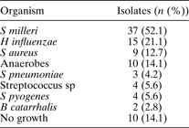
Streptococcus milleri was the commonest organism isolated, being found in 37 of the 71 patients (52.1 per cent; p = 0.0456).
Notably, no bacteria were cultured for 10 patients. This may be related to antibiotic treatment prior to admission.
All patients underwent a surgical procedure; some had combined ENT and neurosurgical procedures. Five patients underwent dental extractions, suggesting dental caries to be the source of infection.
A total of 84 ENT surgical procedures was performed in the 71 patients, an average of 1.2 procedures per patient. The ENT procedures included antral washout, incision and drainage of lid abscess, drainage of subperiosteal abscess, drainage of pus associated with Pott's puffy tumour, and trephination of the frontal sinus(es). The 30 neurosurgical procedures performed, on 14 patients, essentially involved evacuation of pus via either burr holes or craniotomy. Redrainage of pus via the original operative site was frequently required, and the average number of operations per patient was 2.14.
Patients' lengths of hospital stay ranged from two to 50 days (average 9.8 days). Patients from whom S milleri was isolated stayed an average of seven days, while those with other organisms stayed an average of five days, a marginally significant difference (p = 0.055). Patients with intracranial complications, who required multiple operative procedures, stayed longest in hospital, particularly paediatric patients. One adult male patient was admitted twice in two months with orbital cellulitis. One female paediatric patient was admitted twice, a year apart, with acute rhinosinusitis. Streptococcus milleri was isolated in both patients.
Discussion
Acute rhinosinusitis comprises acute bacterial infection of the nose and sinuses. It is commonly associated with upper respiratory tract infection due to viruses such as adenovirus, rhinovirus, influenza virus and parainfluenza virus. It is not known for certain whether the viral infection precedes or is concurrent with the bacterial infection. The mechanisms whereby virus infection predisposes to sinusitis may involve: microbial synergy; induction of local inflammation that blocks the sinus ostia; increase of bacterial attachment to the epithelial cells; and disruption of local immune defences.Reference Brook2, Reference Brook and Frazier3 Sinusitis can be precipitated by apicitis of maxillary teeth, with infection first involving the maxillary sinus and then spreading to other sinuses if not controlled. The common symptoms are headache, purulent nasal discharge, fever, and facial pain with or without swelling. The common clinical signs are pyrexia, facial tenderness with or without periorbital cellulitis, fetor, and identification of pus in the middle meatus.
In recent articles by BrookReference Brook2 and Brook and Frazier,Reference Brook and Frazier3 the causative organisms usually implicated in acute rhinosinusitis were H influenzae, S pneumoniae, Moraxella catarrhalis, S aureus and S pyogenes, and in recurrent acute rhinosinusitis were S pneumoniae, followed by H influenzae, then M catarrhalis. In these reports, S milleri was not mentioned.
In a 1997 review of sinusitis complications by Giannoni et al.,Reference Giannoni, Stewart and Alfred4 of 203 patients seen over a 10-year period, 19 suffered intracranial complications: cerebral abscess (five patients), epidural abscess (five), meningitis (five) and subdural abscess (four). The organisms isolated were streptococcus sp, S aureus, H influenzae, Escherichia coli, Proteus mirabilis, enterobacter and anaerobes. Streptococcus milleri was not isolated, but the streptococcus species were not differentiated, and four patients with these as the isolated organism had recurrence of intracranial abscesses.
In a recent report of intracranial complications of rhinosinusitis by Jones et al.,Reference Jones, Walker, Bassi, Jones and Punt5S milleri was the most common organism, being implicated in 33 of 47 patients. In 11 patients, it was found in association with other organisms: bacteroides and anaerobes (five patients), eikenella and bacteroides (one), S epidermidis (one), Gram-positive cocci (two), S aureus (one), and a microaerophilic streptococcus (one). No organisms were cultured in eight cases.
In the present series, the most commonly isolated organism was S milleri (37/71; 52.1 per cent; p = 0.046, i.e. marginally significant), followed by H influenzae and then S aureus. In the two patients with recurrent sinusitis, S milleri was isolated on both occasions. Anaerobes were isolated in 10 patients (14 per cent), being commonly found together with other organisms.
• Although Streptococcus milleri has been known to cause sepsis in other parts of the body, it has infrequently been reported as a cause of sinus infection
• Streptococcus milleri was a significant pathogen in this series of patients with acute rhinosinusitis with complications, being isolated in 52.1 per cent of cases
• These patients tended to require multiple surgical procedures, both ENT and neurosurgical
The intracranial complications occurring in this series (see Table II) are comparable with those reported by Giannoni et al. Reference Giannoni, Stewart and Alfred4Streptococcus milleri was isolated in nine of the 14 patients, both from the sinuses and the intracranial sepsis. Streptococcus milleri was found significantly more frequently than other organisms (p = 0.0144). For three of the four remaining patients, the organisms isolated were H influenzae with S pyogenes (one patient), group C streptococcus with an anaerobic bacillus (one) and S aureus with S pyogenes (one); in the fourth patient (with meningitis), lumbar puncture results were suggestive of tuberculous meningitis. Culture from the sinuses of this patient showed no growth; however, the patient had received empirical antibiotics.
It is not clear why S milleri was the commonest organism isolated in this series of patients. Factors may have included previous antibiotic treatment, immunisation against H influenzae, improved laboratory techniques (with speciation of streptococcus) or a change in the pathogens involved in acute rhinosinusitis.
Despite in vitro S milleri sensitivity to the antibiotics given, pus tended to recollect, especially intracranially. The reasons for this are not clear. Possibly, it may be due to the virulence of S milleri in relation to intracranial tissues; at present, the degree of such virulence is unknown. On admission, all our patients were commenced on empirical antibiotics (ampicillin, cloxacillin and metronidazole); if intracranial sepsis was present, a third generation cephalosporin (cefuroxime) was added. Antibiotics were altered according to culture and sensitivity results.
Five of our patients required dental extractions as part of their sinusitis treatment; in these cases, dental sepsis may have been the origin of the sinusitis. In dental caries, enamel is affected first; infection then spreads to the pulp, resulting in damage to the pulp vessels. If this remains untreated, infection extends through the roots of decayed teeth into the alveolar bone, then into the sinus. These infections can extend to the orbit, but spread to the buccal area is more common.Reference Mehra and Murad6
Conclusion
Although S milleri has been known to cause sepsis in other parts of the body, it has infrequently been reported as a cause of sinus infection. Streptococcus milleri was a significant pathogen in our series of patients with acute rhinosinusitis with complications, being isolated in 52.1 per cent of cases. Such patients were significantly more likely to require multiple surgical procedures, both ENT (p = 0.0176) and neurosurgical (p = 0.0144). Patients with intracranial complications required longer hospitalisation (up to 50 days); hospital stay was longest in those who required multiple surgical procedures.


