Hostname: page-component-76fb5796d-vvkck Total loading time: 0 Render date: 2024-04-25T19:01:55.584Z Has data issue: false hasContentIssue false

Cellular-Resolution Imaging of Microstructures in Rat Bone using Laboratory Propagation-Based Phase-Contrast X-ray Tomography

Published online by Cambridge University Press:  10 August 2018

Ilian Haggmark*
Affiliation:
Dept. of Applied Physics, KTH Royal Institute of Technology, Stockholm, Sweden
Jenny Romell
Affiliation:
Dept. of Applied Physics, KTH Royal Institute of Technology, Stockholm, Sweden
Susanne Lewin
Affiliation:
Dept. of Engineering Sciences, Uppsala University, Uppsala, Sweden
Caroline Ohman
Affiliation:
Dept. of Engineering Sciences, Uppsala University, Uppsala, Sweden
Hans M. Hertz
Affiliation:
Dept. of Applied Physics, KTH Royal Institute of Technology, Stockholm, Sweden
*
*Corresponding author, ilian.haggmark@biox.kth.se

Abstract

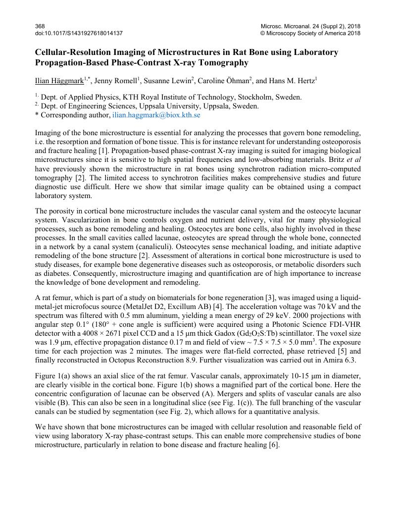
Image of the first page of this content. For PDF version, please use the ‘Save PDF’ preceeding this image.'
Type
Abstract
Copyright
© Microscopy Society of America 2018 

References

[1] Maggiano, I S, et al, J. Anat. 228 2016) p. 719.Google Scholar
[2] Britz, H M, et al, Bone 51 2012) p. 913.CrossRefGoogle Scholar
[3] Hulsart-Billstrom, G, et al, J. Tissue Eng Regen Med 9 2012) p. 799.CrossRefGoogle Scholar
[4] Hemberg, O, et al, Appl. Phys. Lett. 83 2003) p. 1483.Google Scholar
[5] Paganin, D, et al, J. Microsc. 207 2002) p. 33.Google Scholar
[6] The authors acknowledge funding from the Swedish Research Council and the Wallenberg Foundation.Google Scholar