


No CrossRef data available.
Article contents
Correlative Site-Specific Sample Preparation for Atom Probe Tomography on Complex Microstructures
Published online by Cambridge University Press: 03 June 2021
Abstract
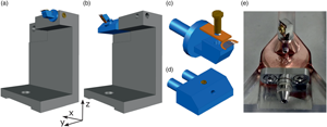
Site-specific specimen preparation for atom probe tomography (APT) is a challenging task. Small features need to be located using a suitable imaging technique and captured within a volume of less than 0.01 μm3. Correlative microscopy has shown to be helpful for target preparation as well as to gain complementary information about the material. Current strategies developed in that direction can be highly time-consuming and not always ensure the correct site extraction in complex microstructures. In this work, we present a methodology to study grain boundaries and interfaces in martensitic steels by combining electron backscattered diffraction, transmission Kikuchi diffraction (TKD), and APT. Furthermore, we include the design of a sample holder that allows to perform TKD and scanning transmission electron microscopy on the specimen during preparation without breaking the vacuum of the scanning electron microscope/focused ion beam workstation. We show a case study where a prior austenite grain boundary is traced from the bulk material to the apex of the APT specimen. The presence of contamination due to the specimen exposure to the electron beam and the use of plasma cleaning to minimize it are discussed.
Keywords
- Type
- Development and Computation
- Information
- Copyright
- Copyright © The Author(s), 2021. Published by Cambridge University Press on behalf of the Microscopy Society of America
References
Araullo-Peters, VJ, Gault, B, Shrestha, SL, Yao, L, Moody, MP, Ringer, SP & Cairney, JM (2012). Atom probe crystallography: Atomic-scale 3-D orientation mapping. Scr Mater 66, 907–910. doi:10.1016/j.scriptamat.2012.02.022.CrossRefGoogle Scholar
Arslan, I, Marquis, EA, Homer, M, Hekmaty, MA & Bartelt, NC (2008). Towards better 3-D reconstructions by combining electron tomography and atom-probe tomography. Ultramicroscopy 108, 1579–1585.CrossRefGoogle ScholarPubMed
Babinsky, K, De Kloe, R, Clemens, H & Primig, S (2014). A novel approach for site-specific atom probe specimen preparation by focused ion beam and transmission electron backscatter diffraction. Ultramicroscopy 144, 9–18.CrossRefGoogle ScholarPubMed
Baik, SI, Isheim, D & Seidman, DN (2018). Systematic approaches for targeting an atom-probe tomography sample fabricated in a thin TEM specimen: Correlative structural, chemical and 3-D reconstruction analyses. Ultramicroscopy 184, 284–292.CrossRefGoogle Scholar
Bhadeshia, H & Honeycombe, R (2017). Steels: Microstructure and Properties, 4th ed. Kidlington, UK: Butterworth-Heinemann.Google Scholar
CAMECA (2019). Technical Note TN-09 Transmission EBSD of APT Specimens. Available at https://www.edax.com/-/media/ametekedax/files/ebsd/technical_notes/cameca_tn_transmission_ebsd_of_apt_specimens.pdf?dmc=1&la=en.Google Scholar
Cayron, C (2007). ARPGE: A computer program to automatically reconstruct the parent grains from electron backscatter diffraction data. J Appl Crystallogr 40, 1183–1188.CrossRefGoogle ScholarPubMed
Danoix, F, Sauvage, X, Huin, D, Germain, L & Gouné, M (2016). A direct evidence of solute interactions with a moving ferrite/austenite interface in a model Fe-C-Mn alloy. Scr Mater 121, 61–65. doi:10.1016/j.scriptamat.2016.04.038.CrossRefGoogle Scholar
Diercks, DR & Gorman, BP (2018). Self-consistent atom probe tomography reconstructions utilizing electron microscopy. Ultramicroscopy 195, 32–46. doi:10.1016/j.ultramic.2018.08.019.CrossRefGoogle ScholarPubMed
Diercks, DR, Tong, J, Zhu, H, Kee, R, Baure, G, Nino, JC, O'Hayre, R & Gorman, BP (2016). Three-dimensional quantification of composition and electrostatic potential at individual grain boundaries in doped ceria. J Mater Chem A 4, 5167–5175.CrossRefGoogle Scholar
Dmitrieva, O, Ponge, D, Inden, G, Millán, J, Choi, P, Sietsma, J & Raabe, D (2011). Chemical gradients across phase boundaries between martensite and austenite in steel studied by atom probe tomography and simulation. Acta Mater 59, 364–374.CrossRefGoogle Scholar
Egerton, RF, Li, P & Malac, M (2004). Radiation damage in the TEM and SEM. Micron 35, 399–409.CrossRefGoogle ScholarPubMed
Ennos, AE (1954). The sources of electron-induced contamination in kinetic vacuum systems. Br J Appl Phys 5, 27–31.CrossRefGoogle Scholar
Felfer, PJ, Alam, T, Ringer, SP & Cairney, JM (2012). A reproducible method for damage-free site-specific preparation of atom probe tips from interfaces. Microsc Res Tech 75, 484–491.CrossRefGoogle ScholarPubMed
Felfer, PJ, Ringer, SP & Cairney, JM (2011). Shaping the lens of the atom probe: Fabrication of site specific, oriented specimens and application to grain boundary analysis. Ultramicroscopy 111, 435–439. doi:10.1016/j.ultramic.2010.12.005.CrossRefGoogle ScholarPubMed
Furuhara, T, Morito, S & Maki, T (2003). Morphology, substructure and crystallography of lath martensite in Fe-C alloys. J Phys, IV 112, 255–258.Google Scholar
Gault, B, Loi, ST, Araullo-Peters, VJ, Stephenson, LT, Moody, MP, Shrestha, SL, Marceau, RKW, Yao, L, Cairney, JM & Ringer, SP (2011). Dynamic reconstruction for atom probe tomography. Ultramicroscopy 111, 1619–1624.CrossRefGoogle ScholarPubMed
Gault, B, Moody, MP, Cairney, JM & Ringer, SP (2012). Atom probe crystallography. Mater Today 15, 378–386. doi:10.1016/S1369-7021(12)70164-5.CrossRefGoogle Scholar
Gault, B, Moody, MP, De Geuser, F, La Fontaine, A, Stephenson, LT, Haley, D & Ringer, SP (2010). Spatial resolution in atom probe tomography. Microsc Microanal 16, 99–110.CrossRefGoogle ScholarPubMed
Gault, B, Moody, MP, De Geuser, F, Tsafnat, G, La Fontaine, A, Stephenson, LT, Haley, D & Ringer, SP (2009). Advances in the calibration of atom probe tomographic reconstruction. J Appl Phys 105, 034913.CrossRefGoogle Scholar
Gorman, BP, Diercks, D, Salmon, N, Stach, E, Amador, G & Hartfield, C (2008). Hardware and techniques for cross-correlative TEM and atom probe analysis. Microsc Today 16, 42–47.CrossRefGoogle Scholar
Griffiths, AJV & Walther, T (2010). Quantification of carbon contamination under electron beam irradiation in a scanning transmission electron microscope and its suppression by plasma cleaning. J Phys: Conf Ser 241, 012017.Google Scholar
Haley, D, Petersen, T, Ringer, SP & Smith, GDW (2011). Atom probe trajectory mapping using experimental tip shape measurements. J Microsc 244, 170–180. doi:10.1111/j.1365-2818.2011.03522.x.CrossRefGoogle ScholarPubMed
Hartshorne, MI, Isheim, D, Seidman, DN & Taheri, ML (2014). Specimen preparation for correlating transmission electron microscopy and atom probe tomography of mesoscale features. Ultramicroscopy 147, 25–32. doi:10.1016/j.ultramic.2014.05.005.CrossRefGoogle ScholarPubMed
Herbig, M, Choi, P & Raabe, D (2015). Combining structural and chemical information at the nanometer scale by correlative transmission electron microscopy and atom probe tomography. Ultramicroscopy 153, 32–39. doi:10.1016/j.ultramic.2015.02.003.CrossRefGoogle ScholarPubMed
Herbig, M & Kumar, A (2020). Removal of hydrocarbon contamination and oxide films from atom probe specimens. Microsc Res Tech 84(2), 291–297.CrossRefGoogle ScholarPubMed
Hutchinson, B, Hagström, J, Karlsson, O, Lindell, D, Tornberg, M, Lindberg, F & Thuvander, M (2011). Microstructures and hardness of as-quenched martensites (0.1-0.5%C). Acta Mater 59, 5845–5858.CrossRefGoogle Scholar
Jenkins, BM, Douglas, JO, Gardner, H, Tweddle, D, Kareer, A, Karamached, PS, Riddle, N, Hyde, JM, Bagot, PAJ, Robert, G & Moody, MP (2020). A more holistic characterisation of internal interfaces in a variety of materials via complementary use of transmission Kikuchi diffraction and atom probe tomography. Appl Surf Sci 528, 147011. doi:10.1016/j.apsusc.2020.147011.CrossRefGoogle Scholar
Kelly, TF & Miller, MK (2007). Invited review article: Atom probe tomography. Rev Sci Instrum 78 (3), 031101.CrossRefGoogle ScholarPubMed
Kuzmina, M, Ponge, D & Raabe, D (2015). Grain boundary segregation engineering and austenite reversion turn embrittlement into toughness: Example of a 9 wt% medium Mn steel. Acta Mater 86, 182–192.CrossRefGoogle Scholar
Lefebvre-Ulrikson, W, Vurpillot, F & Sauvage, X (Eds.) (2016). Atom Probe Tomography: Put Theory Into Practice. London, UK: Academic Press.Google Scholar
Marquis, EA, Choi, P-P, Danoix, F, Kruska, K, Lozano-Perez, S, Ponge, D, Raabe, D & Williams, CA (2012). New insights into the atomic-scale structures and behavior of steels. Microsc Today 20, 44–48.CrossRefGoogle Scholar
Marquis, EA, Geiser, BP, Prosa, TJ & Larson, DJ (2011). Evolution of tip shape during field evaporation of complex multilayer structures. J Microsc 241, 225–233.CrossRefGoogle ScholarPubMed
Marquis, EA & Hyde, JM (2010). Applications of atom-probe tomography to the characterisation of solute behaviours. Mater Sci Eng R 69, 37–62. doi:10.1016/j.mser.2010.05.001.CrossRefGoogle Scholar
Miller, MK, Russell, KF & Thompson, GB (2005). Strategies for fabricating atom probe specimens with a dual beam FIB. Ultramicroscopy 102, 287–298.CrossRefGoogle ScholarPubMed
Mitchell, DRG (2015). Contamination mitigation strategies for scanning transmission electron microscopy. Micron 73, 36–46. doi:10.1016/j.micron.2015.03.013.CrossRefGoogle ScholarPubMed
Moody, MP, Gault, B, Stephenson, LT, Haley, D & Ringer, SP (2009). Qualification of the tomographic reconstruction in atom probe by advanced spatial distribution map techniques. Ultramicroscopy 109, 815–824.CrossRefGoogle ScholarPubMed
Morsdorf, L, Tasan, CC, Ponge, D & Raabe, D (2015). 3D structural and atomic-scale analysis of lath martensite: Effect of the transformation sequence. Acta Mater 95, 366–377. doi:10.1016/j.actamat.2015.05.023.CrossRefGoogle Scholar
Prosa, TJ & Larson, DJ (2017). Modern focused-ion-beam-based site-specific specimen preparation for atom probe tomography. Microsc Microanal 23, 194–209.CrossRefGoogle ScholarPubMed
Prosa, TJ, Olson, D, Geiser, B, Larson, DJ, Henry, K & Steel, E (2013). Analysis of implanted silicon dopant profiles. Ultramicroscopy 132, 179–185. doi:10.1016/j.ultramic.2012.10.005.CrossRefGoogle ScholarPubMed
Rice, KP, Chen, Y, Prosa, TJ & Larson, DJ (2016). Implementing transmission electron backscatter diffraction for atom probe tomography. Microsc Microanal 22, 583–588.CrossRefGoogle ScholarPubMed
Seidman, DN (2007). Three-dimensional atom-probe tomography: Advances and applications. Annu Rev Mater Res 37, 127–158.CrossRefGoogle Scholar
Stoffers, A, Barthel, J, Liebscher, CH, Gault, B, Cojocaru-Mirédin, O, Scheu, C & Raabe, D (2017). Correlating atom probe tomography with atomic-resolved scanning transmission electron microscopy: Example of segregation at silicon grain boundaries. Microsc Microanal 23, 291–299.CrossRefGoogle ScholarPubMed
Thompson, K, Lawrence, D, Larson, DJ, Olson, JD, Kelly, TF & Gorman, B (2007). In situ site-specific specimen preparation for atom probe tomography. Ultramicroscopy 107, 131–139.CrossRefGoogle ScholarPubMed
Vurpillot, F, Cerezo, A, Blavette, D & Larson, DJ (2004). Modeling image distortions in 3DAP. Microsc Microanal 10, 384–390.CrossRefGoogle ScholarPubMed
Zhang, J, Morsdorf, L & Tasan, CC (2016). Multi-probe microstructure tracking during heat treatment without an in-situ setup: Case studies on martensitic steel, dual phase steel and β-Ti alloy. Mater Charact 111, 137–146. doi:10.1016/j.matchar.2015.11.019.CrossRefGoogle Scholar
Campo Schneider et al. supplementary material
Campo Schneider et al. supplementary material 1

PDF
33.6 KB
Campo Schneider et al. supplementary material
Campo Schneider et al. supplementary material 2
PDF
276.4 KB
Campo Schneider et al. supplementary material
Campo Schneider et al. supplementary material 3
PDF
565.2 KB
Campo Schneider et al. supplementary material
Campo Schneider et al. supplementary material 4
PDF
581.5 KB
Campo Schneider et al. supplementary material
Campo Schneider et al. supplementary material 5
PDF
677.8 KB