Hostname: page-component-76fb5796d-wq484 Total loading time: 0 Render date: 2024-04-28T00:11:24.466Z Has data issue: false hasContentIssue false

Electron Crystallographic Investigation of Crystals on the Mesostructural Scale

Published online by Cambridge University Press:  30 June 2021

Wenting Mao
Affiliation:
School of Chemistry and Chemical Engineering, State Key Laboratory of Metal Matrix Composites, Shanghai Key Laboratory for Molecular Engineering of Chiral Drugs, Shanghai Jiao Tong University, Shanghai200240, China
Chao Bao
Affiliation:
School of Chemistry and Chemical Engineering, State Key Laboratory of Metal Matrix Composites, Shanghai Key Laboratory for Molecular Engineering of Chiral Drugs, Shanghai Jiao Tong University, Shanghai200240, China
Lu Han*
Affiliation:
School of Chemical Science and Engineering, Tongji University, Shanghai200092, China
*
*Corresponding author: Lu Han, E-mail: luhan@tongji.edu.cn
Get access

Abstract

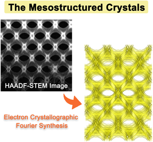
The precise structural solution of crystals on a mesostructural scale is challenging due to the difficulties in obtaining electron diffraction and the complicated relationship between the crystal structure factors (CSFs) and the conventional underfocus phase-contrast transmission electron microscopy (TEM) images due to the large unit cell and the complex structures. Here, we present the structural investigation of mesostructured crystals via the combination of electron crystallographic Fourier synthesis and high-angle annular dark-field scanning transmission electron microscopy (HAADF-STEM) that only relies on the mass-thickness contrast. The three-dimensional electrostatic potential is reconstructed from the amplitudes and phases extracted from the Fourier transforms of the corresponding HAADF-STEM images and merged into a set of CSFs. This method is verified on silica scaffolds following a shifted double-diamond surface network with space group I41/amd. The results indicate that electron crystallography reconstruction by HAADF-STEM images is more suitable and accurate in determining the structure in comparison with conventional TEM electron crystallography reconstruction. This approach transfers the contrast of mesostructured crystals to images more accurately and the relationship between the Fourier transforms of HAADF-STEM images and the CSFs is more intuitive. It shows great advantages for the structural solution of crystals on the mesostructural scale.

Type
Materials Science Applications
Copyright
Copyright © The Author(s), 2021. Published by Cambridge University Press on behalf of the Microscopy Society of America

Access options

Get access to the full version of this content by using one of the access options below. (Log in options will check for institutional or personal access. Content may require purchase if you do not have access.)

References

Alvarez, J, Saudino, G, Musteata, V, Madhavan, P, Genovese, A, Behzad, AR, Sougrat, R, Boi, C, Peinemann, KV & Nunes, SP (2019). 3D analysis of ordered porous polymeric particles using complementary electron microscopy methods. Sci Rep-Uk 9(1), 13987.CrossRefGoogle ScholarPubMed
Cao, X, Xu, DP, Yao, Y, Han, L, Terasaki, O & Che, SN (2016). Interconversion of triply periodic constant mean curvature surface structures: From double diamond to single gyroid. Chem Mater 28(11), 36913702.CrossRefGoogle Scholar
Carlsson, A, Kaneda, M, Sakamoto, Y, Terasaki, O, Ryoo, R & Joo, SH (1999). The structure of MCM-48 determined by electron crystallography. J Electron Microsc 48(6), 795798.CrossRefGoogle Scholar
Cheng, CF, Chen, YM, Zou, F, Yang, KC, Lin, TY, Liu, K, Lai, CH, Ho, RM & Zhu, Y (2018). Nanoporous gyroid Ni/NiO/C nanocomposites from block copolymer templates with high capacity and stability for lithium storage. J Mater Chem A 6(28), 1367613684.CrossRefGoogle Scholar
Chiu, PT, Chien, YC, Georgopanos, P, Sun, YS, Avgeropoulos, A & Ho, RM (2019). Examination of well ordered nanonetwork materials by real- and reciprocal-space imaging. IUCrJ 6(Pt 2), 259266.CrossRefGoogle ScholarPubMed
Chu, CY, Jiang, X, Jinnai, H, Pei, RY, Lin, WF, Tsai, JC & Chen, HL (2015). Real-space evidence of the equilibrium ordered bicontinuous double diamond structure of a diblock copolymer. Soft Matter 11(10), 18711876.CrossRefGoogle ScholarPubMed
Corkery, RW & Tyrode, EC (2017). On the colour of wing scales in butterflies: Iridescence and preferred orientation of single gyroid photonic crystals. Interface Focus 7(4), 20160154.CrossRefGoogle ScholarPubMed
Dong, ZY & Ma, YH (2020). Atomic-level handedness determination of chiral crystals using aberration-corrected scanning transmission electron microscopy. Nat Commun 11(1), 1588.CrossRefGoogle ScholarPubMed
Ercius, P, Alaidi, O, Rames, MJ & Ren, G (2015). Electron tomography: A three-dimensional analytic tool for hard and soft materials research. Adv Mater 27(38), 56385663.CrossRefGoogle ScholarPubMed
Feng, XY, Burke, CJ, Zhuo, MJ, Guo, H, Yang, KQ, Reddy, A, Prasad, I, Ho, RM, Avgeropoulos, A, Grason, GM & Thomas, EL (2019). Seeing mesoatomic distortions in soft-matter crystals of a double-gyroid block copolymer. Nature 575(7781), 175179.CrossRefGoogle ScholarPubMed
Finnemore, AS, Scherer, MRJ, Langford, R, Mahajan, S, Ludwigs, S, Meldrum, FC & Steiner, U (2009). Nanostructured calcite single crystals with gyroid morphologies. Adv Mater 21(38–39), 39283932.CrossRefGoogle Scholar
Firth, DS, Morris, SA, Wheatley, PS, Russell, SE, Slawin, AMZ, Dawson, DM, Mayoral, A, Opanasenko, M, Položij, M, Čejka, J, Nachtigall, P & Morris, RE (2017). Assembly–disassembly–organization–reassembly synthesis of zeolites based on cfi-yype layers. Chem Mater 29(13), 56055611.CrossRefGoogle Scholar
Galusha, JW, Jorgensen, MR & Bartl, MH (2010). Diamond-structured yitania photonic-bandgap crystals from biological templates. Adv Mater 22(1), 107110.CrossRefGoogle ScholarPubMed
Galusha, JW, Richey, LR, Gardner, JS, Cha, JN & Bartl, MH (2008). Discovery of a diamond-based photonic crystal structure in beetle scales. Phys Rev E 77(5), 050904.CrossRefGoogle ScholarPubMed
Gandy, PJF, Bardhan, S, Mackay, AL & Klinowski, J (2001). Nodal surface approximations to the P, G, D and I-WP triply periodic minimal surfaces. Chem Phys Lett 336(3-4), 187195.CrossRefGoogle Scholar
Gao, CB, Sakamoto, Y, Sakamoto, K, Terasaki, O & Che, SN (2006). Synthesis and characterization of mesoporous silica AMS-10 with bicontinuous cubic Pn-3 m symmetry. Angew Chem Int Edit 45(26), 42954298.CrossRefGoogle Scholar
Goris, B, Roelandts, T, Batenburg, KJ, Heidari Mezerji, H & Bals, S (2013). Advanced reconstruction algorithms for electron tomography: From comparison to combination. Ultramicroscopy 127, 4047.CrossRefGoogle ScholarPubMed
Goris, B, Van den Broek, W, Batenburg, KJ, Heidari Mezerji, H & Bals, S (2012). Electron tomography based on a total variation minimization reconstruction technique. Ultramicroscopy 113, 120130.CrossRefGoogle Scholar
Han, L, Miyasaka, K, Terasaki, O & Che, SN (2011). Evolution of packing parameters in the structural changes of silica mesoporous crystals: Cage-type, 2D cylindrical, bicontinuous diamond and gyroid, and lamellar. J Am Chem Soc 133(30), 1152411533.CrossRefGoogle ScholarPubMed
Han, L, Ohsuna, T, Liu, Z, Alfredsson, V, Kjellman, T, Asahina, S, Suga, M, Ma, YH, Oleynikov, P, Miyasaka, K, Mayoral, A, Diaz, I, Sakamoto, Y, Stevens, SM, Anderson, MW, Xiao, CH, Fujita, N, Garcia-Bennett, A, Yoon, KB, Che, SN & Terasaki, O (2014 a). Structures of silica-based nanoporous materials revealed by microscopy. Z Anorg Allg Chem 640(3-4), 521536.CrossRefGoogle Scholar
Han, L, Xu, DP, Liu, Y, Ohsuna, T, Yao, Y, Jiang, C, Mai, YY, Cao, YY, Duan, YY & Che, SN (2014 b). Synthesis and characterization of macroporous photonic structure that consists of azimuthally shifted double-diamond silica frameworks. Chem Mater 26(24), 70207028.CrossRefGoogle Scholar
Han, Y, Zhang, DL, Chng, LL, Sun, JL, Zhao, L, Zou, XD & Ying, JY (2009). A tri-continuous mesoporous material with a silica pore wall following a hexagonal minimal surface. Nat Chem 1(2), 123127.CrossRefGoogle ScholarPubMed
Hovmöller, S (1992). CRISP: Crystallographic image processing on a personal computer. Ultramicroscopy 41(1), 121135.CrossRefGoogle Scholar
Hsueh, HY, Ling, YC, Wang, HF, Chien, LYC, Hung, YC, Thomas, EL & Ho, RM (2014). Shifting networks to achieve subgroup symmetry properties. Adv Mater 26(20), 32253229.CrossRefGoogle ScholarPubMed
Hu, JJ, Li, FH & Fan, HF (1992). Crystal structure determination of K2O⋅7Nb2O5 by combining high-resolution electron microscopy and electron diffraction. Ultramicroscopy 41(4), 387397.CrossRefGoogle Scholar
Hua, W, Chen, H, Yu, ZB, Zou, XD, Lin, JH & Sun, JL (2014). A germanosilicate structure with 11(11(12-ring channels solved by electron crystallography. Angew Chem Int Edit 53(23), 58685871.CrossRefGoogle Scholar
Jiang, Y, Hanwell, MD, Padgett, E, Waldon, S, Muller, DA & Hovden, R (2016). Advanced platform for 3D visualization, reconstruction, and segmentation with electron tomography. Micros Microanal 22(S3), 20702071.CrossRefGoogle Scholar
Jinnai, H, Higuchi, T, Zhuge, XD, Kumamoto, A, Batenburg, KJ & Ikuhara, Y (2017). Three-dimensional visualization and characterization of polymeric self-assemblies by transmission electron microtomography. Accounts Chem Res 50(6), 12931302.CrossRefGoogle ScholarPubMed
Kapaca, E, Zou, XD & Willhammar, T (2017). Detailed structural survey of the zeolite ITQ-39 by electron crystallography. Cryst Growth Des 17(4), 19101917.CrossRefGoogle Scholar
Kim, E, Vaynzof, Y, Sepe, A, Guldin, S, Scherer, M, Cunha, P, Roth, SV & Steiner, U (2014). Gyroid-structured 3D ZnO networks made by atomic layer deposition. Adv Funct Mater 24(6), 863872.CrossRefGoogle Scholar
Kübel, C, Voigt, A, Schoenmakers, R, Otten, M, Su, D, Lee, T-C, Carlsson, A & Bradley, J (2005). Recent advances in electron tomography: TEM and HAADF-STEM tomography for materials science and semiconductor applications. Microsc Microanal 11(5), 378400.CrossRefGoogle ScholarPubMed
La, YJ, Song, J, Jeong, MG, Cho, A, Jin, SM, Lee, E & Kim, KT (2018). Templated synthesis of cubic crystalline single networks having large open-space lattices by polymer cubosomes. Nat Communs 9(1), 5327.CrossRefGoogle ScholarPubMed
Li, H, Liu, Y, Cao, X, Han, L, Jiang, C & Che, S (2017). A shifted double-diamond titania scaffold. Angew Chem Int Edit 56(3), 806811.CrossRefGoogle ScholarPubMed
Lin, ZX, Zhou, JJ, Cortez-Jugo, C, Han, YY, Ma, YT, Pan, SJ, Hanssen, E, Richardson, JJ & Caruso, F (2020). Ordered mesoporous metal-phenolic network particles. J Am Chem Soc 142(1), 335341.CrossRefGoogle ScholarPubMed
Linton, P, Hernandez-Garrido, JC, Midgley, PA, Wennerstrom, H & Alfredsson, V (2009). Morphology of SBA-15-directed by association processes and surface energies. Phys Chem Chem Phys 11(46), 1097310982.CrossRefGoogle ScholarPubMed
Liu, GS, House, SD, Kacher, J, Tanaka, M, Higashida, K & Robertson, IM (2014). Electron tomography of dislocation structures. Maters Charact 87, 111.CrossRefGoogle Scholar
Liu, LM, Chen, ZJ, Wang, JJ, Zhang, DL, Zhu, YH, Ling, SL, Huang, KW, Belmabkhout, Y, Adil, K, Zhang, YX, Slater, B, Eddaoudi, M & Han, Y (2019). Imaging defects and their evolution in a metal-organic framework at sub-unit-cell resolution. Nat Chem 11(7), 622628.CrossRefGoogle Scholar
Mao, WT, Cao, X, Sheng, QQ, Han, L & Che, SA (2017). Silica scaffold with shifted “plumber's nightmare” networks and their interconversion into diamond networks. Angew Chem Int Edit 56(36), 1067010675.CrossRefGoogle ScholarPubMed
Mayoral, A, Anderson, PA & Diaz, I (2015). Zeolites are no longer a challenge: Atomic resolution data by aberration-corrected STEM. Micron 68, 146151.CrossRefGoogle ScholarPubMed
Mayoral, A, Carey, T, Anderson, PA, Lubk, A & Diaz, I (2011). Atomic resolution analysis of silver ion-exchanged zeolite A. Angew Chem Int Edit 50(47), 1123011233.CrossRefGoogle ScholarPubMed
Midgley, PA & Dunin-Borkowski, RE (2009). Electron tomography and holography in materials science. Nat Mater 8(4), 271280.CrossRefGoogle ScholarPubMed
Midgley, PA & Weyland, M (2003). 3D electron microscopy in the physical sciences: The development of Z-contrast and EFTEM tomography. Ultramicroscopy 96(3), 413431.CrossRefGoogle ScholarPubMed
Mille, C, Tyrode, EC & Corkery, RW (2011). Inorganic chiral 3-D photonic crystals with bicontinuous gyroid structure replicated from butterfly wing scales. Chem Commun 47(35), 98739875.CrossRefGoogle ScholarPubMed
Mille, C, Tyrode, EC & Corkery, RW (2013). 3D titania photonic crystals replicated from gyroid structures in butterfly wing scales: Approaching full band gaps at visible wavelengths. Rsc Adv 3(9), 31093117.CrossRefGoogle Scholar
Momma, K & Izumi, F (2011). VESTA 3 for three-dimensional visualization of crystal, volumetric and morphology data. J Appl Crystallogr 44, 12721276.CrossRefGoogle Scholar
Ohsuna, T, Sakamoto, Y, Terasaki, O & Kuroda, K (2011). TEM image simulation of mesoporous crystals for structure type identification. Solid State Sci 13(4), 736744.CrossRefGoogle Scholar
Oleynikov, P (2011). Emap and eSlice: A software package for crystallographic computing. Cryst Res Technol 46(6), 569579.CrossRefGoogle Scholar
Pryor, A, Yang, Y, Rana, A, Gallagher-Jones, M, Zhou, JH, Lo, YH, Melinte, G, Chiu, W, Rodriguez, JA & Miao, JW (2017). GENFIRE: A generalized Fourier iterative reconstruction algorithm for high-resolution 3D imaging. Sci Rep-Uk 7, 10409.CrossRefGoogle ScholarPubMed
Saghi, Z & Midgley, PA (2012). Electron tomography in the (S)TEM: From nanoscale morphological analysis to 3D atomic imaging. Annu Rev Mater Res 42(1), 5979.CrossRefGoogle Scholar
Sakamoto, Y, Kim, TW, Ryoo, R & Terasaki, O (2004). Three-dimensional structure of large-pore mesoporous cubic Ia-3d silica with complementary pores and its carbon replica by electron crystallography. Angew Chem Int Edit 43(39), 52315234.CrossRefGoogle Scholar
Saranathan, V, Osuji, CO, Mochrie, SGJ, Noh, H, Narayanan, S, Sandy, A, Dufresne, ER & Prum, RO (2010). Structure, function, and self-assembly of single network gyroid (I4132) photonic crystals in butterfly wing scales. Proc Natl Acad Sci USA 107(26), 1167611681.CrossRefGoogle ScholarPubMed
Scott, MC, Chen, C-C, Mecklenburg, M, Zhu, C, Xu, R, Ercius, P, Dahmen, U, Regan, BC & Miao, J (2012). Electron tomography at 2.4-ångström resolution. Nature 483(7390), 444447.CrossRefGoogle ScholarPubMed
Sundaramoorthi, G, Hadwiger, M, Ben-Romdhane, M, Behzad, AR, Madhavan, P & Nunes, SP (2016). 3D membrane imaging and porosity visualization. Ind Eng Chem Res 55(12), 36893695.CrossRefGoogle Scholar
Terasaki, O, Ohsuna, T, Alfredson, V, Bovin, JO, Watanabe, D & Tsuno, K (1991). The study of zeolites by HVHREM. Ultramicroscopy 39(1), 238246.CrossRefGoogle Scholar
Van den Broek, W, Rosenauer, A, Goris, B, Martinez, GT, Bals, S, Van Aert, S & Van Dyck, D (2012). Correction of non-linear thickness effects in HAADF STEM electron tomography. Ultramicroscopy 116, 812.CrossRefGoogle Scholar
Wang, B, Rhauderwiek, T, Inge, AK, Xu, HY, Yang, TM, Huang, ZH, Stock, N & Zou, XD (2018). A porous cobalt tetraphosphonate metal-organic framework: Accurate structure and guest molecule location determined by continuous-rotation electron diffraction. Chem-Eur J 24(66), 1742917433.CrossRefGoogle ScholarPubMed
Wang, DN, Hovmöller, S, Kihlborg, L & Sundberg, M (1988). Structure determination and correction for distortions in HREM by crystallographic image processing. Ultramicroscopy 25(4), 303316.CrossRefGoogle Scholar
Wang, YC, Wakabayashi, M, Hasegawa, H & Takenaka, M (2017). 3D-TEM study on the novel bicontinuous microdomain structure. Soft Matter 13(46), 88248828.CrossRefGoogle Scholar
Wen, T, Wang, HF, Georgopanos, P, Avgeropoulo, A & Ho, RM (2019). Three-dimensional visualization of phase transition in polystyrene-block-polydimethylsiloxane thin film. Polymer 167, 209214.CrossRefGoogle Scholar
Werner, JG, Scherer, MRJ, Steiner, U & Wiesner, U (2014). Gyroidal mesoporous multifunctional nanocomposites via atomic layer deposition. Nanoscale 6(15), 87368742.CrossRefGoogle ScholarPubMed
Wikander, K, Hungria, AB, Midgley, PA, Palmqvist, AEC, Holmberg, K & Thomas, JM (2007). Incorporation of platinum nanoparticles in ordered mesoporous carbon. J Colloid Interf Sci 305(1), 204208.CrossRefGoogle ScholarPubMed
Willhammar, T, Mayoral, A & Zou, X (2014). 3D reconstruction of atomic structures from high angle annular dark field (HAADF) STEM images and its application on zeolite silicalite-1. Dalton T 43(37), 1415814163.CrossRefGoogle ScholarPubMed
Willhammar, T, Sun, JL, Wan, W, Oleynikov, P, Zhang, DL, Zou, XD, Moliner, M, Gonzalez, J, Martinez, C, Rey, F & Corma, A (2012). Structure and catalytic properties of the most complex intergrown zeolite ITQ-39 determined by electron crystallography. Nat Chem 4(3), 188194.CrossRefGoogle ScholarPubMed
Wilts, BD, Michielsen, K, Kuipers, J, De Raedt, H & Stavenga, DG (2012). Brilliant camouflage: Photonic crystals in the diamond weevil, Entimus imperialis. P Roy Soc B-Biol Sci 279(1738), 25242530.Google ScholarPubMed
Wilts, BD, Zubiri, BA, Klatt, MA, Butz, B, Fischer, MG, Kelly, ST, Spiecker, E, Steiner, U & Schroder-Turk, GE (2017). Butterfly gyroid nanostructures as a time-frozen glimpse of intracellular membrane development. Sci Adv 3(4), e1603119.CrossRefGoogle ScholarPubMed
Winter, B, Butz, B, Dieker, C, Schroder-Turk, GE, Mecke, K & Spiecker, E (2015). Coexistence of both gyroid chiralities in individual butterfly wing scales of callophrys rubi. Proc Natl Acad Sci USA 112(42), 1291112916.CrossRefGoogle ScholarPubMed
Wu, L, Wang, W, Zhang, W, Su, H, Gu, J, Liu, Q, Zhang, D, Pantelić, D & Jelenković, B (2018). Optical performance study of gyroid-structured TiO2 photonic crystals replicated from natural templates using a sol-gel method. Adv Opt Mater 6(21), 1800064.CrossRefGoogle Scholar
Yang, K-C, Yao, C-T, Huang, L-Y, Tsai, J-C, Hung, W-S, Hsueh, H-Y & Ho, R-M (2019). Single gyroid-structured metallic nanoporous spheres fabricated from double gyroid-forming block copolymers via templated electroless plating. Npg Asia Mater 11(1), 9.CrossRefGoogle Scholar
Yates, TJV, Thomas, JM, Fernandez, JJ, Terasaki, O, Ryoo, R & Midgley, PA (2006). Three-dimensional real-space crystallography of MCM-48 mesoporous silica revealed by scanning transmission electron tomography. Chem Phys Lett 418(4-6), 540543.CrossRefGoogle Scholar
Zhang, YS, Zhu, C & Xia, Y (2017). Inverse opal scaffolds and their biomedical applications. Adv Mater 29(33), 1701115.CrossRefGoogle ScholarPubMed
Zhou, J, Yang, Y, Yang, Y, Kim, DS, Yuan, A, Tian, X, Ophus, C, Sun, F, Schmid, AK, Nathanson, M, Heinz, H, An, Q, Zeng, H, Ercius, P & Miao, J (2019). Observing crystal nucleation in four dimensions using atomic electron tomography. Nature 570(7762), 500503.CrossRefGoogle ScholarPubMed
Zou, X, Hovmöller, S & Oleynikov, P (2011). Electron Crystallography : Electron Microscopy and Electron Diffraction. Oxford: Oxford University Press.CrossRefGoogle Scholar
Supplementary material: File

Mao et al. supplementary material

Mao et al. supplementary material

Download Mao et al. supplementary material(File)
File 5.3 MB