Hostname: page-component-848d4c4894-pftt2 Total loading time: 0 Render date: 2024-05-19T23:39:00.158Z Has data issue: false hasContentIssue false

Low-Cost Fluorescent Microvascular Visualization in Ambystoma mexicanum

Published online by Cambridge University Press:  30 July 2021

Luke Bollinger
Affiliation:
Towson University, Towson, Maryland, United States
Renee Dickie
Affiliation:
Towson University, Towson, Maryland, United States

Abstract

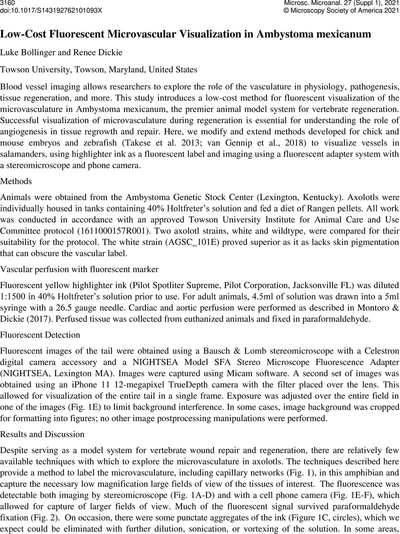
Image of the first page of this content. For PDF version, please use the ‘Save PDF’ preceeding this image.'
Type
Microscopy and Microanalysis for Real World Problem Solving
Copyright
Copyright © The Author(s), 2021. Published by Cambridge University Press on behalf of the Microscopy Society of America

References

MONTORO, R. & DICKIE, R. (2017). Comparison of tissue processing methods for microvascular visualization in axolotls. MethodsX 4:265-273Google ScholarPubMed
TAKAHASHI, T., TAKASE, Y., YOSHINO, T., SAITO, D., TADOKORO, R. & TAKAHASHI, Y. (2015). Angiogenesis in the developing spinal cord: Blood vessel exclusion from neural progenitor region is mediated by VEGF and its antagonists. PLoS ONE 10(1):e0116119Google ScholarPubMed
TAKASE, Y., TADOKORO, R. & TAKAHASHI, Y. (2013). Low cost labeling with highlighter ink efficiently visualizes developing blood vessels in avian and mouse embryos. Development Growth and Differentiation 55(9):792-801Google ScholarPubMed
VAN GENNIP, J. L. M., BOSWELL, C. W. & CIRUNA, B. (2018). Neuroinflammatory signals drive spinal curve formation in zebrafish models of idiopathic scoliosis. Science Advances 4(12):eaav1781CrossRefGoogle ScholarPubMed