


No CrossRef data available.
Article contents
Low-Voltage Electron-Probe Microanalysis of Uranium
Published online by Cambridge University Press: 18 March 2021
Abstract
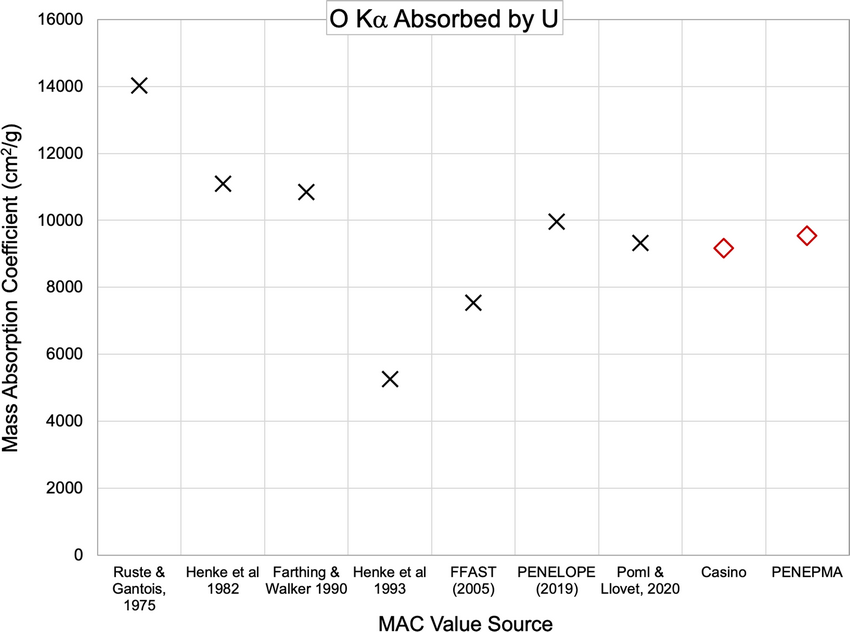
Electron-probe microanalysis of uranium and uranium alloys poses several problems, such as rapid oxidation, large poorly constrained correction factors, and a large number of characteristic x-ray lines. We show that U-metal can grow 10 nm of oxide within ~20 s of air exposure, increasing to 15–20 nm within a few minutes, which can produce a 30% quantification error at 5 kV. A 15 nm carbon coating on the UO2 reference material also produces an ~30% quantification error of the uncoated but surface oxidized U sample at 5 kV. Correcting for both the coating and oxide improved the analysis accuracy to better than ±1% down to 7 kV and ~2% at 5 kV, but the error increases strongly below this. The measurement of C in U identified a previously unreported U N6–O4 line interference on the C Kα peak, which can produce over 1% error in the analysis total. Oxide stoichiometry was demonstrated to have only a small impact on quantification. The measurement of the O Kα and U Mα mass absorption coefficients in U as 9,528 and 798 cm2/g, respectively, shows good agreement with recently published values and also produces small differences in a quantification error.
- Type
- Materials Science Applications
- Information
- Copyright
- Copyright © The Author(s), 2021. Published by Cambridge University Press on behalf of the Microscopy Society of America
References
Allen, GC, Crofts, JA & Griffiths, AJ (1976). Infrared spectroscopy of the uranium/oxygen system. J Nucl Mater 62, 273–281.CrossRefGoogle Scholar
Bastin, GF & Heijligers, HJM (1991). Nonconductive specimens in the electron probe microanalyzer—a hitherto poorly discussed problem. In Electron Probe Quantitation, Heinrich, KFJ & Newbury, DE (Eds.), pp. 163–175. New York, NY: Plenum Press.CrossRefGoogle Scholar
Bastin, GF & Heijligers, HJM (1992). Quantitative EPMA of the ultra-light elements boron through oxygen. In Electron Microbeam Analysis: Mikrochimica Acta Supplement, Boekestein, A & Pavicevic, MK (Eds.), pp. 19–36. Vienna: Springer.CrossRefGoogle Scholar
Bearden, JA (1967). X-ray wavelengths and x-ray atomic energy levels. Rev Mod Phys 39, 78–124.CrossRefGoogle Scholar
Bearden, JA & Burr, AF (1967). Reevaluation of x-ray atomic energy levels. Rev Mod Phys 39, 125–142.CrossRefGoogle Scholar
Bera, S, Sali, SK, Sampath, S, Narasimhan, SV & Venugopal, V (1998). Oxidation state of uranium: An XPS study of alkali and alkaline earth uranates. J Nucl Mater 255, 26–33.CrossRefGoogle Scholar
Bowles, JFW (1978). Quantitative microprobe analysis of uranium minerals. Microscope 26, 55–67.Google Scholar
Buse, B, Kearns, SL, Clapham, C & Hawley, D (2016). Decontamination in the electron probe microanalysis with a Peltier-cooled cold finger. Microsc Microanal 22, 981–986.CrossRefGoogle ScholarPubMed
Busker, G (2002). Solution and Migration of Impurity Ions in UO2, U3O8 and Y2O3. London: Imperial College. Available at http://abulafia.mt.ic.ac.uk/publications/theses/busker/.Google Scholar
Chernia, Z, Ben-Eliyahu, Y, Kimmel, G, Braun, G & Sariel, J (2006). The initial stage of uranium oxidation: Mechanism of UO2 scale formation in the presence of a native lateral stress field. J Phys Chem B 110, 23041–23051.CrossRefGoogle Scholar
Colby, JW (1963). Electron Microprobe Examination of Uranium.Cincinnati.National Lead Company of Ohio Inc.Google Scholar
Colby, JW (1966). The applicability of theoretically calculated intensity corrections in microprobe analysis. In The Electron Microprobe, McKinley, T, Heinrich, KFJ & Wittry, DB (Eds.), pp. 95–188. New York, NY: John Wiley and Sons, Inc.Google Scholar
Drouin, D, Couture, AR, Joly, D, Tastet, X, Aimez, V & Gauvin, R (2007). CASINO v2.42—a fast and easy-to-use modeling tool for scanning electron microscopy and microanalysis users. Scanning 29, 92–101.CrossRefGoogle ScholarPubMed
Duane, W & Hunt, FL (1915). On x-ray wave-lengths. Phys Rev 6, 166–172. doi:10.1103/PhysRev.6.166.Google Scholar
Farthing, IR & Walker, CT (1990). Heinrichs Mass Absorption Coefficients (for the K, L and M X-Ray Lines). Technical Note: K0290140. Karlsruhe: Commission of the European Communities, Joint Research Centre.Google Scholar
Hakkila, EA, Waterbury, GR & Metz, CF (1964). Electron microprobe examination of delta-stabilized plutonium. Los Alamos Report LA-3125.Google Scholar
Heinrich, KFJ (1986). Mass absorption coefficients for electron probe microanalysis. In 11th International Congress on X-ray Optics and Microanalysis, Brown, JD & Packwood, RH (Eds.), pp. 67–119. London, Canada: University Western Ontario.Google Scholar
Henke, BL, Gullikson, EM & Davis, JC (1993). X-ray interactions: Photoabsorption, scattering, transmission, and reflection at E=50–30,000 eV, Z=1–92. At Data Nucl Data Tables 54, 181–342.CrossRefGoogle Scholar
Henke, BL, Lee, P, Tanaka, TJ, Shimabukuro, RL & Fujikawa, BK (1982). Low-energy x-ray interaction coefficients: Photoabsorption, scatering and reflection. At Data Nucl Data Tables 27, 1–144.CrossRefGoogle Scholar
Holley, C (1965). Thermodynamics and transport properties of uranium dioxide and related phases. Vienna. Available at: http://www.iaea.org/inis/collection/NCLCollectionStore/_Public/24/071/24071477.pdf.Google Scholar
Jeffery, BM (1967). Microanalysis of inclusions in irradiated UO2. J Nucl Mater 22, 33–40.CrossRefGoogle Scholar
Joy, DC & Luo, S (1989). An empirical stopping power relationship for low-energy electrons. Scanning 11, 176–180.CrossRefGoogle Scholar
Kerrick, DM, Eminhizer, LB & Villaume, JF (1973). The role of carbon film thickness in electron microprobe analysis. Am Mineral 58, 920–925.Google Scholar
Kitamura, K, Sakane, K & Shunsaku, K (1982). X-ray microanalysis of uranium on fibrous amidoxime-type adsorbent. Bull Chem Soc Jpn 55, 2305–2306.CrossRefGoogle Scholar
Kleykamp, H (1981). Wavelengths of the M X-ray spectra of uranium, neptunium, plutonium, and americium. Zeitschr Naturforsch A 36, 1388–1390.CrossRefGoogle Scholar
Llovet, X & Salvat, F (2016). PENEPMA: A Monte Carlo programme for the simulation of X-ray emission in EPMA. IOP Conf Ser: Mater Sci Eng 109, 012009.CrossRefGoogle Scholar
Love, G, Cox, MGC & Scott, VD (1974). Electron probe microanalysis using oxygen x-rays: II. Absorption correction models. J Phys D: Appl Phys 7, 2142–2155.CrossRefGoogle Scholar
Matthews, MB (2016). Performance characteristics of WDS and EDS detectors. In 12th EMAS Regional Workshop on Electron Probe Microanalysis of Materials Today, May 8–11, 2016, Podor R (Ed.), pp. 83–114. Bagnols-sur-Cèze: EMAS and CEA Marcoule.Google Scholar
Matthews, MB, Buse, B & Kearns, SL (2019). Electron probe microanalysis through coated oxidized surfaces. Microsc Microanal 25, 1112–1129.CrossRefGoogle ScholarPubMed
Matthews, MB, Kearns, SL & Buse, B (2018 a). The accuracy of Al and Cu film thickness determinations and the implications for electron probe microanalysis. Microsc Microanal 24, 83–92.CrossRefGoogle ScholarPubMed
Matthews, MB, Kearns, SL & Buse, B (2018 b). Electron beam-induced carbon erosion and the impact on electron probe microanalysis. Microsc Microanal 24, 612–622.CrossRefGoogle ScholarPubMed
McEachern, RJ & Taylor, P (1998). A review of the oxidation of uranium dioxide at temperatures below 400 °C. J Nucl Mater 254, 87–121.CrossRefGoogle Scholar
McSwiggen, P (2014). Characterisation of sub-micrometre features with the FE-EPMA. IOP Conf Ser: Mater Sci Eng 55, 1–12.CrossRefGoogle Scholar
McSwiggen, P, Mori, N, Takakura, M & Nielsen, C (2011). Improving analytical spatial resolution with the JEOL field emission electron microprobe. Microsc Microanal 17, 624–625.CrossRefGoogle Scholar
Merlet, C (1998). Quantification procedures in EPMA. In Electron Probe Microanalysis Today: Practical Aspects. Proceedings 3rd Regional Workshop EMAS, Llovet X, Merlet C & Salvat F (Eds.), pp. 176–191. Barcelona: Universitat de Barcelona.Google Scholar
Pinard, PT (2016). Electron Probe Microanalysis of Carbon Containing Steels at a High Spatial Resolution. RWTH Aachen University. Available at: http://publications.rwth-aachen.de/record/673259/files/673259.pdf.Google Scholar
Pöml, P & Llovet, X (2020). Determination of mass attenuation coefficients of Th, U, Np, and Pu for oxygen Kα X-rays using an electron microprobe. Microsc Microanal 26, 194–203.CrossRefGoogle ScholarPubMed
Pouchou, J-L & Pichoir, F (1988). Determination of mass absorption coefficients for soft x-rays by use of the electron microprobe. In Microbeam Analysis 1988: Proceedings of the 23rd Annual Conference of the Microbeam Analysis Society, Milwaukee, Wisconsin, August 8–12, 1988, Newbury DE (Ed.), pp. 319–324. San Francisco: San Francisco Press.Google Scholar
Pouchou, J-L & Pichoir, F (1990). Surface film x-ray microanalysis. Scanning 12, 212–224.CrossRefGoogle Scholar
Ranzetta, GVT & Scott, VD (1964). Electron-probe micoanalysis of low atomic number elements. Br J Appl Phys 15, 263–274.CrossRefGoogle Scholar
Reed, SJB (1975). Electron Microprobe Analysis. Cambridge: Cambridge University Press.Google Scholar
Ritchie, NWM (2009). Spectrum simulation in DTSA-II. Microsc Microanal 15, 454–468.CrossRefGoogle ScholarPubMed
Romig, AD Jr. (1984). Quantitative x-ray microanalysis of uranium alloys with the analytical electron microscope. J Microsc 135, 191–202.CrossRefGoogle Scholar
Ruste, J & Gantois, M (1975). A quantitative analysis of very light elements by the electron probe microanalyser. J Phys D: Appl Phys 8, 872–890.CrossRefGoogle Scholar
Saloman, EB, Hubbell, JH & Scofield, JH (1988). X-ray attenuation cross sections for energies 100 eV to 100 keV and elements Z = 1 to Z = 92. At Data Nucl Data Tables 38, 1–196.CrossRefGoogle Scholar
Salvat, F (2015). PENELOPE-2014: A code system for Monte Carlo simulation of electron and photon transport. Available at: http://www.oecd-nea.org/lists/penelope.html.Google Scholar
Scott, VD (1961). Electron beam micro-analysis of some plutonium-iron alloys. J Nucl Mater 2, 284–293.CrossRefGoogle Scholar
Scott, VD & Ranzetta, GVT (1961). Quantitative analysis of the plutonium-iron system. J Inst Metals 90, 160–167.Google Scholar
Senanayake, SD, Rousseau, R, Colegrave, D & Idriss, H (2005). The reaction of water on polycrystalline UO2: Pathways to surface and bulk oxidation. J Nucl Mater 342, 179–187.CrossRefGoogle Scholar
Skomurski, FN, Wang, JW, Ewing, RC & Becker, U (2013). Charge distribution and oxygen diffusion in hyperstoichiometric uranium dioxide UO2+x (x ≤ 0.25). J Nucl Mater 434, 422–433. doi:10.1016/j.jnucmat.2011.09.003.CrossRefGoogle Scholar
Waldo, RA (1988). An iteration procedure to calculate film compositions and thicknesses in electron-probe microanalysis. In Microbeam Analysis, Newbury, DE (Ed.), pp. 310–314. San Francisco: San Francisco Press.Google Scholar
Walker, CT (1999). Assessment of the radial extent and completion of recrystallisation in high burn-up UO2 nuclear fuel by EPMA. J Nucl Mater 275, 56–62.CrossRefGoogle Scholar
Younes, CM, Allen, GC & Embong, Z (2007). Auger electron spectroscopic study of the surface oxidation of uranium-niobium alloy {U-6 wt% Nb} in a UHV environment containing primarily H2, H2O and CO. Surf Sci 601, 3207–3214.CrossRefGoogle Scholar