


No CrossRef data available.
Article contents
Morphological and Ultrastructural Studies of Pecten in the Eurasian Tree Sparrow
Part of:
Micrographia Collection
Published online by Cambridge University Press: 18 October 2021
Abstract
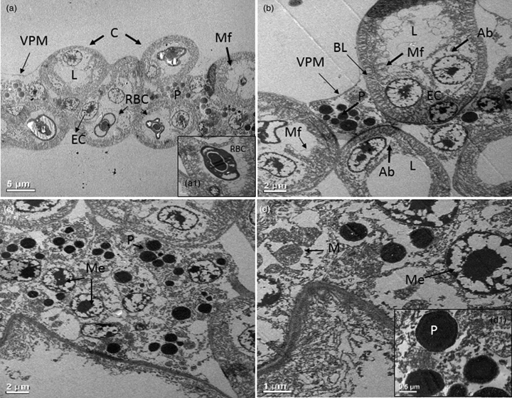
We studied the fine histological structures of pecten oculi of the Eurasian tree sparrow using various microscopy techniques. The pecten of the tree sparrow was found to be of a pleated type comprising of pleats, bridges, and base. The light microscopic study revealed further that the pleats consist of capillaries of varying sizes, blood vessels, and numerous pigmented cells that give them a black color. Histochemical studies of pecten showed a large deposition of lipid droplets, which were more abundant in the basal area. The transmission electron microscopy displayed capillaries and blood vessels that remain surrounded by a thick fibrous basal membrane. They are formed of endothelial cells having a large lumen and abluminal area with microfolds. Interstitial spaces were found filled with rounded melanocytes, electron-dense pigment granules, and mitochondria. Observations under the scanning electron microscope revealed the presence of a dense vascular network of capillaries and vessels. In addition, large hyalocytes were also observed on the surface of the pleats. The above observations suggest that the histological structure of the pecten of the tree sparrow resembles those present in the pecten of other diurnal birds. However, further investigation is required to ascertain its functional role in birds.
Keywords
- Type
- Micrographia
- Information
- Copyright
- Copyright © The Author(s), 2021. Published by Cambridge University Press on behalf of the Microscopy Society of America
References
Amemiya, T (1985). Constituents of connective tissue around the capillary of chick pecten oculi. Acta Anat 122, 235–238.CrossRefGoogle ScholarPubMed
Barlow, HB & Ostwald, TS (1972). Pecten of the pigeon's eye as an intraocular eye shade. Nature 236, 88–90.Google Scholar
Bawa, SR & YashRoy, RC (1972). Effect of dark and light adaptation on the retina and pecten of chicken. Exp Eye Res 13, 92–97.CrossRefGoogle ScholarPubMed
Bawa, SR & YashRoy, RC (1974). Structure and function of vulture pecten. Acta Anat 89, 473–480.CrossRefGoogle ScholarPubMed
Bhattacharjee, J (1993). A presumably astroglial cell in the retinopecteneal junction in Gallus domesticus, demonstrated by nonspecific esterase staining. Eur J Morphol 31, 169–173.Google Scholar
Brach, V (1975). The effect of intraocular ablation of the pecten oculi of the chicken. Invest Ophthalmol 14, 166–168.Google ScholarPubMed
Brach, V (1977). The functional significance of the avian pecten: A review. Condor 79, 321–327.CrossRefGoogle Scholar
Braekevelt, CR (1984). Electron microscopic observations on the pecten of the night hawk (Chordieles minor). Ophthalmologica 189, 211–220.CrossRefGoogle Scholar
Braekevelt, CR (1988). Fine structure of the pecten of the pigeon (Columba livia). Ophthalmologica 196(3), 151–159.CrossRefGoogle Scholar
Braekevelt, CR (1990). Fine structure of the pecten oculi of the mallard (Anas platyrhynchos). Can J Zool 68, 427–432.CrossRefGoogle Scholar
Braekevelt, CR (1991). Electron microscopic observations on the pecten of the great blue heron (Ardea herodias). Histol Histopathol 6, 345–351.Google Scholar
Braekevelt, CR (1994). Fine structure of the pecten oculi in the American crow (Corvus brachyrhyncho). Anat Histol Embryol 23, 357–366.CrossRefGoogle Scholar
Braekevelt, CR & Richardson, KC (1996). Fine structure of the pecten oculi in the Australian galah (Eolophus roseicapillus) (Aves). Histol Histopathol 11, 565–571.Google Scholar
Corona, M, Scala, G & Perrella, A (2004). Angioarchitecture of the duck pecten. Biomed Res 15, 19–25.Google Scholar
Crozier, WJ & Wolf, E (1944). Theory and measurement of visual mechanisms. X. Modifications of the flicker response contour, and the significance of the avian pecten. J Gen Physiol 27, 287–313.CrossRefGoogle ScholarPubMed
Dayan, MO & Ozaydın, T (2013). A comparative morphometrical study of the pecten oculi in different avian species. Sci World J. doi:10.1155/2013/968652.CrossRefGoogle ScholarPubMed
Demirkan, AC, Turkmenoglu, I, Demirkan, I, Akosman, MS & Akalan, MA (2018). Morphology and volume measurement of pecten oculi by stereology in merlin (Falco columbarius). Kocatepe Vet J 11, 309–315.Google Scholar
Dey, S, Baul, TB, Roy, B & Dey, D (1989). A new rapid method of air-drying for scanning electron microscopy using tetramethylsilane. J Microsc 156(2), 259–261.CrossRefGoogle Scholar
Dieterich, CE, Dieterich, HJ, Spycher, MA & Pfautsch, M (1973). Fine structural observations of the pecten oculi capillaries of the chicken. Freeze-etching, scanning and transmission electron microscopic investigations. Z Zellforsch 146, 473–489.CrossRefGoogle ScholarPubMed
Dixit, AS, Byrsat, S & Singh, NS (2020). Circadian rhythm in photoperiodic expressions of GnRH-I and GnIH regulating seasonal reproduction in the Eurasian tree sparrow, Passer montanus. J Photochem Photobiol B 211, 111993.CrossRefGoogle ScholarPubMed
Dixit, AS & Singh, NS (2012). Seasonal variation in the sensitivity of the photoperiodic response system and the termination of photorefractoriness in the subtropical tree sparrow (Passer montanus). J Exp Zool 317A, 488–498.CrossRefGoogle Scholar
Dixit, AS & Singh, NS (2016). Seasonality in circadian locomotor activity and serum testosterone level in the subtropical tree sparrow (Passer montanus). J Photochem Photobiol B 158, 61–68.CrossRefGoogle Scholar
Dixit, AS, Singh, NS & Byrsat, S (2017). Role of GnIH in photoperiodic regulation of seasonal reproduction in the Eurasian tree sparrow. J Exp Biol 220(20), 3742–3750.Google ScholarPubMed
Gerhardt, H, Schuck, J & Wolburg, H (1999). Differentiation of a unique macroglial cell type in the pecten oculi of the chicken. Glia 28(3), 201–214.3.0.CO;2-M>CrossRefGoogle ScholarPubMed
Goodman, G & Bercovich, D (2008). Melanin directly converts light for vertebrate metabolic use: Heuristic thoughts on birds, Icarus and dark human skin. Med Hypotheses 71(2), 190–202.CrossRefGoogle ScholarPubMed
Gultiken, ME, Yildiz, D, Onuk, B & Karayigit, MO (2012). The morphology of the pecten oculi in the common buzzard (Buteo buteo). Vet Ophthalmol 15, 72–76.CrossRefGoogle Scholar
Jasinski, A (1973). Fine structure of capillaries in the pecten oculi of the sparrow, Passer domesticus. Z Zellforsch Mikrosk Anat 146(2), 281–292.CrossRefGoogle ScholarPubMed
Jones, MP, Pierce, KE Jr & Ward, D (2007). Avian vision: A review of form and function with special consideration to birds of prey. J Exo Pet Med 16(2), 69–87.CrossRefGoogle Scholar
Kiama, SG, Bhattacharjee, J, Maina, JN & Weyrauch, KD (1994). A scanning electron microscope study of the pecten oculi of the black kite (Milvus migrans): Possible involvement of melanosomes in protecting the pecten against damage by ultraviolet light. J Anat 185(3), 637–642.Google ScholarPubMed
Kiama, SG, Maina, JN, Bhattacharjee, J, Mwangi, DK, Macharia, RG & Weyrauch, KD (2006). The morphology of the pecten oculi of the ostrich, Struthio camelus. Ann Anat 188, 519–528.CrossRefGoogle ScholarPubMed
Kiama, SG, Maina, JN, Bhattacharjee, J & Weyrauch, KD (2001). Functional morphology of the pecten oculi in the nocturnal spotted eagle owl (Bubo bubo africanus), and the diurnal black kite (Milvus migrans) and domestic fowl (Gallus gallus var. domesticus): A comparative study. J Zool 254(4), 521–528.CrossRefGoogle Scholar
Kiama, SG, Maina, JN, Bhattacharjee, J, Weyrauch, KD & Gehr, P (1998). A scanning electron microscope study of the luminal surface specializations in the blood vessels of the pecten oculi in a diurnal bird, the black kite (Milvus migrans). Ann Anat 180, 455–460.CrossRefGoogle Scholar
King, AS & McLelland, J (1984). Birds, Their Structure and Function, 2nd edn. St. Annes Road: Bailliere Tindall.Google Scholar
Korkmaz, D & Kum, S (2016). Investigation of the antigen recognition and presentation capacity of pecteneal hyalocytes in the chicken (Gallus gallus domesticus). Biotechnic Histochem 91(3), 212–219.CrossRefGoogle Scholar
Liebner, S, Gerhardt, H & Wolburg, H (1997). Maturation of the blood retina barrier in the developing pecten oculi of the chicken. Dev Brain Res 100, 205–219.CrossRefGoogle ScholarPubMed
Llombart, C, Nacher, V & Ramos, D (2009). Morphological characterization of pecteneal hyalocytes in the developing quail retina. J Anat 215, 280–291.CrossRefGoogle ScholarPubMed
Manorama, M, Ramanujam, SN & Dey, S (2014). Scanning electron microscopy of some vital structures of Puntius shalynius Yazdani and Talukdar 1975, an endemic hill-stream fish of North East India. J Adv Microsc Res 9(1), 22–28.CrossRefGoogle Scholar
Meyer, DB (1977). The avian eye. In Handbook of Sensory Physiology, Crescentelli, F (Ed.), pp. 549–612. Berlin, Germany: Springer Verlog.Google Scholar
Micali, A, Pisani, A, Ventrici, C, Puzzolo, D, Roszkowska, AM, Spinella, R & Aragona, P (2012). Morphological and morphometric study of the pecten oculi in the budgerigar (Melopsitta cusundulatus). Anat Rec 295, 540–550.CrossRefGoogle Scholar
Moselhy, AA & El-Hady, E (2019). Gross, histochemical and electron microscopical characterization of the pecten oculi of Baladi ducks (Anas boschas domesticus). J Adv Vet Anim Res 6(4), 456.CrossRefGoogle Scholar
Nguyen, J, Delaville, A & Coujard, R (1967). L’ endothelium capillaire du peigne de I’ oeil des oiseaux. Z Micr Anat Forsch 77, 432–441.Google Scholar
Orhan, O, Ekim, O & Bayraktaroglu, AG (2011). Morphological investigation of the pecten oculi in quail (Coturnix coturnix japonica). Ankara Univ Vet Fak Derg 58, 5–10.Google Scholar
Perrault, C (1676). Description anatomique d'une grande Tortue des Indes, Mémoires de. l'Academie Royale des Sciences Paris 3, 395–422.Google Scholar
Pettigrew, JD, Wallman, J & Wildsoet, CF (1990). Saccadic oscillations facilitate ocular perfusion from the avian pecten. Nature 343, 362–363.CrossRefGoogle ScholarPubMed
Rahman, ML, Lee, E, Aoyama, M & Sugita, S (2010). Light and electron microscopy study of the pecten oculi of the jungle crow (Corvus macrorhynchos). Okajimas Folia Anat Jpn 87, 75–83.CrossRefGoogle Scholar
Rajab, JM (2012). Morphological and histological description of the pecten oculi in the sparrow hawk (Accipiter nisus). Diy J Pur Sci 8(1), 8–19.Google Scholar
Rodriguez-Peralta, LA (1975). Hematic and fluid barriers of the retina and vitreous body. J Comp Neurol 132, 109–124.CrossRefGoogle Scholar
Scala, G, Corona, M, Mirabella, N, Perrella, A & Pelagalli, GV (2002). Microvasculature of the pecten oculi in Anas platyrhynchos. Ital J Anat Embryol 107, 65–75.Google Scholar
Schuck, J, Gerhardt, H & Wolburg, H (2000). The peripapillary glia of the optic nerve head in the chicken retina. Anat Rec 259(3), 263–275.3.0.CO;2-W>CrossRefGoogle ScholarPubMed
Schwab, IR & Maggs, IR (2007). An eye for the land. Br J Ophthalmol 91(7), 855.CrossRefGoogle Scholar
Seaman, AR & Himelfarb, TM (1963). Correlated ultrafine structural changes of the avian pecten oculi and ciliary body of Gallus domesticus. Am J Opthalmol 56, 278–296.CrossRefGoogle ScholarPubMed
Segovia, Y, Victory, N, Navarro-Sempere, A, Pinilla, V & García, M (2020). A comparative ultrastructural study of the pecten oculi in adult, juvenile, and nestling yellow-legged gulls, Larus michahellis (Naumann, 1840). Vet Ophthalmol 23(1), 113–122.CrossRefGoogle Scholar
Sibley, CG & Monroe, BL Jr (1990). Distribution and Taxonomy of Birds of the World. New Haven: Yale University Press.Google Scholar
Singh, NS & Dixit, AS (2014). Morphology and ultrastructural studies of pineal organ of the tree sparrow (Passer montanus). Micron 58, 9–14.CrossRefGoogle Scholar
Singh, NS, Dixit, AS & Sougrakpam, R (2011). A report on the presence of three avian lice (Insecta: Phthiraptera) in different regions of North-East India. J Bombay Nat Hist Soc 108(1), 65–67.Google Scholar
Thomson, A (1929). The pecten considered from an environmental point of view. Ibis 12, 608–639.Google Scholar
Tucker, R (1975). The surface of the pecten oculi in the pigeon. Cell Tissue Res 157, 457–465.CrossRefGoogle ScholarPubMed
Uehara, M, Imagawa, T & Kitagawa, H (1996). Morphological studies of the hyalocytes in the chicken eye: Scanning electron microscopy and inflammatory response after the intra vitreous injection of carbon particles. J Anat 188(3), 661–669.Google Scholar
Uehara, M, Oomori, S, Kitagawa, H & Ueshima, T (1990). The development of the pecten oculi in the chick. Jpn J Vet Sci 52(3), 503–512.CrossRefGoogle ScholarPubMed
Walls, GL (1942). The Vertebrate Eye and Its Adaptive Radiation. Bloomfield Hills, Michigan: The Carnbrook Press.Google Scholar
Wolburg, H, Libner, S, Reichenbach, A & Gerhardt, H (1999). The pecten oculi of the chicken: A model system for vascular differentiation and barrier maturation. Int Rev Cytol 187, 111–159.CrossRefGoogle Scholar
Wood, HP (1917). The chicken mite: its life history and habits (No. 553). US Department of Agriculture.CrossRefGoogle Scholar
Yilmaz, B, Korkmaz, D, Aydın, A, Demircioğlu, İ, Akbulut, Y & Cağdaş, O (2017). Light and scanning electron microscopic structure of the pecten oculi in the common barn owl (Tyto alba). Kafkas Univ Vet Fak Derg 23(6), 973–979.Google Scholar