


No CrossRef data available.
Article contents
Morphometric Analysis of Lysosomes in the Renal Tubule in Monoclonal Gammopathy Using Transmission Electron Microscopy: “Mottled Appearance” and Beyond
Published online by Cambridge University Press: 19 April 2022
Abstract
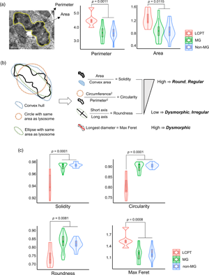
Lysosomal “mottled appearance”, or uneven electron-dense content related to monoclonal gammopathy (MG), has been mainly described in light chain proximal tubulopathy (LCPT). We aimed to determine the ultrastructural characteristics of lysosomal mottled appearance in kidney biopsies and its association with LCPT and MG. Seventy-seven biopsies were grouped into LCPT (n = 5), MG conditions other than LCPT (n = 43), and non-MG conditions (n = 29). The mottled lysosomes in the renal tubules were evaluated using transmission electron microscopy and morphometric analysis. Mottled lysosomes were more prevalent (% of present cases) and frequent (no. of mottled lysosomes/20,000× ultramicroscopic field) in the LCPT group (100% and 8.20 ± 4.15/field) than in the MG (41.9% and 1.13 ± 2.05/field) and non-MG (37.9% and 0.80 ± 1.44/field) groups. In morphometric analysis of all mottled lysosomes (n = 520) detected from the 34 biopsies (5 LCPT, 18 MG, and 11 non-MG), we found that mottled lysosomes were larger, more irregular, and more electron-dense for the LCPT group than for the MG and non-MG groups. Therefore, mottled lysosomes can be present in disorders other than LCPT or even without MG. The morphological characteristics of mottled lysosomes could provide objective guidance for the diagnosis of LCPT.
Keywords
- Type
- Biological Applications
- Information
- Copyright
- Copyright © The Author(s), 2022. Published by Cambridge University Press on behalf of the Microscopy Society of America
References
Brealey, JK, Tran, Y, Ninnes, R & Abeyaratne, A (2018). Ultrastructural identification of a proximal tubulopathy without crystals in a relapsed multiple myeloma patient. Ultrastruct Pathol 42, 458–463.CrossRefGoogle Scholar
Bridoux, F, Leung, N, Hutchison, CA, Touchard, G, Sethi, S, Fermand, JP, Picken, MM, Herrera, GA, Kastritis, E, Merlini, G, Roussel, M, Fervenza, FC, Dispenzieri, A, Kyle, RA, Nasr, SH & International Kidney and Monoclonal Gammopathy Research Group (2015). Diagnosis of monoclonal gammopathy of renal significance. Kidney Int 87, 698–711.CrossRefGoogle ScholarPubMed
Cheng, M, Gu, X, Turbat-Herrera, EA & Herrera, GA (2019). Tubular injury and dendritic cell activation are integral components of light chain-associated acute tubulointerstitial nephritis. Arch Pathol Lab Med 143, 1212–1224.CrossRefGoogle ScholarPubMed
Dimopoulos, MA, Sonneveld, P, Leung, N, Merlini, G, Ludwig, H, Kastritis, E, Goldschmidt, H, Joshua, D, Orlowski, RZ, Powles, R, Vesole, DH, Garderet, L, Einsele, H, Palumbo, A, Cavo, M, Richardson, PG, Moreau, P, San Miguel, J, Rajkumar, SV, Durie, BG & Terpos, E (2016). International Myeloma Working Group recommendations for the diagnosis and management of myeloma-related renal impairment. J Clin Oncol 34, 1544–1557.CrossRefGoogle Scholar
Doshi, M, Lahoti, A, Danesh, FR, Batuman, V, Sanders, PW & American Society of Nephrology Onco-Nephrology Forum (2016). Paraprotein-related kidney disease: Kidney injury from paraproteins-what determines the site of injury? Clin J Am Soc Nephrol 11, 2288–2294.CrossRefGoogle ScholarPubMed
El Hamel, C, Thierry, A, Trouillas, P, Bridoux, F, Carrion, C, Quellard, N, Goujon, JM, Aldigier, JC, Gombert, JM, Cogne, M & Touchard, G (2010). Crystal-storing histiocytosis with renal Fanconi syndrome: Pathological and molecular characteristics compared with classical myeloma-associated Fanconi syndrome. Nephrol Dial Transplant 25, 2982–2990.CrossRefGoogle ScholarPubMed
Herrera, GA (2014). Proximal tubulopathies associated with monoclonal light chains: The spectrum of clinicopathologic manifestations and molecular pathogenesis. Arch Pathol Lab Med 138, 1365–1380.CrossRefGoogle ScholarPubMed
Jung, M, Lee, Y, Lee, H & Moon, KC (2020). Clinicopathological characteristics of light chain proximal tubulopathy in Korean patients and the diagnostic usefulness of immunohistochemical staining for immunoglobulin light chain. BMC Nephrol 21, 146.CrossRefGoogle ScholarPubMed
Kousios, A & Roufosse, C (2019). An update on paraprotein-related renal pathology. Diagn Histopathol 25, 408–421.CrossRefGoogle Scholar
Larsen, CP, Bell, JM, Harris, AA, Messias, NC, Wang, YH & Walker, PD (2011). The morphologic spectrum and clinical significance of light chain proximal tubulopathy with and without crystal formation. Mod Pathol 24, 1462–1469.CrossRefGoogle ScholarPubMed
Leung, N, Bridoux, F, Batuman, V, Chaidos, A, Cockwell, P, D'Agati, VD, Dispenzieri, A, Fervenza, FC, Fermand, JP, Gibbs, S, Gillmore, JD, Herrera, GA, Jaccard, A, Jevremovic, D, Kastritis, E, Kukreti, V, Kyle, RA, Lachmann, HJ, Larsen, CP, Ludwig, H, Markowitz, GS, Merlini, G, Mollee, P, Picken, MM, Rajkumar, VS, Royal, V, Sanders, PW, Sethi, S, Venner, CP, Voorhees, PM, Wechalekar, AD, Weiss, BM & Nasr, SH (2019). The evaluation of monoclonal gammopathy of renal significance: A consensus report of the International Kidney and Monoclonal Gammopathy Research Group. Nat Rev Nephrol 15, 45–59.CrossRefGoogle ScholarPubMed
Luzio, JP, Hackmann, Y, Dieckmann, NM & Griffiths, GM (2014). The biogenesis of lysosomes and lysosome-related organelles. Cold Spring Harb Perspect Biol 6, a016840.CrossRefGoogle ScholarPubMed
Nasr, SH, Galgano, SJ, Markowitz, GS, Stokes, MB & D'Agati, VD (2006). Immunofluorescence on pronase-digested paraffin sections: A valuable salvage technique for renal biopsies. Kidney Int 70, 2148–2151.CrossRefGoogle ScholarPubMed
Reich, HN, Troyanov, S, Scholey, JW, Cattran, DC & Toronto Glomerulonephritis Registry (2007). Remission of proteinuria improves prognosis in iga nephropathy. J Am Soc Nephrol 18, 3177–3183.CrossRefGoogle ScholarPubMed
Schneider, CA, Rasband, WS & Eliceiri, KW (2012). NIH image to ImageJ: 25 years of image analysis. Nat Methods 9, 671–675.CrossRefGoogle ScholarPubMed
Sethi, S, Rajkumar, SV & D'Agati, VD (2018). The complexity and heterogeneity of monoclonal immunoglobulin-associated renal diseases. J Am Soc Nephrol 29, 1810–1823.CrossRefGoogle ScholarPubMed
Sirac, C, Bridoux, F, Carrion, C, Devuyst, O, Fernandez, B, Goujon, JM, El Hamel, C, Aldigier, JC, Touchard, G & Cogne, M (2006). Role of the monoclonal kappa chain V domain and reversibility of renal damage in a transgenic model of acquired Fanconi syndrome. Blood 108, 536–543.CrossRefGoogle Scholar
Stokes, MB, Valeri, AM, Herlitz, L, Khan, AM, Siegel, DS, Markowitz, GS & D'Agati, VD (2016). Light chain proximal tubulopathy: Clinical and pathologic characteristics in the modern treatment era. J Am Soc Nephrol 27, 1555–1565.CrossRefGoogle ScholarPubMed
Jung et al. supplementary material
Figures S1 and S2

PDF
297.7 KB