


No CrossRef data available.
Article contents
Standardized User-Independent Confocal Microscopy Image Acquisition and Analysis for Thickness Measurements of Microscale Collagen Scaffolds
Published online by Cambridge University Press: 31 March 2021
Abstract
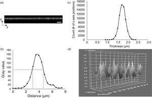
The ability to accurately and precisely measure the thickness of biomaterial constructs is critical for characterizing both specific dimensional features and related mechanical properties. However, in the absence of a standardized approach for thickness measurements, a variety of imaging modalities have been employed, which have been associated with varying limits of accuracy, particularly for ultrathin hydrated structures. Electron microscopy (EM), a commonly used modality, yields thickness values for extensively processed and nonhydrated constructs, potentially resulting in overestimated mechanical properties, including elastic modulus and ultimate tensile strength. Confocal laser scanning microscopy (CLSM) has often been used as a nondestructive imaging alternative. However, published CLSM-derived image analysis protocols use arbitrary signal intensity cutoffs and provide minimal information regarding thickness variability across imaged surfaces. To address the aforementioned limitations, we present a standardized, user-independent CLSM image acquisition and analysis approach developed as a custom ImageJ macro and validated with collagen-based scaffolds. In the process, we also quantify thickness discrepancies in collagen-based scaffolds between CLSM and EM techniques, further illustrating the need for improved strategies. Employing the same image acquisition protocol, we also demonstrate that this approach can be used to estimate the surface roughness of the same scaffolds without the use of specialized instrumentation.
- Type
- Software and Instrumentation
- Information
- Copyright
- Copyright © The Author(s), 2021. Published by Cambridge University Press on behalf of the Microscopy Society of America
References
Bhushan, B (2000). 2.1 The nature of surfaces. In Modern Tribology Handbook, vol. 1, Bhushan, B. & Marti, O. (Eds.), pp. 49–120. Boca Raton, FL: CRC Press.Google Scholar
Caves, JM, Cui, W, Wen, J, Kumar, VA, Haller, CA & Chaikof, EL (2011). Elastin-like protein matrix reinforced with collagen microfibers for soft tissue repair. Biomaterials 32(23), 5371–5379.CrossRefGoogle ScholarPubMed
Dahl, SL, Kypson, AP, Lawson, JH, Blum, JL, Strader, JT, Li, Y, Manson, RJ, Tente, WE, DiBernardo, L, Hensley, MT, Carter, R, Williams, TP, Prichard, HL, Dey, MS, Begelman, KG & Niklason, LE (2011). Readily available tissue-engineered vascular grafts. Sci Transl Med 3(68), 68ra9.CrossRefGoogle ScholarPubMed
Dahl, SL, Rhim, C, Song, YC & Niklason, LE (2007). Mechanical properties and compositions of tissue engineered and native arteries. Ann Biomed Eng 35(3), 348–355.CrossRefGoogle ScholarPubMed
Dapson, RW, Fagan, C, Kiernan, JA & Wickersham, TW (2011). Certification procedures for sirius red F3B (CI 35780, direct red 80). Biotech Histochem 86(3), 133–139.CrossRefGoogle Scholar
Dzhoyashvili, NA, Thompson, K, Gorelov, AV & Rochev, YA (2016). Film thickness determines cell growth and cell sheet detachment from spin-coated poly(N-isopropylacrylamide) substrates. ACS Appl Mater Interfaces 8(41), 27564–27572.CrossRefGoogle ScholarPubMed
Eskandarinia, A, Kefayat, A, Agheb, M, Rafienia, M, Amini Baghbadorani, M, Navid, S, Ebrahimpour, K, Khodabakhshi, D & Ghahremani, F (2020). A novel bilayer wound dressing composed of a dense polyurethane/propolis membrane and a biodegradable polycaprolactone/gelatin nanofibrous scaffold. Sci Rep 10, 3063.CrossRefGoogle Scholar
Fotticchia, A, Musson, D, Lenardi, C, Demirci, E & Liu, Y (2018). Anisotropic cytocompatible electrospun scaffold for tendon tissue engineering elicits limited inflammatory response in vitro. J Biomater Appl 33(1), 127–139.CrossRefGoogle ScholarPubMed
Gremare, A, Guduric, V, Bareille, R, Heroguez, V, Latour, S, L'Heureux, N, Fricain, JC, Catros, S & Le Nihouannen, D (2018). Characterization of printed PLA scaffolds for bone tissue engineering. J Biomed Mater Res A 106(4), 887–894.CrossRefGoogle ScholarPubMed
Gremare, A, Jean-Gilles, S, Musqui, P, Magnan, L, Torres, Y, Fenelon, M, Brun, S, Fricain, JC & L'Heureux, N (2019). Cartography of the mechanical properties of the human amniotic membrane. J Mech Behav Biomed Mater 99, 18–26.CrossRefGoogle ScholarPubMed
Jiang, Z, Xi, Y, Lai, K, Wang, Y, Wang, H & Yang, G (2017). Laminin-521 promotes rat bone marrow mesenchymal stem cell sheet formation on light-induced cell sheet technology. Biomed Res Int 2017, 9474573.CrossRefGoogle ScholarPubMed
Kaiser, NJ, Kant, RJ, Minor, AJ & Coulombe, KLK (2019). Optimizing blended collagen-fibrin hydrogels for cardiac tissue engineering with human iPSC-derived cardiomyocytes. ACS Biomater Sci Eng 5(2), 887–899.CrossRefGoogle ScholarPubMed
Konig, G, McAllister, TN, Dusserre, N, Garrido, SA, Iyican, C, Marini, A, Fiorillo, A, Avila, H, Wystrychowski, W, Zagalski, K, Maruszewski, M, Jones, AL, Cierpka, L, de la Fuente, LM & L'Heureux, N (2009). Mechanical properties of completely autologous human tissue engineered blood vessels compared to human saphenous vein and mammary artery. Biomaterials 30(8), 1542–1550.CrossRefGoogle ScholarPubMed
Kumar, VA, Caves, JM, Haller, CA, Dai, E, Liu, L, Grainger, S & Chaikof, EL (2013). Acellular vascular grafts generated from collagen and elastin analogs. Acta Biomater 9(9), 8067–8074.CrossRefGoogle ScholarPubMed
Kuypers, LC, Decraemer, WF, Dirckx, JJ & Timmermans, JP (2005). A procedure to determine the correct thickness of an object with confocal microscopy in case of refractive index mismatch. J Microsc 218(Pt 1), 68–78.CrossRefGoogle ScholarPubMed
Lawrence, BD, Wharram, S, Kluge, JA, Leisk, GG, Omenetto, FG, Rosenblatt, MI & Kaplan, DL (2010). Effect of hydration on silk film material properties. Macromol Biosci 10(4), 393–403.CrossRefGoogle ScholarPubMed
Li, JJ, Roohani-Esfahani, SI, Kim, K, Kaplan, DL & Zreiqat, H (2017). Silk coating on a bioactive ceramic scaffold for bone regeneration: Effective enhancement of mechanical and in vitro osteogenic properties towards load-bearing applications. J Tissue Eng Regen Med 11(6), 1741–1753.CrossRefGoogle ScholarPubMed
Luo, J, Qin, L, Zhao, L, Gui, L, Ellis, MW, Huang, Y, Kural, MH, Clark, JA, Ono, S, Wang, J, Yuan, Y, Zhang, SM, Cong, X, Li, G, Riaz, M, Lopez, C, Hotta, A, Campbell, S, Tellides, G, Dardik, A, Niklason, LE & Qyang, Y (2020). Tissue-engineered vascular grafts with advanced mechanical strength from human iPSCs. Cell Stem Cell 26(2), 251–261.e8.CrossRefGoogle ScholarPubMed
Malladi, S, Miranda-Nieves, D, Leng, L, Grainger, SJ, Tarabanis, C, Nesmith, AP, Kosaraju, R, Haller, CA, Parker, KK, Chaikof, EL & Guenther, A (2020). Continuous formation of ultrathin, strong collagen sheets with tunable anisotropy and compaction. ACS Biomater Sci Eng 6(7), 4236–4246.CrossRefGoogle ScholarPubMed
Neal, RA, Jean, A, Park, H, Wu, PB, Hsiao, J, Engelmayr, GC Jr., Langer, R & Freed, LE (2013). Three-dimensional elastomeric scaffolds designed with cardiac-mimetic structural and mechanical features. Tissue Eng Part A 19(5–6), 793–807.CrossRefGoogle ScholarPubMed
Pisani, S, Croce, S, Chiesa, E, Dorati, R, Lenta, E, Genta, I, Bruni, G, Mauramati, S, Benazzo, A, Cobianchi, L, Morbini, P, Caliogna, L, Benazzo, M, Avanzini, MA & Conti, B (2020). Tissue engineered esophageal patch by mesenchymal stromal cells: Optimization of electrospun patch engineering. Int J Mol Sci 21(5), 1764.CrossRefGoogle ScholarPubMed
Quan, BD & Sone, ED (2013). Cryo-TEM Analysis of Collagen Fibrillar Structure, 1st ed. New York, NY: Elsevier Inc. http://dx.doi.org/10.1016/B978-0-12-416617-2.00009-6.CrossRefGoogle ScholarPubMed
Rodriguez, M, Kluge, JA, Smoot, D, Kluge, MA, Schmidt, DF, Paetsch, CR, Kim, PS & Kaplan, DL (2020). Fabricating mechanically improved silk-based vascular grafts by solution control of the gel-spinning process. Biomaterials 230, 119567.CrossRefGoogle ScholarPubMed
Sang, Y, Li, M, Liu, J, Yao, Y, Ding, Z, Wang, L, Xiao, L, Lu, Q, Fu, X & Kaplan, DL (2018). Biomimetic silk scaffolds with an amorphous structure for soft tissue engineering. ACS Appl Mater Interfaces 10(11), 9290–9300.CrossRefGoogle ScholarPubMed
Sileika, TS, Barrett, DG, Zhang, R, Lau, KH & Messersmith, PB (2013). Colorless multifunctional coatings inspired by polyphenols found in tea, chocolate, and wine. Angew Chem Int Ed Engl 52(41), 10766–10770.CrossRefGoogle ScholarPubMed
Silver, FH & Trelstad, RL (1980). Type I collagen in solution. Structure and properties of fibril fragments. J Biol Chem 255(19), 9427–9433.CrossRefGoogle ScholarPubMed
Soletti, L, Hong, Y, Guan, J, Stankus, JJ, El-Kurdi, MS, Wagner, WR & Vorp, DA (2010). A bilayered elastomeric scaffold for tissue engineering of small diameter vascular grafts. Acta Biomater 6(1), 110–122.CrossRefGoogle ScholarPubMed
Stylianou, A, Kontomaris, SB, Kyriazi, M & Yova, D (2010. Surface characterization of collagen films by atomic force microscopy. In IFMBE Proceedings, vol. 29. pp. 612–615. Chennai, India: Scientific Publishing Services Pvt.Ltd.Google Scholar
Stylianou, A & Yova, D (2013). Surface nanoscale imaging of collagen thin films by atomic force microscopy. Mater Sci Eng C 33(5), 2947–2957.CrossRefGoogle ScholarPubMed
Stylianou, A, Yova, D & Alexandratou, E (2014). Investigation of the influence of UV irradiation on collagen thin films by AFM imaging. Mater Sci Eng C 45, 455–468.CrossRefGoogle ScholarPubMed
Versteegden, LR, van Kampen, KA, Janke, HP, Tiemessen, DM, Hoogenkamp, HR, Hafmans, TG, Roozen, EA, Lomme, RM, van Goor, H, Oosterwijk, E, Feitz, WF, van Kuppevelt, TH & Daamen, WF (2017). Tubular collagen scaffolds with radial elasticity for hollow organ regeneration. Acta Biomater 52, 1–8.CrossRefGoogle ScholarPubMed
Wise, SG, Byrom, MJ, Waterhouse, A, Bannon, PG, Weiss, AS & Ng, MK (2011). A multilayered synthetic human elastin/polycaprolactone hybrid vascular graft with tailored mechanical properties. Acta Biomater 7(1), 295–303.CrossRefGoogle ScholarPubMed
Zhang, HR, Egerton, RF & Malac, M (2012). Local thickness measurement through scattering contrast and electron energy-loss spectroscopy. Micron 43(1), 8–15.CrossRefGoogle ScholarPubMed
Zhao, HL, Zhang, CP, Zhu, H, Jiang, YF & Fu, XB (2017). Autofluorescence of collagen fibres in scar. Skin Res Technol 23(4), 588–592.CrossRefGoogle ScholarPubMed
Zhao, L, Sundaram, S, Le, AV, Huang, AH, Zhang, J, Hatachi, G, Beloiartsev, A, Caty, MG, Yi, T, Leiby, K, Gard, A, Kural, MH, Gui, L, Rocco, KA, Sivarapatna, A, Calle, E, Greaney, A, Urbani, L, Maghsoudlou, P, Burns, A, DeCoppi, P & Niklason, LE (2016). Engineered tissue-stent biocomposites as tracheal replacements. Tissue Eng Part A 22(17–18), 1086–1097.CrossRefGoogle ScholarPubMed
Zhou, J, Ying, H, Wang, M, Su, D, Lu, G & Chen, J (2018). Dual layer collagen-GAG conduit that mimic vascular scaffold and promote blood vessel cells adhesion, proliferation and elongation. Mater Sci Eng C Mater Biol Appl 92, 447–452.CrossRefGoogle ScholarPubMed

Tarabanis et al. supplementary material
Tarabanis et al. supplementary material

File
24.9 KB