Hostname: page-component-848d4c4894-r5zm4 Total loading time: 0 Render date: 2024-06-19T14:24:57.021Z Has data issue: false hasContentIssue false

Structural Analysis of Gliding Motility of a Bacteroidetes Bacterium by Correlative Light and Scanning Electron Microscopy (CLSEM)

Published online by Cambridge University Press:  02 February 2022

Devanshi Khare
Affiliation:
Molecular Biology Division, Bhabha Atomic Research Centre, Trombay, Mumbai400085, India Homi Bhabha National Institute, Anushakti Nagar, Mumbai400094, India
Pallavi Chandwadkar
Affiliation:
Molecular Biology Division, Bhabha Atomic Research Centre, Trombay, Mumbai400085, India
Celin Acharya*
Affiliation:
Molecular Biology Division, Bhabha Atomic Research Centre, Trombay, Mumbai400085, India Homi Bhabha National Institute, Anushakti Nagar, Mumbai400094, India
*
*Corresponding author: Celin Acharya, E-mail: celin@barc.gov.in
Get access

Abstract

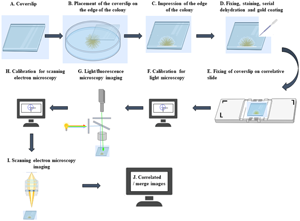
The members of the Bacteroidetes phylum move on surfaces by gliding motility in the absence of external motility appendages, leading to the formation of spreading colonies. Here, the structural features of the spreading colony were assessed in a uranium-tolerant Bacteroidetes bacterium, Chryseobacterium sp. strain PMSZPI, by using correlative light and scanning electron microscopy (CLSEM). We developed a simple and convenient workflow for CLSEM using a shuttle and find software module and a correlative sample holding slide designed to transport samples between the light/fluorescence microscope (LM/FM) and the scanning electron microscope (SEM) to image spreading colony edges. The datasets from the CLSEM studies allowed convenient examination of the colonial organization by LM/FM followed by ultrastructural analysis by SEM. The regions of interest (ROIs) of the spreading colony edges that were observed in LM/FM in the absence and presence of uranium could be re-identified in the SEM quickly without prolonged searching. Perfect correlation between LM and SEM could be achieved with minimum preparation steps. Subsequently, imaging of the correlated regions was done at higher resolution in SEM to obtain more comprehensive information. We further showed the association of uranium with the gliding PMSZPI cells by energy-dispersive X-ray spectroscopy (EDS) attached to SEM.

Type
Biological Applications
Copyright
Copyright © The Author(s), 2022. Published by Cambridge University Press on behalf of the Microscopy Society of America

Access options

Get access to the full version of this content by using one of the access options below. (Log in options will check for institutional or personal access. Content may require purchase if you do not have access.)

Footnotes

The first two authors contributed equally to this work.

References

Dang, H & Lovell, CR (2016). Microbial surface colonization and biofilm development in marine environments. Microbiol Mol Biol Rev 80, 91138.CrossRefGoogle ScholarPubMed
De Boer, P, Hoogenboom, JP & Giepmans, BN (2015). Correlated light and electron microscopy: Ultrastructure lights up!. Nat Methods 12, 503513.CrossRefGoogle ScholarPubMed
Giepmans, BNG, Deerinck, TJ, Smarr, BL, Jones, YZ & Ellisman, MH (2005). Correlated light and electron microscopic imaging of multiple endogenous proteins using quantum dots. Nat Methods 2, 743749.CrossRefGoogle ScholarPubMed
Halary, S, Duperron, S & Boudier, T (2011). Direct image-based correlative microscopy technique for coupling identification and structural investigation of bacterial symbionts associated with metazoans. Appl Environ Microbiol 12, 41724179.CrossRefGoogle Scholar
Jarrell, KF & McBride, MJ (2008). The surprisingly diverse ways that prokaryotes move. Nat Rev Microbiol 6, 466476.CrossRefGoogle ScholarPubMed
Johansen, VE, Catón, L, Hamidjaja, R, Oosterink, E, Wilts, BD, Rasmussen, TS, Sherlock, MM, Ingham, CJ & Vignolini, S (2018). Genetic manipulation of structural color in bacterial colonies. Proc Natl Acad Sci 115, 26522657.CrossRefGoogle ScholarPubMed
Khare, D, Kumar, R & Acharya, C (2020). Genomic and functional insights into the adaptation and survival of Chryseobacterium sp. strain PMSZPI in uranium enriched environment. Ecotoxicol Environ Saf 191, 110217.CrossRefGoogle ScholarPubMed
Kientz, B, Agogué, H, Lavergne, C, Marié, P & Rosenfeld, E (2013). Isolation and distribution of iridescent Cellulophaga and other iridescent marine bacteria from the Charente-Maritime coast, French Atlantic. Syst Appl Microbiol 36, 244251.CrossRefGoogle ScholarPubMed
Kientz, B, Ducret, A, Luke, S, Vukusic, P, Mignot, T & Rosenfeld, E (2012 a). Glitter-like iridescence within the Bacteroidetes especially Cellulophaga spp.: Optical properties and correlation with gliding motility. PLoS One 7, e52900.CrossRefGoogle ScholarPubMed
Kientz, B, Vukusic, P, Luke, S & Rosenfeld, E (2012 b). Iridescence of a marine bacterium and classification of prokaryotic structural colors. Appl Environ Microbiol 7, 20922099.CrossRefGoogle Scholar
Kommnick, C, Lepper, A & Hensel, M (2019). Correlative light and scanning electron microscopy (CLSEM) for analysis of bacterial infection of polarized epithelial cells. Sci Rep 9, 17079.CrossRefGoogle ScholarPubMed
Kumar, R, Nongkhlaw, M, Acharya, C & Joshi, SR (2013). Uranium (U)-tolerant bacterial diversity from U ore deposit of domiasiat in north-east India and its prospective utilisation in bioremediation. Microbes Environ 28, 3341.CrossRefGoogle ScholarPubMed
McBride, MJ (2001). Bacterial gliding motility: Multiple mechanisms for cell movement over surfaces. Annu Rev Microbiol 55, 4975.CrossRefGoogle ScholarPubMed
McBride, MJ & Nakane, D (2015). Flavobacterium gliding motility and the type IX secretion system. Curr Opin Microbiol 28, 7277.CrossRefGoogle ScholarPubMed
McBride, MJ & Zhu, Y (2013). Gliding motility and Por secretion system genes are widespread among members of the phylum Bacteroidetes. J Bacteriol 195, 270278.CrossRefGoogle ScholarPubMed
Miyata, M (2010). Unique centipede mechanism of Mycoplasma gliding. Annu Rev Microbiol 64, 519537.CrossRefGoogle ScholarPubMed
Nakane, D, Sato, K, Wada, H, McBride, MJ & Nakayama, K (2013). Helical flow of surface protein required for bacterial gliding motility. Proc Natl Acad Sci 110, 1114511150.CrossRefGoogle ScholarPubMed
Nan, B & Zusman, DR (2011). Uncovering the mystery of gliding motility in the myxobacteria. Annu Rev Genet 45, 2139.CrossRefGoogle ScholarPubMed
Peddie, CJ, Blight, K, Wilson, E, Melia, C, Marrison, J, Carzaniga, R, Domart, MC, O'Toole, P, Larijani, B & Collinson, LM (2014). Correlative and integrated light and electron microscopy of in-resin GFP fluorescence, used to localise diacylglycerol in mammalian cells. Ultramicroscopy 100, 314.CrossRefGoogle Scholar
Sato, K, Naito, M, Yukitake, H, Hirakawa, H, Shoji, M, McBride, MJ, Rhodes, RG & Nakayama, K (2010). A protein secretion system linked to bacteroidete gliding motility and pathogenesis. Proc Natl Acad Sci 107, 276281.CrossRefGoogle ScholarPubMed
Sato, K, Naya, M, Hatano, Y, Kondo, Y, Sato, M, Narita, Y, Nagano, K, Naito, M, Nakayama, K & Sato, C (2021). Colony spreading of the gliding bacterium Flavobacterium johnsoniae in the absence of the motility adhesin SprB. Sci Rep 11, 967.CrossRefGoogle ScholarPubMed
Shrivastava, A & Berg, HC (2015). Towards a model for Flavobacterium gliding. Curr Opin Microbiol 28, 9397.CrossRefGoogle Scholar