Hostname: page-component-848d4c4894-nr4z6 Total loading time: 0 Render date: 2024-05-01T05:58:49.128Z Has data issue: false hasContentIssue false

Structural and Ultrastructural Characteristics of the Red-Rumped Agouti (Dasyprocta leporina—Linnaeus, 1758) Palatine Epithelium

Published online by Cambridge University Press:  23 April 2021

André Neri Tomiate
Affiliation:
Laboratório de Anatomia – LAMAF, Instituto de Biociências, Universidade Estadual Paulista-UNESP, 13506-752, Rio Claro-SP, Brasil
Gabriela Klein Barbosa
Affiliation:
Laboratório de Anatomia – LAMAF, Instituto de Biociências, Universidade Estadual Paulista-UNESP, 13506-752, Rio Claro-SP, Brasil
Lara Caetano Rocha
Affiliation:
Laboratório de Anatomia – LAMAF, Instituto de Biociências, Universidade Estadual Paulista-UNESP, 13506-752, Rio Claro-SP, Brasil
Sonia Regina Yokomizo de Almeida
Affiliation:
Departamento de Anatomia, Instituto de Ciências Biomédicas - ICBIII, Universidade de São Paulo-USP, 05508-900, São Paulo-SP, Brasil
Moacir Franco de Oliveira
Affiliation:
Departamento de Ciências Animais, Universidade Federal do Semi-Árido, UFERSA, 59625-900, Mossoró-RN, Brasil
Ii-sei Watanabe
Affiliation:
Departamento de Anatomia, Instituto de Ciências Biomédicas - ICBIII, Universidade de São Paulo-USP, 05508-900, São Paulo-SP, Brasil Departamento de Cirurgia, Faculdade de Medicina Veterinária e Zootecnia-FMVZ, Universidade de São Paulo-USP, 05508-270, São Paulo-SP, Brasil
Adriano Polican Ciena*
Affiliation:
Laboratório de Anatomia – LAMAF, Instituto de Biociências, Universidade Estadual Paulista-UNESP, 13506-752, Rio Claro-SP, Brasil
*
*Author for correspondence: Adriano Polican Ciena, E-mail: adriano.ciena@unesp.br
Get access

Abstract

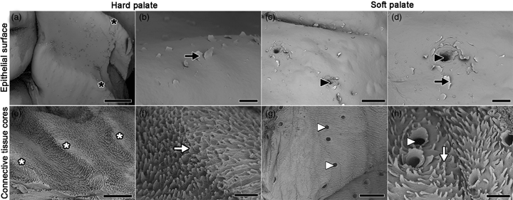
The palate is a fundamental region in food swallowing and presents different adaptations in species. This research aimed to describe structural and ultrastructural characteristics of the palatine epithelium and the connective tissue cores (CTCs) of ten red-rumped agoutis (Dasyprocta leporina—Linnaeus, 1758) using macroscopic, light microscopy, scanning electron microscopy, and transmission electron microscopy. We found nine palatine ridges in the diastema and hard palate, and a smooth surface in the soft palate. Stratified squamous keratinized epithelium with projections of lamina propria and soft palate had gland clusters. Epithelial removal revealed CTCs with a conical shape with high density in the hard palate and the sides of the soft palate. Near the CTCs were nerve fibers in the hard palate, and the soft palate had muscular tissue below the gland clusters. The structural and ultrastructural characteristics enable stability of the hard palate and fixation to the soft palate sides, while the soft palate center has greater mobility thus assisting in food swallowing. We concluded that structural characteristics are similar to other mammals, although the morphology of agouti's palate differs in the amount and disposition of palatine ridges, and the conical CTC's morphology.

Type
Micrographia
Copyright
Copyright © The Author(s), 2021. Published by Cambridge University Press on behalf of the Microscopy Society of America

Access options

Get access to the full version of this content by using one of the access options below. (Log in options will check for institutional or personal access. Content may require purchase if you do not have access.)

References

Ari, HH & Uslu, S (2019). Morphology and histology of the anatolian bobcat (Lynx lynx) hard palate. Anat Histol Embryol 48, 290295.CrossRefGoogle ScholarPubMed
Barbosa, GK, Jacob, CS, Pimentel Neto, J, Oliveira, MF, Rici, REG, Watanabe, I & Ciena, AP (2020). Structural and ultrastructural characteristics of the tongue of the collared peccary (Pecari tajacu, Linnaeus, 1758). Anat Histol Embryol. doi:10.1111/ahe.12557.CrossRefGoogle Scholar
Ciena, AP, Bolina, CS, Almeida, SRY, Rici, REG, Oliveira, MF, Silva, MCP, Miglino, MA & Watanabe, I (2013). Structural and ultrastructural features of the agouti tongue (Dasyprocta aguti Linnaeus, 1766). J Anat 223, 152158.CrossRefGoogle Scholar
Ciena, AP, Santos, AC, Vasconcelos, BG, Rici, REG, Assis Neto, AC, Almeida, SRY, Miglino, MA & Watanabe, I (2019). Morphological characteristics of the papillae and lingual epithelium of Guinea pig (Cavia porcellus). Acta Zool 100(1), 5360.CrossRefGoogle Scholar
De Aro, MM, Santos, AC, Silveira, EE, Silva Lisboa Neto, AF, Oliveira, MF & Assis Neto, AC (2019). Morphological tools to evaluate the digestory apparatus in rocky cavy (Kerodon rupestris). Microsc Res Tech 82(6), 696708.CrossRefGoogle Scholar
Eisentraut, VM (1975). Das Gaumenfaltenmuster bei afrikanischen. Zeitschrifte für Säugetierkunde 40, 133141.Google Scholar
Halata, Z & Baumann, KI (1999). Sensory nerve endings in the hard palate and papilla incisiva of the rhesus monkey. Anat Embryol 199, 427437.CrossRefGoogle ScholarPubMed
Halata, Z, Brian, YC, Baumann, KI, Schwegmann, C & Friedman, RM (1999). Sensory nerve endings in the hard palate and papila incisiva of the goat. Exp Brain Res 129, 219228.CrossRefGoogle Scholar
Jones, KR, Lall, KR & Garcia, GW (2019). Omnivorous behaviour of the agouti (Dasyprocta leporina): A neotropical rodent with the potential for domestication. Scientifica 2019, 15.CrossRefGoogle ScholarPubMed
Kutuzov, H & Sicher, H (1952). Anatomy and function of the palate in the white rat. Anat Rec 114, 6784.CrossRefGoogle ScholarPubMed
Mahdy, MA, Abdalla, KE & Mohamed, SA (2018). Morphological study of the hard palate in the Egyptian goats (Capra hircus): A scanning electron microscopic study. Anat, Histol, Embryol 47(5), 391397.CrossRefGoogle ScholarPubMed
Martinez, M, Martinez, FE & Watanabe, I (1998). Light and scanning electron microscopic study of the palatine mucosa of nine-banded armadillo (Dasypus novemcinctus). Eur J Morphol 36, 97104.CrossRefGoogle Scholar
Martinez, M, Watanabe, I, Martinez, FE, Bass, NA, Ribeiro, O & Gregório, EA (1997). Scanning electron microscopic study of the hard palatine mucosa of opossum (Didelphis albiventris). Rev Chil Anat 15, 1318.CrossRefGoogle Scholar
McMillan, MD (1979). The complementary structure of the superficial and deep surfaces of the cells of the stratum corneum of the hard palate in the rat. J Periodontol Res 14, 429502.CrossRefGoogle ScholarPubMed
Mustapha, OA, Ayoade, OE, Ogunbunmi, TK & Olude, MA (2015). Morphology of the oral cavity of the African giant rat (Cricetomys gambianus, waterhouse). Bulg J Vet Med 18, 1930.CrossRefGoogle Scholar
Nakano, T (1992). NaOH cell maceration/scanning electron microscopic studies on the architecture of the lamina propria of the mouse palate. Auris Nasus Larynx (Tokyo) 19, 133142.CrossRefGoogle ScholarPubMed
Pantalacci, S, Prochazka, J, Martin, A, Rothova, M, Lambert, A, Bernard, L, Charles, C, Viriot, L, Peterkova, R & Laudet, V (2008). Patterning of palatal rugae through sequential addition reveals an anterior/posterior boundary in palatal development. BMC Dev Biol 8, 116.CrossRefGoogle ScholarPubMed
Sa, G, Xiong, X, Wu, T, Yang, J, He, S & Zhao, Y (2016). Histological features of oral epithelium in seven animal species: As a reference for selecting animal models. Eur J Pharm Sci 81, 1017.CrossRefGoogle ScholarPubMed
Sakai, H, Morino, S & Inoué, T (2001). Scanning electron-microscopic view of the oral and basal epithelial surfaces of the rat soft palate. Cells Tissues Organs 1, 158164.CrossRefGoogle Scholar
Silva, DCB, Fagundes, NCF, Teixeira, FB, Penha, NEA, Santana, LNS, Mendes-Oliveira, AC & Lima, RR (2013). Anatomical and histological characteristics of teeth in agouti (Dasyprocta prymnolopha Wagler, 1831). Pesqui Vet Bras 33, 5157.CrossRefGoogle Scholar
Teófilo, TS, Morais, MRPT, Dias, GF, Diniz, AN, Chaves, HSA & Fontanele-Neto, JD (2014). Histology of palate and soft palate tonsil of collared peccary (Tayassu tajacu). Anat, Histol, Embryol 43(5), 361368.CrossRefGoogle Scholar