


Introduction
The first time Australia hosted the International Microscopy Congress was in 1974 in Canberra, the capital of Australia. In 2018, Australia again had the opportunity to host the global microscopy community, this time in Sydney. The congress attracted over 2,500 delegates to the Sydney International Convention Centre (ICC), centrally located in the Darling Harbour precinct adjacent the Harbour Bridge and Sydney Opera House.
As part of this event, The Australian Microscopy and Microanalysis Society (AMMS) and the conference organizers were pleased to welcome 570 students (aged between 12–18 years) and accompanying staff to a dedicated Outreach Learning Space. Over four days of the conference, classes from secondary schools across the state of New South Wales were invited to experience the latest in microscopy technology from light microscopy to virtual reality and 3D printing of micrographs. This program presented a rare opportunity to promote STEM education and vocations in Australia. The primary goals of the outreach space were to energize inquisitive minds and to show students that a career in STEM is something that they could aspire to (Figure 1). Committee organizers focused heavily on promoting the event to schools populated by students from low socio-economic backgrounds, who ultimately made up more than half the program attendees. Classes were generally made up of science students from a single-year group or gifted and talented students across multiple years.
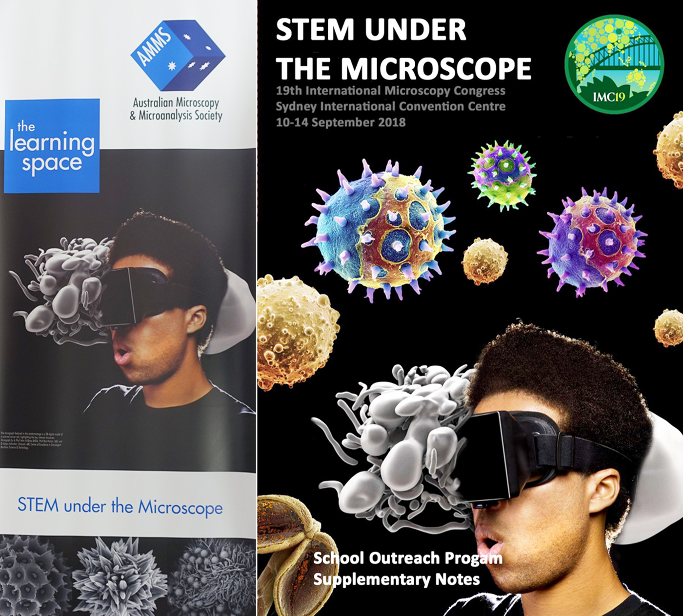
Figure 1: IMC19 Outreach Learning Space welcome banner (left) greeting classes on arrival, and booklet provided to each class summarizing the industrial applications of a range of microscopy techniques (right). Note: Grayscale 3D model is a 3D VR-ready digital model of a scanned cancer cell highlighting the key internal structures. Credit: A/Prof. John McGhee (UNSW), Prof. Rob Parton UQ - (CBNS Research) [Reference McGhee and Parton1].
A common challenge facing outreach programs concerns the ongoing engagement of students and teachers through the existing education system, once a program has been completed. A crucial outcome for the AMMS was to ensure that the final outreach program would have a legacy as an ongoing resource and form an integral part of the Australian national curriculum in future years. To do this, we first sought to develop strong themes for the Outreach Learning Space that reflect our context and the future direction of microscopy research. These themes included: (a) uniquely Australian materials and challenges, (b) correlative and multidisciplinary research, and (c) microscopy datasets coming to life.
To make these themes come alive, we worked with industry suppliers who generously donated their instruments for the four-day program. These included a range of stereo and compound microscopes and four scanning electron microscopes (SEMs) for the students’ hands-on work, a digital microscope, a virtual reality system, and a number of 3D printing machines.
Lesson Plans
In situations where students only have an hour to experience technology they may never have dealt with before, we felt it was crucial that they were introduced to these themes, their wider context, and the theory and practice of microscopy before they arrived at the Outreach Learning Space. Equally, it was important to our committee that classes were provided with follow-up work that would continue to challenge students to grapple with the themes and samples they would observe. We engaged the Australian Science Teacher's Association (ASTA) in producing a set of lesson plans that would complement activities that students undertook on-site. Our committee settled on four major topics of investigation. These topics, known as the “Storylines,” were:
– Unique Australian Flora: Oil glands are a special feature of Eucalyptus leaves, but why are they there? And how does the leaf structure accommodate them? Microscopy can show you these structures in a new light. Inspired by the work of Prof. Margaret Barbour [Reference Barbour2] from the University of Sydney.
– Corals in Competition; The International Year of the Reef: How are corals even possible? It's thanks to many tiny species living together in a symbiotic relationship. But life is crowded on the reef, and only the most robust survive. See how the structures of stony and soft polyp corals help them take advantage of the reef environment and how bleaching of the reef leads to new competition and challenges. Inspired by work from the Future Reefs [Reference Suggett3] collaborative research center at the University of Technology, Sydney.
– Clean Your Teeth: Your body puts a lot of work into generating 32 hard, rigid teeth to tear and crush your next meal. Yet both organic and metallic contaminants can slowly undermine their tough enamel barrier. Take an atomic-resolution look at this amazing composite structure, its strength, and its weaknesses. Inspired by tooth enamel research [Reference Hollick4] at the Australian Centre for Microscopy and Microanalysis [5] at the University of Sydney.
– Titanic Rusticles: The wrecks of the HMAS Sydney and the RMS Titanic are disappearing. Complex structures, known as rusticles, cover their surface, slowly feeding off iron supplied by the steel cladding. Researchers have noticed that rusticles are colonized by a range of bacteria, surviving at pressures 400 times greater than on land. By looking at the growth and structure of rusticles at the microscale, you can be helping researchers at the Maritime Museum predict how quickly underwater support structures might degrade so engineers can take preventative action. Inspired by work at the Australian Maritime Museum and the Microbiologically Induced Corrosion (MIC) [Reference Suarez6] research team at Curtin University.
These samples were intended to represent a cross section of complex materials, both organic and inorganic. While larger organic samples are often used exclusively in outreach situations, our committee wanted to make the full scope of microscopy research accessible to visiting classes. Each sample was drawn from actual research projects either currently underway or recently completed around Australia.
Implementation
A panel of outstanding science teachers from across Australia volunteered their time to develop a set of pre-work, on-the-day, and post-work activity sheets that explored each of these issues and introduced students to concepts in light, electron, X-ray, and atom probe microscopy (depending on level of education). Each visiting class was broken up into four groups specializing in one of the major themes introduced in their pre-work. After visiting the outreach space, students were able to share what they learned about their individual topics. It was intended that this format would promote greater discussion and critical thinking.
The physical outreach space was designed specifically to accommodate the Storyline framework and the themes flagged in the students’ pre-work activities. Although segregated from the main conference thoroughfares, the space was nevertheless centrally located within the convention center, directly outside one of the main entrances to the plenary auditorium. This allowed for delegates to share in the experience casually, while helping students to feel that they were intimately involved in the events of the conference.
For a typical hour-long session, visiting classes were welcomed by ICC and conference staff and guided through an art exhibition that presented a series of indigenous artworks inspired by light and electron micrographs (for more information, see “Stories and Structures” [Reference Vasquez7]). They were then led into an introductory exhibition room where students were officially welcomed to the program and introduced to the instruments they would be using. It was seen as crucial that students had some guided practice on the instruments before going on to complete their activities. The room showcased stereo and compound light microscopes (Figure 2), a rotating digital light microscope, a freely available online SEM simulator [Reference Vasquez8] from the national research facility Microscopy Australia [9], a micrograph display showing individual atoms of gallium nitride imaged with an aberration-corrected transmission electron microscope (TEM), and a video display demonstrating how virtual reality can give a new perspective on 3D data. The online simulator was especially useful in getting each class up to speed with how to operate an SEM instrument. Most students (and teachers) were impressed with the micrograph display showing images of individual atoms. Inevitably, the question arose as to whether we can image subatomic particles. The introduction room gave a brief opportunity to show both students and teachers what is possible with digital connectivity and microscope imaging. As each student operated a microscope, other students were able to view the live imaging on their mobile devices via an app available from the microscope supplier and then capture an image to keep.

Figure 2: Students from ages 12 to 18 years were heavily engaged in the storyline investigations.
The layout of the larger learning space was geared toward the Storyline arrangement. Each Storyline was allocated a separate bay, which contained two compound light microscopes, two stereo microscopes, one SEM, and one additional monitor displaying a 3D dataset (Figure 3). After their 15-minute introduction, each class separated into their four Storyline groups at these bays. With the assistance of volunteers and industry staff, students worked through an activity sheet, exploring each of the microscopes and what they revealed about the structure of their samples. For each microscope, a student needed to complete a quick sketch of the main features he or she observed and answer two questions about the sample and the instrument.

Figure 3: A large number of academic and technical staff volunteered to facilitate Storyline investigation activities.
The final dataset in each Storyline was chosen from a range of techniques. For the tooth enamel sample, two 3D videos were on display. A micro-computed X-ray tomography (MicroCT) dataset showing a tooth cross section was displayed in rotation, with students asked to point out the different parts of the tooth from their preparation. An atom probe tomography dataset was also displayed, showing a rotating atomic reconstruction of tooth enamel, where parallel planes indicated areas of magnesium concentration. For the Eucalyptus sample, a University of Sydney volunteer generously produced a complete 3D reconstruction from SEM datasets of multiple leaf cross sections, allowing students to visualize the trade-offs that occur in different leaves based on pore space, as well as the size, shape, and distribution of chloroplasts. For the rusticle sample, a Macquarie University team captured and reconstructed a 3D image of a single rusticle that could then be rotated and translated by students to observe smaller features. Lastly, both a video and a MicroCT dataset of the coral samples were displayed. We provided a link to some excellent work by an Israeli–US research team pioneering underwater live microscopic imaging [Reference Mullen10] and recording of coral polyp interactions. These really brought the coral skeletons that were on show to life for the students. This was supported by MicroCT through a section of branching coral, highlighting the density of polyps.
Virtual Reality and 3D
Once students had completed their worksheets and explored each instrument and dataset, they moved over to the 3D space. With the help of Professor John McGhee [Reference McGhee11] from UNSW Art and Design, students were able to experience an immersive virtual reality (VR) environment. As soon as students put the VR headset and handsets on, they were put into a space that contained only a TEM. As they navigated over to the TEM and looked down into the chamber view, they were drawn into the microscopic world of the sample—a metastasizing breast cancer cell (Figure 4). Navigating in this space, designed by Prof. McGhee and his team, one can observe the intrusion of cancer cells into the breast tissue and even uncover pop-up information about specific objects in the field of view, since each significant object was labeled for convenience. This special setup gave students a great insight into how interpreting microscopic features can be assisted by VR reconstruction.

Figure 4: A student comes to grips with the interactive virtual environment inside a metastasizing breast cancer cell. Annotations pop up throughout the VR space to guide users in their exploration. Interactive Display: Journey to Centre of Cell VR experience. An immersive virtual cancer cell environment modeled from electron microscopy data. Credit: A/Prof. John McGhee (UNSW), Mr. John Bailey (UNSW), Prof. Rob Parton (UQ), Dr. Angus Johnston (Monash) - (CBNS Research) [Reference McGhee and Parton1].
This experience was followed up by a demonstration of the applications of 3D printing in microscopy. Each class watched as the samples they had just imaged with light and electron microscopes were printed at high magnification. The samples included MicroCT scans of tooth cross sections and coral. As shown in Figure 5, tooth and coral samples were printed at up to 5 × magnification based on MicroCT datasets. This allowed students to explore details of the samples they would not otherwise be able to visualize in 3D. Students were then able to take the samples back to their school for further analysis and discussion. We felt this was a great opportunity to show how we can use technology to produce scaled-up versions of our samples, which can then be used for teaching and demonstration long-term. With more time available, a much larger, higher-resolution sample could also be produced, but printing at this low magnification allowed classes to observe the process from beginning to end. This process also gave students ideas about how we might be able to scale up micro-structural features to perform macro-structural functions. Each class was seen off with sample replicates and gifts from the conference. It was really pleasing to hear students still talking enthusiastically about what they saw and asking their teacher questions as they walked back down the stairs after their visit.

Figure 5: 3D printed tooth and coral samples given to students, highlighting the ease with which microstructural aspects can be brought into the macro-world.
Program Evaluation
Without feedback, it is hard to know for sure how well these events went.
To get some data on this, surveys were issued to both teachers and students who attended. Whilst teachers can give you very constructive feedback, students can give the brutal assessment you require to make genuine improvements. Although the feedback from teachers was more positive than that of students (Table 1), the overall feedback indicates that the experience motivated students to consider a future in STEM.
Table 1: Teacher and Student Surveys (selected results)*

* Response rate: 12 out of approx. 38 visiting teachers, 16 out of approx. 570 students
Mrs. Diane Fairweather, Head of Science at Riverstone Highschool, said the program allowed students to access cutting-edge technology: “This was a great STEM initiative by IMC19 and an amazing experience for the students who came. We don't have these microscopes at our school, and without the IMC19 Outreach Program, most of our students wouldn't be able to use this equipment. Our students are engaged; they are learning directly from experts on things like using the lens to focus—it's a real hands-on experience.”
Vendor and Academic Volunteers
To plan and execute such a detailed program required a significant effort. However, with significant outlays typically required to coordinate delegates, vendors, registration, catering, venue booking and setup, it is often difficult to commit to an outreach program that would rival the immersive experience of a science and technology museum. Nevertheless, the Outreach Learning Space was by any measure a successful and stimulating venture. This would not have been possible without the commitment of more than 20 individuals who voluntarily took time out of their research and industry roles to bring this experience together. To help classes make a full day (or week) of their visit, surrounding attractions also provided discounted and specialized programs to school and delegate visitors.
Also instrumental in the program's success were the involvement of Zeiss, Leica, NewSpec, AXT, Coherent, ATA, UNSW Art & Design, me3D, and Keepad Interactive for their pro-bono contributions of staff, instrumentation, and time to this landmark outreach program. This included producing a take-home booklet on microscopy techniques for each class. Arrangement of the space and coordination of venue volunteers would not have been possible without the excellent assistance and organization of Arinex Pty Ltd, who were the conference coordinators, and the ICC, who were the venue liaisons. An IMC19 outreach webpage was contributed by Arinex staff, and a media team from Microscopy Australia created eye-catching school advertisements, promotional materials, and pull-up banners.
The central organizing committee for the IMC19 outreach program included researchers and staff from The University of Sydney, Macquarie University, UNSW Sydney, and UNSW Art & Design, and industry representatives from Zeiss, AXT, and Leica. Thanks to these individual volunteer efforts, the performance of the program and conference coordination has been recognized [Reference Channon12] with the International Association of Professional Congress Organisers (IAPCO) Collaboration Award for conference organization. Given the success of this program and the conference, the AMMS looks forward to hosting international visitors when Australia again wins the bid to host IMC31 in 52 years’ time.


