


EDAX Launches New Clarity Direct Electron Detector for EBSD
EDAX, Inc., a leader in X-ray microanalysis and electron diffraction instrumentation, has launched the world's first commercially available direct electron detector designed for electron backscatter diffraction (EBSD). The Clarity™ system provides zero noise and zero distortion collection of EBSD patterns by directly measuring the incident electrons onto the detector. This approach removes the traditional phosphor and optics from the collection path, resulting in sharper EBSD patterns with higher sensitivity.
EDAX
SSE Balance Enclosure Workstations
The SSE balance enclosure workstations are designed for an island or peninsula location with two-sided access for student labs and light duty procedures. Offered in 24”, 36”, and 48” widths to accommodate an analytical balance and other small-scale lab processes, the workstation is constructed of chemical-resistant metal framing, 1/4” thick clear acrylic side panels, and 15-degree viewing sash. Designed with airfoil and bypass, it directs contaminates to baffled exhaust providing superior air flow.
Hemco Corporation

Wasatch Photonics Introduces a Compact Raman Spectroscopy System
Wasatch's Raman spectroscopy system is comprised of three new components. The innovative WP-785-ER-L combines a high-throughput, high-resolution, f/1.3 Raman spectrometer with an onboard excitation laser in a single, compact instrument, forming the basis of this semi-integrated Raman system. The high-efficiency spectrometer features a comprehensive measurement range of 100–3600 cm−1. It is designed with a smaller footprint to lower the cost of modular Raman spectroscopy while allowing flexibility in the probe or sampling optics used, making it ideal for scientific and industrial research.
Wasatch Photonics

Correlative Software for AFM and EM
Relate is a new software package for correlative imaging with Asylum Research AFMs, electron microscopes, and Oxford Instruments NanoAnalysis EDS and EBSD detectors. Relate makes it simple to correlate, visualize, and analyze the multitude of image data types generated by these techniques. The software matches regions of interest between AFM, EM, EDS, and EBSD detectors, prepares composite 2D and 3D renderings from the layered images, and allows true quantitative analysis of the combined datasets.
Oxford Instruments

DXR3 Raman Microscope from Thermo Fisher Scientific
The Thermo Scientific™ DXR3 Raman microscope provides point-and-shoot Raman microscopy with exceptional sensitivity and spatial resolution for working applications. The system completely redefines the Raman microscope so that it meets the demands of busy laboratories. Without sacrificing performance, the Raman instrument family provides reliability, confidence, and no-expertise-required usability. New automatic x-axis calibration reduces drift providing additional data reliability.
Thermo Fisher Scientific

CODEX® Powers Spatial Biology Studies
The Akoya CODEX® multiplex imaging system uses an antibody conjugated to proprietary Akoya Barcodes, comprised of a unique oligonucleotide sequence. The CODEX Assay targets specific barcodes with a dye-labeled reporter for highly specific detection. The system can image 40+ markers across large tissue sections providing single-cell spatial resolution. Both FFPE samples and cell suspensions can be imaged with similar workflows over large regions of interest.
Akoya Biosciences

PrismaXRM Microscope from Sigray
Sigray announces PrismaXRM, a tri-contrast 3D X-ray microscope, providing new capabilities over leading X-ray microscopes. The system achieves non-destructive imaging at sub-0.5 µm resolution with powerful contrast mechasnisms for seeing information that was previously inaccessible. Quantitative Phase™ and Subresolution Darkfield™ enable quantifying material composition and visualizing subresolution cracks/voids, respectively. The system holds major advantages for in situ experiments, CFRPs, battery research, and more.
Sigray, Inc.

The New Leica Aperio GT 450
The Aperio GT 450 high-throughput slide scanner enables histo-technicians to complete scanning tasks quickly and with confidence by leveraging a 32-second scan speed. The GT 450 can scan up to 81 slides/hour at 40 × while delivering high-quality images with Leica optics and an IT architecture that is secure and scalable. From the pathology lab to the IT room, the Aperio GT 450 is designed to scale up digital pathology operations.
Leica Biosystems

EVOS M7000 Imaging System
The EVOS M7000 Imaging System is a high-performance, fully automated, inverted, multi-channel fluorescence and transmitted light imaging system. It was developed to meet today's expectations for image quality, user interface interactions, rapid data generation and flexibility, and end-user value. The powerful and time-saving acquisition tools enable the generation of publication-quality images and data, with enhanced autofocus algorithms and automated routines for microwell plate assays, time-lapse live cell imaging, area scanning in time lapse and Z-stacks in scan mode, and a superb optomechanical design. All of this is done with just a few clicks when and where needed.
Thermo Fisher Scientific

DHM® Cameras are a Cost-Effective Alternative to Purchasing New Camera Systems
Upgrade all microscopes with a DHM® Camera (DHM-C). DHM-C systems can enhance all existing inverted microscope capabilities by adding quantitative phase imaging (QPI) with a simple module mounted on the side port. DHM-C benefits include imaging at up to 194 frames per second for fast cell dynamics and label-free imaging for multi-day time-lapse imaging without phototoxicity.
Lyncée Tec

Laser Glass Cutting Optical Module Solution for Single-Pulse Cut with DeepCleave™ by Holo/Or Ltd.
Holo/Or, the world-leading diffractive optical elements manufacturer, is proud to present its new DeepCleave™ module for ultra-short pulse IR laser glass cutting equipment. DeapCleave™ transforms a Gaussian laser beam into constant peak power along the entire depth of focus. DeapCleave™ is optimized to increase throughput by preventing energy waste below the process threshold and enabling full-depth glass cutting from a single pulse. DeepCleave™ is designed to easily integrate with any existing opto-mechanics.
Holo/Or Ltd
www.holoor.co.il/glass-cutting-solutions

JEOL's New Ultrahigh Resolution Field Emission SEM
The new JSM-IT800 is a top-of-the-line, ultrahigh spatial resolution imaging microscope with analysis at the nanoscale. The IT800 provides magnifications up to 2,000,000 × and an accelerating voltage range of 0.01 to 30kV, making it possible to acquire stunning details of nanostructures and comprehensive analysis of materials and biological specimens. It incorporates a Schottky field emission gun, an embedded energy dispersive X-ray (EDS) detector, and a hybrid objective lens that combines electromagnetic and electrostatic lenses with a through-the-lens electron detection system.
JEOL USA

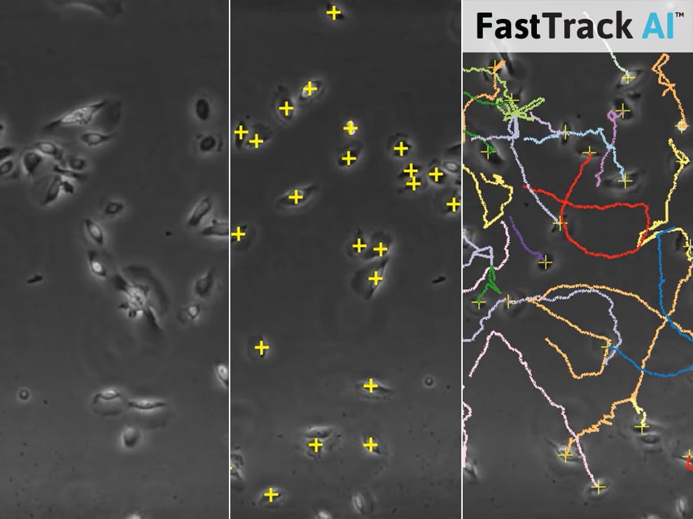
Save Time Using Automated Analysis of Chemotaxis Assays
In cooperation with MetaVi Labs, ibidi is launching FastTrack AI, an automated software solution that tracks and analyzes chemotaxis data. FastTrack AI helps save time on chemotaxis assays—no need for both manual tracking and fluorescence labeling. After sample preparation and live cell imaging (for example, using bright-field or phase contrast microscopy), scientists can simply upload their raw image data to the cloud and get objective and reproducible results within minutes.
ibidi
https://ibidi.com/img/cms/about_us/press/ibidi_PR_2020_07_Chemotaxis_AI.jpg

Scientific Volume Imaging Announces Huygens Software 20.04
Enjoy new features and options in a new version of Huygens 20.04, such as Huygens Everywhere, the ultimate flexible solution to use Huygens Essential or Localizer. Huygens can be used on any computer by installing Huygens and logging in with Huygens Everywhere account credentials at www.svi.nl.
Scientific Volume Imaging
https://svi.nl/Huygens-Essential

MuviCyte Provides Live Cell Imaging and Analysis
The MuviCyte live cell imaging system is used by researchers to analyze spheroid proliferation, scratch assays and migration, transfection rates, stem cell differentiation, embryogenesis, and more. The system operates inside an incubator to maintain cells under optimal conditions, keeping them healthy for weeks at a time. PC-controlled, MuviCyte allows observation of cells remotely, which helps to keep the culture chamber at optimum levels of temperature, CO2, and humidity.
Perkin Elmer
www.perkinelmer.com/product/muvicyte-live-cell-imaging-kit-hh40000000

AirFlow Monitor
The Hemco airflow monitor continuously monitors face velocity air flow for a fume hood. The monitor can be calibrated to the desired feet per minute velocity set point. If the hood face velocity falls below the set point, an audible alarm will sound, and a visual red indicator light will appear. Air flow alarms can be factory or field installed for improved laboratory safety.
Hemco Corporation

