Skip to main content Accessibility help
×
  • Cited by 1
Publisher:
Cambridge University Press
Online publication date:
April 2015
Print publication year:
2000
Online ISBN:
9781316167359

Book description

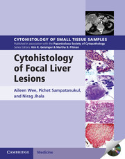
Each volume in this richly illustrated series, published in association with the Papanicolaou Society of Cytopathology, provides an organ-based approach to the cytologic and histologic diagnosis of small tissue samples. Benign, pre-malignant and malignant entities are presented in a well-organized and standardized format, with high-resolution color photomicrographs, tables, and lists of key specific morphologic criteria. Example vignettes allow the reader to assimilate the diagnostic principles in a case-based format. This volume provides comprehensive coverage of both surgical pathology and cytopathology of focal liver lesions. Extensively illustrated throughout, it contains key cytologic and histologic features, practical points, radiologic and morphologic pictures, flow charts, and tabulated summaries for easy comprehensive overview and quick reference and provides a pragmatic algorithmic approach to cytohistologic diagnosis. With over 700 printed photomicrographs and a CD-ROM offering all images in a downloadable format, this is an important resource for all anatomic pathologists.

Refine List

Actions for selected content:

Select all | Deselect all
  • View selected items
  • Export citations
  • Download PDF (zip)
  • Save to Kindle
  • Save to Dropbox
  • Save to Google Drive

Save Search

You can save your searches here and later view and run them again in "My saved searches".

Please provide a title, maximum of 40 characters.
×

Contents

Metrics

Full text views

Total number of HTML views: 0
Total number of PDF views: 0 *
Loading metrics...

Book summary page views

Total views: 0 *
Loading metrics...

* Views captured on Cambridge Core between #date#. This data will be updated every 24 hours.

Usage data cannot currently be displayed.