We use cookies to distinguish you from other users and to provide you with a better experience on our websites. Close this message to accept cookies or find out how to manage your cookie settings.
To save content items to your account,
please confirm that you agree to abide by our usage policies.
If this is the first time you use this feature, you will be asked to authorise Cambridge Core to connect with your account.
Find out more about saving content to .
To save content items to your Kindle, first ensure no-reply@cambridge.org
is added to your Approved Personal Document E-mail List under your Personal Document Settings
on the Manage Your Content and Devices page of your Amazon account. Then enter the ‘name’ part
of your Kindle email address below.
Find out more about saving to your Kindle.
Note you can select to save to either the @free.kindle.com or @kindle.com variations.
‘@free.kindle.com’ emails are free but can only be saved to your device when it is connected to wi-fi.
‘@kindle.com’ emails can be delivered even when you are not connected to wi-fi, but note that service fees apply.
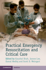
Acute resuscitation and care of unstable and critically ill patients can be a daunting experience for all trainees in the emergency department or the intensive care unit. The practical, easy-to-read and evidence-based information in Practical Emergency Resuscitation and Critical Care will help all physicians understand and begin management of these patients. This book offers the collaborative expertise of dozens of critical care physicians from different specialities, including but not limited to: emergency medicine, surgery, medicine and anaesthesia. Divided into sections by medical entities, it covers essential topics that are likely to be encountered in the emergency department where critical care often begins. The portable format and bullet point style content allows all practitioners instant access to the principle information that is necessary for the diagnosis and management of critical care patients.
This chapter discusses the diagnosis, evaluation and management of neutropenic fever. The initial presentation of the critically ill with neutropenic fever may be overt with a clinical presentation similar to that of septic shock and including hypotension, respiratory failure, or any other major organ dysfunction. It may also be cryptogenic with isolated confusion, coagulopathy, or cardiac arrhythmias. Elderly patients and those taking steroids may present as hypothermic or euthermic. Any unexplained acute clinical deterioration should be considered a fever equivalent. Critically ill patients with neutropenic fever will most frequently present with common infections. However, their immunocompromised state places them at risk for more complex disease processes of almost any organ system. The pathophysiology of the decompensating patient with neutropenic fever is similar to that of a patient in septic shock. Those patients should be resuscitated similarly by the rapid and aggressive administration of crystalloids.
This chapter discusses the management of trauma. The primary survey for a trauma patient is performed with regard to airway, breathing and circulation. Several airway adjuncts are available to assist in endotracheal intubation, including the gum elastic bougie, supraglottic airway devices, videolaryngoscopy, or fiberoptic scopes. The breathing evaluation include visualization of chest rise, auscultation of breath sounds, palpation of the chest wall feeling for crepitus or flail segments, and assuring that the trachea is midline. The patient is examined for signs of hemorrhage, including all compartments that can hold life-threatening amounts of blood loss. Evaluation for disability in the primary survey should include Glasgow Coma Scale (GCS), neurological examination to rule out neurological deficit, and pupil examination for signs of intracranial injury. Emergency department thoracotomy (EDT) is a resuscitative procedure that has low survival rate and should be performed in unique circumstances.
This chapter discusses the diagnosis, evaluation and management of rhabdomyolysis. Physical examination of a patient with rhabdomyolysis may reveal muscle swelling and tenderness, with occasional skin changes including discoloration, induration, and blistering. It is possible for rhabdomyolysis to present without any of these signs or symptoms, making serum markers essential to the diagnosis. Severe cases may present with hypovolemic shock, acute kidney injury (AKI), metabolic acidosis, disseminated intravascular coagulation (DIC), compartment syndrome, hyperkalemia, and cardiac arrhythmias. Compartment syndrome occurs due to swelling and edema of the injured muscle: classic physical examination findings include pain, paresthesias, paralysis, pallor, and pulselessness. The cornerstone of management includes discontinuation of inciting factors and aggressive management of fluid and electrolyte abnormalities. Intravenous fluids enhance renal perfusion and increase urinary flow in order to prevent AKI and increase potassium excretion.
This chapter discusses the diagnosis, evaluation and management of renal emergencies including metabolic acidosis, metabolic alkalosis, respiratory acidosis and respiratory alkalosis. The symptoms depend on the severity and etiology of the underlying acidosis, and are often nonspecific. Altered mental status, weakness, nausea, and abdominal pain are common. Hyperkalemia is often present due to transcellular shift of K-plus out of cells and H-plus into cells. Kussmaul respirations are classically associated with diabetic ketoacidosis (DKA), and refer to rapid, deep breathing. The critical presentation includes extreme acidemia that leads to neurological dysfunction (severe obtundation, coma, and seizures) as well as cardiovascular complications (arrhythmias, decreased cardiac contractility, arteriolar vasodilation, and decreased responsiveness to catecholamines). Profound hypotension and shock can result, which can complicate management since hypotension and shock are often the cause of the acidosis. If acidosis is due to DKA, treatment requires insulin and IV fluid resuscitation.
This chapter discusses the diagnosis, evaluation and management of bradyarrhythmias. Sinus bradycardia occurs in 15-20% of patients with acute myocardial infarction secondary to ischemia of the sinoatrial (SA) node. Syncope may result from primary dysrhythmia or from reduced cardiac output. A 12-lead electrocardiogram (ECG) is essential for the diagnosis of bradycardia and to differentiate between the different types of bradyarrhythmias. History should focus particularly on symptoms of ischemic heart disease, and on medications such as nodal blockers. As the bradycardia worsens, cardiac output decreases as well. This results in hypotension and hypoperfusion that need to be corrected using medications or a pacemaker. In addition, a decrease in cardiac output can result in pulmonary edema. Even with the development of pulmonary edema, the bradycardia is the first thing that needs to be addressed. Treatment of the cardiogenic pulmonary edema can be instituted afterward.
This chapter discusses the diagnosis, evaluation and management of neck trauma. It presents special considerations with regard to immobilization and the safety of removing the cervical collar for penetrating neck trauma. High-resolution computed tomography angiography (CTA) is the initial diagnostic study of choice in the stable patient with penetrating neck trauma or blunt neck trauma when blunt cerebrovascular injury is suspected. CTA can be the initial diagnostic study of choice regardless of zone of injury. CTA is particularly useful for zone I and III penetrating injuries, which are more difficult to evaluate by physical examination. Unstable patients with penetrating injuries require immediate surgical consultation and exploration in the OR. Unstable patients include those patients with hard signs: clear airway injury (air bubbling through wound), hemodynamic instability despite resuscitation, uncontrolled bleeding (including expanding hematoma), or evolving neurological deficit.
This chapter discusses the diagnosis, evaluation and management of shock. It presents special circumstances which make diagnosis and management of shock difficult in pediatric and pregnant patients. Shock should be suspected when patients present with a constellation of signs including ill-appearance, tachycardia, tachypnea, hypotension, and oliguria. The principles of shock management include specific therapy for treating the underlying cause, and general therapy to manage the shock syndrome. Recognition of shock is difficult due to variations in age-dependent vital signs, difficulty in assessing mental status, and the non-specificity of early manifestations of shock such as irritability and poor feeding. Elderly patients experience significantly more morbidity and mortality from all causes of shock due to their limited ability to augment cardiac output and maintain vascular tone. Elderly patients often have multiple comorbidities or use multiple medications that distort the diagnosis and management of shock.
This chapter discusses the diagnosis, evaluation and management of fulminant hepatic failure. Patients can present with hypotension due to generalized systemic inflammatory response, coagulopathy, and encephalopathy with progression to coma and brain herniation. Diagnosis is based on clinical presentation and laboratory findings. Critical care management should be performed for cardiovascular, pulmonary, and infectious complications and other comorbidities. Prognosis and treatment decisions can be based on Rumack-Matthew nomogram. Renal failure occurs in up to 50% of cases, even more frequently in acetaminophen toxicity. In most cases continuous renal replacement therapy is indicated. Intermittent hemodialysis should be avoided as some evidence suggests rapid fluid shifts lead to brain herniation. Patients should be transferred to a transplantation facility as soon as possible. If a transplantation center is not readily available, consider transfer to a center that utilizes molecular adsorbents recirculation system (MARS) or artificial extracorporeal liver support therapy.
This chapter discusses the diagnosis, evaluation and management of asthma. Airflow restriction may be severe, leading to asthma patients presenting in an upright or tripod position, with cyanosis, altered mental status, and respiratory arrest. Asthma exacerbations initially produce tachypnea and a resultant low carbon dioxide level; a normal or elevated carbon dioxide level may indicate fatigue and impending respiratory failure. Laboratory testing and ECG should be used to differentiate asthma exacerbations from alternative etiologies or comorbid conditions. Patients should be placed on supplemental oxygen therapy as needed to maintain adequate oxygen saturations. Patients must be monitored for signs of impending respiratory failure. Constant positive airway pressure (CPAP) and bi-level positive airway pressure (Bi-PAP) may be considered for patients with severe asthma. The goal of ventilator management in asthmatic is to oxygenate and ventilate without worsening hyperinflation, which causes barotrauma and hemodynamic instability.
This chapter discusses the management of trauma. The primary survey for a trauma patient is performed with regard to airway, breathing and circulation. Several airway adjuncts are available to assist in endotracheal intubation, including the gum elastic bougie, supraglottic airway devices, videolaryngoscopy, or fiberoptic scopes. The breathing evaluation include visualization of chest rise, auscultation of breath sounds, palpation of the chest wall feeling for crepitus or flail segments, and assuring that the trachea is midline. The patient is examined for signs of hemorrhage, including all compartments that can hold life-threatening amounts of blood loss. Evaluation for disability in the primary survey should include Glasgow Coma Scale (GCS), neurological examination to rule out neurological deficit, and pupil examination for signs of intracranial injury. Emergency department thoracotomy (EDT) is a resuscitative procedure that has low survival rate and should be performed in unique circumstances.
This chapter discusses the diagnosis, evaluation and management of intracranial hemorrhage. It describes types of intracranial hemorrhage, including subarachnoid hemorrhage (SAH), subdural hemorrhage (SDH), epidural hemorrhage (EDH) and intracerebral hemorrhage (ICH). Emergent non-contrast head CT is the cornerstone for detection of ICH. MRI is equally effective in identifying ICH and better at detecting predisposing underlying parenchymal or vascular anomalies. Emergency department management focuses on protecting cerebral perfusion by balancing the forces of mean arterial pressure (MAP) and intracranial pressure (ICP). Patients should be positioned with the head of the bed elevated to 30 degrees to support cerebral venous drainage to reduce ICP. The main goal of emergency management is to temporize ICP changes, avoid secondary insults (e.g., hypoxia and hypotension) and protect cerebral perfusion pressure (CPP) while expediting neurosurgical evaluation for possible life-saving surgical intervention.
This chapter discusses the diagnosis, evaluation and management of pulmonary embolism (PE). The evaluation for suspected PE is tailored to the level of the clinician's suspicion for this diagnosis based on the patient's history, physical examination, and risk factors. A chest radiograph is rarely diagnostic for PE, but can identify alternative diagnoses. Hampton's hump, a pleural-based, wedge-shaped area of infiltrate, can be seen in pulmonary infarction and is suggestive of PE. Patients diagnosed with PE should be started on anticoagulation unless otherwise contraindicated to prevent clot propagation. Patients with a high clinical probability of PE should be started on anticoagulation therapy while awaiting diagnostic confirmation. The most common causes for sudden decompensation are respiratory and hemodynamic as the result of sudden shift or increase in clot burden. Intubation may be necessary to improve oxygenation/ventilation and establish control of the airway of the patient with PE.