Hostname: page-component-78c5997874-8bhkd Total loading time: 0 Render date: 2024-10-31T23:03:00.189Z Has data issue: false hasContentIssue false

A Bottom-Up Volume Reconstruction Method for Atom Probe Tomography

Published online by Cambridge University Press:  28 January 2022

Yu-Ting Ling*
Affiliation:
Imec Vision Lab, University of Antwerp, Universiteitsplein 1, 2610 Antwerp, Belgium
Siegfried Cools
Affiliation:
Applied Mathematics Group, University of Antwerp, Middelheimlaan 1, 2020 Antwerp, Belgium
Janusz Bogdanowicz
Affiliation:
Imec vzw, Kapeldreef 75, 3001 Heverlee, Belgium
Claudia Fleischmann
Affiliation:
Imec vzw, Kapeldreef 75, 3001 Heverlee, Belgium
Jan De Beenhouwer
Affiliation:
Imec Vision Lab, University of Antwerp, Universiteitsplein 1, 2610 Antwerp, Belgium
Jan Sijbers
Affiliation:
Imec Vision Lab, University of Antwerp, Universiteitsplein 1, 2610 Antwerp, Belgium
Wilfried Vandervorst
Affiliation:
Imec vzw, Kapeldreef 75, 3001 Heverlee, Belgium Quantum Solid-State Physics, KU Leuven, Celestijnenlaan 200D, 3001 Leuven, Belgium
*
*Corresponding author: Yu-Ting Ling, E-mail: yu-ting.ling@uantwerpen.be
Get access

Abstract

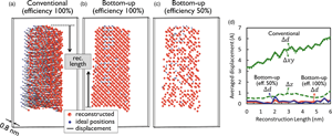
This paper describes a reconstruction method for atom probe tomography based on a bottom-up approach accounting for (i) the final tip morphology (which is frequently induced by inhomogeneous evaporation probabilities across the tip surface due to laser absorption, heat diffusion effects, and inhomogeneous material properties), (ii) the limited (and changing) field of view, and (iii) the detector efficiency. The reconstruction starts from the final tip morphology and reverses the evaporation sequence through the pseudo-deposition of defined small reconstruction volumes, which are then stacked together to create the full three-dimensional (3D) tip. The subdivision in small reconstruction volumes allows the scheme to account for the changing tip shape and field of view as evaporation proceeds. Atoms within the same small reconstruction volume are reconstructed at once by placing atoms back onto their possible lattice sites through a trajectory-matching process involving simulated and experimental hit maps. As the ejected ion trajectories are simulated using detailed electrostatic modeling inside the chamber, no simplifications have been imposed on the shape of the trajectories, projection laws, or tip surface. We demonstrate the superior performance of our approach over the conventional reconstruction method (Bas) for an asymmetrical tip shape.

Type
Development and Computation
Copyright
Copyright © The Author(s), 2022. Published by Cambridge University Press on behalf of the Microscopy Society of America

Access options

Get access to the full version of this content by using one of the access options below. (Log in options will check for institutional or personal access. Content may require purchase if you do not have access.)

References

Baik, SI, Isheim, D & Seidman, DN (2018). Systematic approaches for targeting an atom-probe tomography sample fabricated in a thin TEM specimen: Correlative structural, chemical and 3-D reconstruction analyses. Ultramicroscopy 184, 284292.CrossRefGoogle Scholar
Barnes, J, Grenier, A, Mouton, I, Barraud, S, Audoit, G, Bogdanowicz, J, Fleischmann, C, Melkonyan, D, Vandervorst, W & Duguay, S (2018). Atom probe tomography for advanced nanoelectronic devices: Current status and perspectives. Scr Mater 148, 9197.CrossRefGoogle Scholar
Bas, P, Bostel, A, Deconihout, B & Blavette, D (1995). A general protocol for the reconstruction of 3D atom probe data. Appl Surf Sci 87, 298304.CrossRefGoogle Scholar
Beinke, D, Oberdorfer, C & Schmitz, G (2016). Towards an accurate volume reconstruction in atom probe tomography. Ultramicroscopy 165, 3441.CrossRefGoogle ScholarPubMed
Beinke, D & Schmitz, G (2018). Atom probe reconstruction with a locally varying emitter shape. Microsc Microanal 25(2), 280287.CrossRefGoogle ScholarPubMed
Blavette, D, Cadel, E, Cojocaru-Mirédin, O & Deconihout, B (2010). The investigation of boron-doped silicon using atom probe tomography. In IOP Conference Series: Materials Science and Engineering, p. 012004. IOP Publishing.Google Scholar
Bogdanowicz, J, Gilbert, M, Innocenti, N, Koelling, S, Vanderheyden, B & Vandervorst, W (2013). Light absorption in conical silicon particles. Opt Express 21(3), 38913896.CrossRefGoogle ScholarPubMed
Fleischmann, C, Paredis, K, Melkonyan, D & Vandervorst, W (2018). Revealing the 3-dimensional shape of atom probe tips by atomic force microscopy. Ultramicroscopy 194, 221226.CrossRefGoogle ScholarPubMed
Fletcher, C, Moody, MP & Haley, D (2019). Fast modelling of field evaporation in atom probe tomography using level set methods. J Phys D: Appl Phys 52(43), 435305.CrossRefGoogle Scholar
Fletcher, C, Moody, MP & Haley, D (2020). Towards model-driven reconstruction in atom probe tomography. J Phys D: Appl Phys 53(47), 475303.CrossRefGoogle Scholar
Gault, B, Haley, D, de Geuser, F, Moody, MP, Marquis, EA, Larson, DJ & Geiser, BP (2011). Advances in the reconstruction of atom probe tomography data. Ultramicroscopy 111(6), 448457.CrossRefGoogle ScholarPubMed
Gault, B, Moody, MP, De Geuser, F, Tsafnat, G, La Fontaine, A, Stephenson, LT, Haley, D & Ringer, SP (2009). Advances in the calibration of atom probe tomographic reconstruction. J Appl Phys 105(3), 034913.CrossRefGoogle Scholar
Geiser, BP, Kelly, TF, Larson, DJ, Schneir, J & Roberts, JP (2007). Spatial distribution maps for atom probe tomography. Microsc Microanal 13, 437447.CrossRefGoogle ScholarPubMed
Ghysels, P & Vanroose, W (2014). Hiding global synchronization latency in the preconditioned conjugate gradient algorithm. Parallel Comput 40(7), 224238.CrossRefGoogle Scholar
Giddings, AD, Koelling, S, Shimizu, Y, Estivill, R, Inoue, K, Vandervorst, W & Yeoh, WK (2018). Industrial application of atom probe tomography to semiconductor devices. Scr Mater 148, 8290.CrossRefGoogle Scholar
Gruber, M, Vurpillot, F, Bostel, A & Deconihout, B (2011). Field evaporation: A kinetic Monte Carlo approach on the influence of temperature. Surf Sci 605(23–24), 20252031.CrossRefGoogle Scholar
Haley, D, Moody, MP & Smith, GD (2013). Level set methods for modelling field evaporation in atom probe. Microsc Microanal 19(6), 17091717.CrossRefGoogle ScholarPubMed
Hatzoglou, C, Da Costa, G & Vurpillot, F (2019). Enhanced dynamic reconstruction for atom probe tomography. Ultramicroscopy 197, 7282.CrossRefGoogle ScholarPubMed
Hatzoglou, C & Vurpillot, F (2019). A mesoscopic field evaporation model. Microsc Microanal 25(S2), 286287.CrossRefGoogle Scholar
Hestenes, MR & Stiefel, E (1952). Methods of conjugate gradients for solving linear systems. J Res Natl Bur Stand 49(6), 409436.CrossRefGoogle Scholar
Houard, J, Vella, A, Vurpillot, F & Deconihout, B (2010). Optical near-field absorption at a metal tip far from plasmonic resonance. Phys Rev B 81(12), 125411.CrossRefGoogle Scholar
Kambham, AK, Kumar, A, Gilbert, M & Vandervorst, W (2013). 3D site specific sample preparation and analysis of 3D devices (FinFETs) by atom probe tomography. Ultramicroscopy 132, 6569.CrossRefGoogle ScholarPubMed
Kambham, AK, Mody, J, Gilbert, M, Koelling, S & Vandervorst, W (2011). Atom-probe for FinFET dopant characterization. Ultramicroscopy 111(6), 535539.CrossRefGoogle ScholarPubMed
Kellogg, GL (1984). Measurement of activation energies for field evaporation of tungsten ions as a function of electric field. Phys Rev B 29(8), 43044312.CrossRefGoogle Scholar
Kelly, TF & Larson, DJ (2012). Atom probe tomography 2012. Annu Rev Mater Res 42, 131.CrossRefGoogle Scholar
Kelly, TF, Vella, A, Bunton, JH, Houard, J, Silaeva, EP, Bogdanowicz, J & Vandervorst, W (2014). Laser pulsing of field evaporation in atom probe tomography. Curr Opin Solid State Mater Sci 18(2), 8189.CrossRefGoogle Scholar
Koelling, S, Innocenti, N, Bogdanowicz, J & Vandervorst, W (2013). Optimal laser positioning for laser-assisted atom probe tomography. Ultramicroscopy 132, 7074.CrossRefGoogle ScholarPubMed
Koelling, S, Innocenti, N, Schulze, A, Gilbert, M, Kambham, A & Vandervorst, W (2011). In-situ observation of non-hemispherical tip shape formation during laser-assisted atom probe tomography. J Appl Phys 109(10), 104909.CrossRefGoogle Scholar
Kolli, RP (2018). Atom probe tomography: A review of correlative analysis of interfaces and precipitates in metals and alloys. JOM 70(9), 17251735.CrossRefGoogle Scholar
Kühbach, M, Breen, A, Herbig, M & Gault, B (2019). Building a library of simulated atom probe data for different crystal structures and tip orientations using TAPSim. Microsc Microanal 25(2), 320330.CrossRefGoogle ScholarPubMed
Larson, DJ, Gault, B, Geiser, BP, De Geuser, F & Vurpillot, F (2013). Atom probe tomography spatial reconstruction: Status and directions. Curr Opin Solid State Mater Sci 17(5), 236247.CrossRefGoogle Scholar
Larson, DJ, Geiser, BP, Prosa, TJ, Gerstl, S, Reinhard, DA & Kelly, TF (2011). Improvements in planar feature reconstructions in atom probe tomography. J Microsc 243(1), 1530.CrossRefGoogle ScholarPubMed
Larson, DJ, Geiser, BP, Prosa, TJ & Kelly, TF (2012). On the use of simulated field-evaporated specimen apex shapes in atom probe tomography data reconstruction. Microsc Microanal 18(5), 953963.CrossRefGoogle ScholarPubMed
Lefebvre, W, Hernandez-Maldonado, D, Moyon, F, Cuvilly, F, Vaudolon, C, Shinde, D & Vurpillot, F (2015). HAADF–STEM atom counting in atom probe tomography specimens: Towards quantitative correlative microscopy. Ultramicroscopy 159, 403412.CrossRefGoogle ScholarPubMed
Loi, ST, Gault, B, Ringer, SP, Larson, DJ & Geiser, BP (2013). Electrostatic simulations of a local electrode atom probe: The dependence of tomographic reconstruction parameters on specimen and microscope geometry. Ultramicroscopy 132, 107113.CrossRefGoogle ScholarPubMed
Martin, AJ, Wei, Y & Scholze, A (2018). Analyzing the channel dopant profile in next-generation FinFETs via atom probe tomography. Ultramicroscopy 186, 104111.CrossRefGoogle ScholarPubMed
Menand, A & Kingham, DR (1984). Isotopic variations in field evaporation charge-state of boron ions. J Phys D: Appl Phys 17(1), 203208.CrossRefGoogle Scholar
Menand, A & Kingham, DR (1985). Evidence for the quantum mechanical tunnelling of boron ions. J Phys C: Solid State Phys 18(23), 45394547.CrossRefGoogle Scholar
Miller, M & Forbes, R (2014). Atom-Probe Tomography: The Local Electrode Atom Probe. New York: Springer US.CrossRefGoogle Scholar
Mouton, I, Printemps, T, Grenier, A, Gambacorti, N, Pinna, E, Tiddia, M, Vacca, A & Mula, G (2017). Toward an accurate quantification in atom probe tomography reconstruction by correlative electron tomography approach on nanoporous materials. Ultramicroscopy 182, 112117.CrossRefGoogle ScholarPubMed
Oberdorfer, C, Eich, SM & Schmitz, G (2013). A full-scale simulation approach for atom probe tomography. Ultramicroscopy 128, 5567.CrossRefGoogle ScholarPubMed
Oberdorfer, C & Schmitz, G (2011). On the field evaporation behavior of dielectric materials in three-dimensional atom probe: A numeric simulation. Microsc Microanal 17(1), 1525.CrossRefGoogle ScholarPubMed
Oberdorfer, C, Withrow, T, Yu, LJ, Fisher, K, Marquis, EA & Windl, W (2018). Influence of surface relaxation on solute atoms positioning within atom probe tomography reconstructions. Mater Charact 146, 324335.CrossRefGoogle Scholar
Rolland, N, Larson, DJ, Geiser, BP, Duguay, S, Vurpillot, F & Blavette, D (2015 a). An analytical model accounting for tip shape evolution during atom probe analysis of heterogeneous materials. Ultramicroscopy 159, 195201.CrossRefGoogle ScholarPubMed
Rolland, N, Vurpillot, F, Duguay, S & Blavette, D (2015 b). A meshless algorithm to model field evaporation in atom probe tomography. Microsc Microanal 21(6), 16491656.CrossRefGoogle ScholarPubMed
Rolland, N, Vurpillot, F, Duguay, S, Mazumder, B, Speck, JS & Blavette, D (2017). New atom probe tomography reconstruction algorithm for multilayered samples: Beyond the hemispherical constraint. Microsc Microanal 23(2), 247254.CrossRefGoogle ScholarPubMed
Saad, Y (2003). Iterative Methods for Sparse Linear Systems, 2nd ed. Philadelphia, PA: SIAM.CrossRefGoogle Scholar
Shewchuk, JR (1994). An introduction to the Conjugate Gradient Method Without the Agonizing Pain. Pittsburgh, PA: Carnegie-Mellon University. Department of Computer Science.Google Scholar
Thompson, K, Sebastian, J & Gerstl, S (2007). Observations of Si field evaporation. Ultramicroscopy 107(2–3), 124130.CrossRefGoogle ScholarPubMed
Tu, Y, Takamizawa, H, Han, B, Shimizu, Y, Inoue, K, Toyama, T, Yano, F, Nishida, A & Nagai, Y (2017). Influence of laser power on atom probe tomographic analysis of boron distribution in silicon. Ultramicroscopy 173, 5863.CrossRefGoogle ScholarPubMed
Vella, A (2013). On the interaction of an ultra-fast laser with a nanometric tip by laser assisted atom probe tomography: A review. Ultramicroscopy 132, 518.CrossRefGoogle ScholarPubMed
Vurpillot, F, Bostel, A & Blavette, D (1999). The shape of field emitters and the ion trajectories in three-dimensional atom probes. J Microsc 196, 332336.CrossRefGoogle ScholarPubMed
Vurpillot, F, Gault, B, Geiser, BP & Larson, D (2013). Reconstructing atom probe data: A review. Ultramicroscopy 132, 1930.CrossRefGoogle ScholarPubMed
Vurpillot, F, Houard, J, Vella, A & Deconihout, B (2009). Thermal response of a field emitter subjected to ultra-fast laser illumination. J Phys D: Appl Phys 42(12), 125502.CrossRefGoogle Scholar
Xu, Z, Li, D, Xu, W, Devaraj, A, Colby, R, Thevuthasan, S, Geiser, B & Larson, DJ (2015). Simulation of heterogeneous atom probe tip shapes evolution during field evaporation using a level set method and different evaporation models. Comput Phys Commun 189, 106113.CrossRefGoogle Scholar
Zschiesche, H, Campos, APC, Dominici, C, Roussel, L, Charai, A, Mangelinck, D & Alfonso, C (2019). Correlated TKD/EDS-TEM-APT analysis on selected interfaces of CoSi2 thin films. Ultramicroscopy 206, 112807.CrossRefGoogle ScholarPubMed