Skip to main content Accessibility help
×
Hostname: page-component-848d4c4894-4rdrl Total loading time: 0 Render date: 2024-06-17T12:22:40.347Z Has data issue: false hasContentIssue false

Chapter 7 - Cerebral perfusion imaging by exogenous contrast agents

from Section 1 - Physiological MR techniques

Published online by Cambridge University Press:  05 March 2013

Jonathan H. Gillard
Affiliation:
University of Cambridge
Adam D. Waldman
Affiliation:
Imperial College London
Peter B. Barker
Affiliation:
The Johns Hopkins University School of Medicine
Get access

Summary

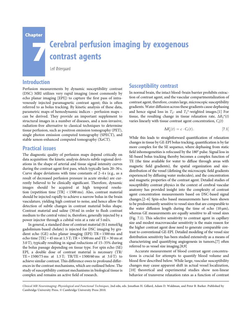
Image of the first page of this content. For PDF version, please use the ‘Save PDF’ preceeding this image.'
Type
Chapter
Information
Clinical MR Neuroimaging
Physiological and Functional Techniques
, pp. 86 - 93
Publisher: Cambridge University Press
Print publication year: 2009

Access options

Get access to the full version of this content by using one of the access options below. (Log in options will check for institutional or personal access. Content may require purchase if you do not have access.)

References

Villringer, A, Rosen, BR, Belliveau, JW, et al. Dynamic imaging with lanthanide chelates in normal brain: contrast due to magnetic susceptibility effects. Magn Reson Med 1988; 6: 164–174.CrossRefGoogle ScholarPubMed
Fisel, CR, Ackerman, JL, Buxton, RB, et al. MR contrast due to microscopically heterogeneous magnetic susceptibility: numerical simulations and applications to cerebral physiology. Magn Reson Med 1991; 17: 336–347.CrossRefGoogle ScholarPubMed
Weisskoff, RM, Zuo, CS, Boxerman, JL, Rosen, BR.Microscopic susceptibility variation and transverse relaxation: theory and experiment. Magn Reson Med 1994; 31: 601–610.CrossRefGoogle ScholarPubMed
Boxerman, JL, Hamberg, LM, Rosen, BR, Weisskoff, RM.MR contrast due to intravascular magnetic susceptibility perturbations. Magn Reson Med 1995; 34: 555–566.CrossRefGoogle ScholarPubMed
Kiselev, VG, Posse, S.Analytical theory of susceptibility induced NMR signal dephasing in a cerebrovascular network. Phys Rev Lett 1998; 81: 5696–5699.CrossRefGoogle Scholar
Kiselev, VG, Posse, S.Analytical model of susceptibility-induced MR signal dephasing: effect of diffusion in a microvascular network. Magn Reson Med 1999; 41: 499–509.3.0.CO;2-O>CrossRefGoogle Scholar
Dennie, J, Mandeville, JB, Boxerman, JL, et al. NMR imaging of changes in vascular morphology due to tumor angiogenesis. Magn Reson Med 1998; 40: 793–799.CrossRefGoogle ScholarPubMed
Kiselev, VG, Strecker, R, Ziyeh, S, Speck, O, Hennig, J.Vessel size imaging in humans. Magn Reson Med 2005; 53: 553–63.CrossRefGoogle ScholarPubMed
Troprès, I, Grimault, S, Vaeth, A, et al. Vessel size imaging. Magn Reson Med 2001; 45: 397–408.3.0.CO;2-3>CrossRefGoogle ScholarPubMed
Rausch, M, Scheffler, K, Rudin, M, Radü, EW.Analysis of input functions from different arterial branches with gamma variate functions and cluster analysis for quantitative blood volume measurements. Magn Reson Imaging 2000; 18: 1235–1243.CrossRefGoogle ScholarPubMed
Kiselev, VG. On the theoretical basis of perfusion measurements by dynamic susceptibility contrast MRI. Magn Reson Med 2001; 46: 1113–1122.CrossRefGoogle ScholarPubMed
van Osch, M, Vonken, E, Viergever, M, van der Grond, J, Bakker, C.Measuring the arterial input function with gradient echo sequences. Magn Reson Med 2003; 49: 1067–1076.CrossRefGoogle ScholarPubMed
Akbudak, E, Kotys, M, Conturo, T.Quadraticity and hematocrit dependence of ΔR2* AIF signals at 3 T: a blood phantom study under physiologic conditions. In The ISMRM workshop on Quantitative Cerebral Perfusion Imaging Using MRI: A Technical Perspective, Venice, 2004, pp 10–11.Google Scholar
Kjølby, BF, Mikkelsen, IK, Pedersen, M, Østergaard, L, Kiselev, VG. Analysis of partial volume effects on arterial input functions using gradient echo: a simulation studyMagn Reson Med 2009; 61: 1300–1309.CrossRefGoogle ScholarPubMed
van Osch, MJ, Vonken, EJ, Bakker, CJ, Viergever, MA.Correcting partial volume artifacts of the arterial input function in quantitative cerebral perfusion MRI. Magn Reson Med 2001; 45: 477–485.3.0.CO;2-4>CrossRefGoogle ScholarPubMed
van Osch, MJP, van der Grond, J, Bakker, CJG.Partial volume effects on arterial input functions: shape and amplitude distortions and their correction. J Magn Reson Imaging 2005; 22: 704–709.CrossRefGoogle ScholarPubMed
Chen, JJ, Smith, MR, Frayne, R.The impact of partial-volume effects in dynamic susceptibility contrast magnetic resonance perfusion imaging. J Magn Reson Imaging 2005; 22: 390–399.CrossRefGoogle ScholarPubMed
Duhamel, G, Schlaug, G, Alsop, DC.Measurement of arterial input functions for dynamic susceptibility contrast magnetic resonance imaging using echoplanar images: comparison of physical simulations with in vivo results. Magn Reson Med 2006; 55: 514–523.CrossRefGoogle ScholarPubMed
Calamante, F, Vonken, EPA, van Osch, MJP.Contrast agent concentration measurements affecting quantification of bolus-tracking perfusion MRI. Magn Reson Med 2007; 58: 544–553.CrossRefGoogle ScholarPubMed
Jochimsen, TH, Newbould, RD, Skare, ST, et al. Identifying systematic errors in quantitative dynamic-susceptibility contrast perfusion imaging by high-resolution multi-echo parallel EPI. NMR Biomed 2007; 20: 429–438.CrossRefGoogle ScholarPubMed
Larsson, HB, Hansen, AE, Berg, HK, Rostrup, E, Haraldseth, O.Dynamic contrast-enhanced quantitative perfusion measurement of the brain using T1-weighted MRI at 3 T. J Magn Reson Imaging 2008; 27: 754–762.CrossRefGoogle Scholar
Rosen, BR, Belliveau, JW, Aronen, HJ, et al. Susceptibility contrast imaging of cerebral blood volume: human experience. Magn Reson Med 1991; 22: 293–299.CrossRefGoogle ScholarPubMed
Rosen, BR, Belliveau, JW, Buchbinder, BR, et al. Contrast agents and cerebral hemodynamics. Magn Reson Med 1991; 19: 285–292.CrossRefGoogle ScholarPubMed
Rosen, BR, Belliveau, JW, Vevea, JM, Brady, TJ.Perfusion imaging with NMR contrast agents. Magn Reson Med 1990; 14: 249–265.CrossRefGoogle ScholarPubMed
Belliveau, JW, Kennedy-, DNJ, McKinstry, RC, et al. Functional mapping of the human visual cortex by magnetic resonance imaging. Science 1991; 254: 716–719.CrossRefGoogle ScholarPubMed
Meier, P, Zierler, KL.On the theory of the indicator-dilution method for measurement of blood flow and volume. J Appl Phys 1954; 6: 731–744.Google ScholarPubMed
Thompson, HK, Starmer, F, Whalen, RE, McIntosh, HD.Indicator transit time considered as a gamma variate. Circ Res 1964; 14: 502–515.CrossRefGoogle ScholarPubMed
Perkiö, JP, Aronen, HJ, Kangasmaki, A, et al. Evaluation of four postprocessing methods for determination of cerebral blood volume and mean transit time by dynamic susceptibility contrast imaging. Magn Reson Med 2002; 47: 973–981.CrossRefGoogle ScholarPubMed
Gobbel, GT, Fike, JR.A deconvolution method for evaluating indicator-dilution curves. Phys Med Biol 1994; 39: 1833–1854.CrossRefGoogle ScholarPubMed
Rempp, KA, Brix, G, Wenz, F, et al. Quantification of regional cerebral blood flow and volume with dynamic susceptibility contrast-enhanced MR imaging. Radiology 1994; 193: 637–641.CrossRefGoogle ScholarPubMed
Valentinuzzi, ME, Montaldo, VE.Discrete deconvolution. Med Biol Eng 1975; 13: 123–125.CrossRefGoogle ScholarPubMed
Bronikowski, TA, Dawson, CA, Linehan, JH.Model-free deconvolution techniques for estimating vascular transport functions. Int J Biomed Comput 1983; 14: 411–429.CrossRefGoogle ScholarPubMed
Østergaard, L, Weisskoff, RM, Chesler, DA, Gyldensted, C, Rosen, BR.High resolution measurement of cerebral blood flow using intravascular tracer bolus passages. Part I: mathematical approach and statistical analysis. Magn Reson Med 1996; 36: 715–725.CrossRefGoogle ScholarPubMed
Liu, HL, Pu, Y, Liu, Y, et al. Cerebral blood flow measurement by dynamic contrast MRI using singular value decomposition with an adaptive threshold. Magn Reson Med 1999; 167–172.3.0.CO;2-Q>CrossRefGoogle ScholarPubMed
Østergaard, L, Sorensen, AG, Kwong, KK, et al. High resolution measurement of cerebral blood flow using intravascular tracer bolus passages. Part II: experimental comparison and preliminary results. Magn Reson Med 1996; 36: 726–736.CrossRefGoogle ScholarPubMed
Østergaard, L, Johannsen, P, Høst-Poulsen, P, et al. Cerebral blood flow measurements by MRI bolus tracking: comparison with [15O]H2O PET in humans. J Cereb Blood Flow Metab 1998; 18: 935–940.CrossRefGoogle Scholar
Calamante, F, Gadian, DG, Connely, A.Delay and dispersion effects in dynamic susceptibility contrast MRI: simulations using singular value decomposition. Magn Reson Med 2000; 44: 466–473.3.0.CO;2-M>CrossRefGoogle ScholarPubMed
Wu, O, Østergaard, L, Koroshetz, WJ, et al. Effects of tracer arrival time on flow estimates in MR perfusion-weighted imaging. Magn Reson Med 2003; 50: 856–64.CrossRefGoogle ScholarPubMed
Wu, O, Østergaard, L, Weisskoff, RM, et al. Tracer arrival timing-insensitive technique or estimating flow in MR perfusion-weighted imaging using singular value decomposition with a block-circulant deconvolution matrix. Magn Reson Med 2003; 50: 100–110.CrossRefGoogle ScholarPubMed
Østergaard, L, Chesler, D, Weisskoff, RM, Sorensen, AG, Rosen, BR.Modeling cerebral blood flow and flow heterogeneity from magnetic resonance residue detection. J Cereb Blood Flow Metab 1999; 19: 690–699.CrossRefGoogle Scholar
Andersen, IK, Szymkowiak, A, Rasmussen, CE, et al. Perfusion quantification using Gaussian process deconvolution. Magn Reson Med 2002; 48: 351–361.CrossRefGoogle ScholarPubMed
Mouridsen, K, Friston, K, Hjort, N, et al. Bayesian estimation of cerebral perfusion using a physiological model of microvasculature. Neuroimage 2006; 33: 570–579.CrossRefGoogle ScholarPubMed
Vonken, EP, Beekman, FJ, Bakker, CJ, Viergever, MA.Maximum likelihood estimation of cerebral blood flow in dynamic susceptibility contrast MRI. Magn Reson Med 1999; 41: 343–350.3.0.CO;2-T>CrossRefGoogle ScholarPubMed
Calamante, F, Gadian, DG, Connelly, A.Quantification of bolus-tracking MRI: improved characterization of the tissue residue function using Tikhonov regularization. Magn Reson Med 2003; 50: 1237–1247.CrossRefGoogle ScholarPubMed
Østergaard, L, Sorensen, AG, Chesler, D, et al. Combined diffusion-weighted and perfusion-weighted flow heterogeneity magnetic resonance imaging in acute stroke. Stroke 2000; 31: 1097–1103.CrossRefGoogle ScholarPubMed
Simonsen, CZ, Røhl, L, Vestergaard-Poulsen, P, et al. Final infarct size after acute stroke: prediction with flow heterogeneity. Radiology 2002; 225: 269–275.CrossRefGoogle ScholarPubMed
Perkiö, J, Soinne, L, Østergaard, L, et al. Abnormal intravoxel cerebral blood flow heterogeneity in human ischemic stroke determined by dynamic susceptibility contrast magnetic resonance imaging. Stroke 2004; 36: 44–49.CrossRefGoogle ScholarPubMed
Weisskoff, RM, Chesler, D, Boxerman, JL, Rosen, BR.Pitfalls in MR measurement of tissue blood flow with intravascular tracers: which mean transit time?Magn Reson Med 1993; 29: 553–558.CrossRefGoogle ScholarPubMed
Mouridsen, K, Christensen, S, Gyldensted, L, Østergaard, L.Automatic selection of arterial input function using cluster analysis. Magn Reson Med 2006; 55: 524–531.CrossRefGoogle ScholarPubMed
Murase, K, Kikuchi, K, Miki, H, Shimizu, T, Ikezoe, J.Determination of arterial input function using fuzzy clustering for quantification of cerebral blood flow with dynamic susceptibility contrast-enhanced MR imaging. J Magn Reson Imaging 2001; 13: 797–806.CrossRefGoogle ScholarPubMed
Calamante, F, Mørup, M, Hansen, LK. Defining a local arterial input function for perfusion MRI using independent component analysis. Magn Reson Med 2004; 52: 789–797.CrossRefGoogle ScholarPubMed
Alsop, DC, Wedmid, A, Schlaug, G.Defining a local input function for perfusion quantification with bolus contrast MRI. In Proceedings of the Annual Meeting of the International Society for Magnetic Resonance in Medicine, New York, 2002, p. 659.Google Scholar
Christensen, S, Calamante, F, Hjort, N, et al. Inferring origin of vascular supply from tracer arrival timing patterns using bolus tracking MRI. J Magn Reson Imaging 2008; 27: 1371–81.CrossRefGoogle ScholarPubMed
Lorenz, C, Benner, T, Lopez, CJ, et al. Effect of using local arterial input functions on cerebral blood flow estimation. J Magn Reson Imaging 2006; 24: 57–65.CrossRefGoogle ScholarPubMed
Aronen, HJ, Gazit, IE, Louis, DN, et al. Cerebral blood volume maps of gliomas: comparison with tumor grade and histologic findings. Radiology 1994; 191: 41–51.CrossRefGoogle ScholarPubMed
Schmainda, KM, Rand, SD, Joseph, AM, et al. Characterization of a first-pass gradient-echo spin-echo method to predict brain tumor grade and angiogenesis. J Am Neuroradiol 2004; 25: 1524–1532.Google ScholarPubMed
Law, M, Young, RJ, Babb, JS, et al. Gliomas: predicting time to progression or survival with cerebral blood volume measurements at dynamic susceptibility-weighted contrast-enhanced perfusion MR imaging. Radiology 2008; 247: 490–498.CrossRefGoogle ScholarPubMed
Emblem, KE, Nedregaard, B, Nome, T, et al. Glioma grading by using histogram analysis of blood volume heterogeneity from MR-derived cerebral blood volume maps. Radiology 2008; 247: 808–817.CrossRefGoogle ScholarPubMed
Weisskoff, RM, Boxerman, JL, Sorensen, AG, et al. Simultaneous blood volume and permeability mapping using a single Gd-based contrast injection. In Proceedings of the Annual Meeting of the Society of Magnetic Resonance in Medicine, San Francisco, 1994; p. 279.Google Scholar
Johnson, G, Wetzel, S, Cha, S, et al. A new approach to measuring blood volume and vascular transfer constant from dynamic, contrast-enhanced MRI. Magn Res Med 2004; 51: 961–968.CrossRefGoogle Scholar
Law, M, Young, R, Babb, J, et al. Comparing perfusion metrics obtained from a single compartment versus pharmacokinetic modeling methods using dynamic susceptibility contrast-enhanced perfusion MR imaging with glioma grade. AJNR Am J Neuroradiol 2006; 27: 1975–82.Google ScholarPubMed
Boxerman, JL, Schmainda, KM, Weisskoff, RM.Relative cerebral blood volume maps corrected for contrast agent extravasation significantly correlate with glioma tumor grade, whereas uncorrected maps do not. AJNR Am J Neuroradiol 2006; 27: 859–867.Google ScholarPubMed
Quarles, CC, Ward, BD, Schmainda, KM.Improving the reliability of obtaining tumor hemodynamic parameters in the presence of contrast agent extravasation. Magnetic Resonance in Medicine, 2005: 53: 1307–16.CrossRefGoogle ScholarPubMed

Save book to Kindle

To save this book to your Kindle, first ensure coreplatform@cambridge.org is added to your Approved Personal Document E-mail List under your Personal Document Settings on the Manage Your Content and Devices page of your Amazon account. Then enter the ‘name’ part of your Kindle email address below. Find out more about saving to your Kindle.

Note you can select to save to either the @free.kindle.com or @kindle.com variations. ‘@free.kindle.com’ emails are free but can only be saved to your device when it is connected to wi-fi. ‘@kindle.com’ emails can be delivered even when you are not connected to wi-fi, but note that service fees apply.

Find out more about the Kindle Personal Document Service.

Available formats
×

Save book to Dropbox

To save content items to your account, please confirm that you agree to abide by our usage policies. If this is the first time you use this feature, you will be asked to authorise Cambridge Core to connect with your account. Find out more about saving content to Dropbox.

Available formats
×

Save book to Google Drive

To save content items to your account, please confirm that you agree to abide by our usage policies. If this is the first time you use this feature, you will be asked to authorise Cambridge Core to connect with your account. Find out more about saving content to Google Drive.

Available formats
×